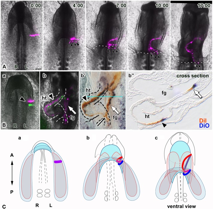
Fig. 5.
The heart tube and foregut coordinately form and lengthen through similar tissue dynamics. (A) Selected images from a time-lapse recording (Movie 7, ventral views), showing movements of a single stripe of labeled cells (magenta) in one heart primordium along with endodermal cells (arrowheads), accidentally marked when DiI was injected into the heart mesoderm. Dashed lines delineate the edge of the LHP. Labeled endodermal cells roll over the AIP into the foregut as the AIP descends, concomitant with folding of the LHP, demonstrating that movements of these two primordia are coupled. Scale bar: 200 µm. (B) Two stripes of labeled cells (magenta, DiI and rhodamine, anterior stripe; green, DiO and fluorescein, posterior stripe) at the time of injection (a, arrowhead) and after heart tube formation (b, arrowhead). Dyes were then immunostained in whole-mount embryos (b′) to enhance and preserve their labeling (rhodamine, brown; fluorescein, dark blue) during subsequent paraffin sectioning (b″). Labeled cells extended anteroposteriorly in both the heart tube (ht; arrowheads) and foregut (fg; white arrow); the black arrow marks an area where labeled cells in the heart and foregut overlap. (b″) Paraffin wax-embedded cross-section at the level shown by the blue line in b′. (C) Model of coordinated formation of the foregut and heart tube based on our results (A,B): the endoderm (blue) overlying the bilateral heart primordia (pink) converges toward the midline while diagonally folding, thereby reorienting its initial mediolateral polarity anterorposteriorly, coordinately with heart tube formation. These tubes elongate posteriorly together through CE. The red and blue lines in b and c represent simultaneously labeled cell stripes (a, purple line) in the heart mesoderm and in the endoderm, respectively.

