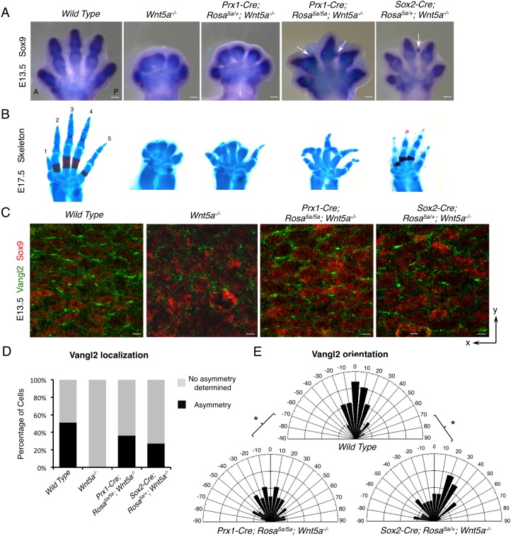
Fig. 4.
Non-graded Wnt5a expression partially rescued the PCP defects of the Wnt5a−/− embryo. (A) Sox9 whole-mount in situ hybridization in E13.5 mouse forelimbs. Arrows point to the ectopic cartilage. (B) Alizarin Red and Alcian Blue staining of forelimbs from E17.5 embryos with the genotypes indicated in A. Digits 1-5 are labeled. (C) Representative images of fluorescence immunostaining of Vangl2 (green) and Sox9 (red). x- and y-axes of the images are defined as shown in Fig. S13A. (D) The percentage of cells with discernible Vangl2 asymmetric localization in each genotype. (E) Schematics summarizing the quantification of orientation angles of Vangl2 in each genotype. x-axis, angle of orientation (−90° to 90°); y-axis, percentage of cells at angle x. Kolmogorov–Smirnov test, *P=0.0466 (wild type versus Prx1-Cre; Rosa5a/5a; Wnt5a−/−) and 0.0285 (wild type versus Sox2-Cre; Rosa5a/+; Wnt5a−/−). (D,E) Number of samples and number of cells analyzed for each genotype: wild type, N=2, n=230; Wnt5a−/−, N=2, n=188; Prx1-Cre; Rosa5a/5a; Wnt5a−/−, N=3, n=271; Sox2-Cre; Rosa5a/+; Wnt5a−/−, N=5, n=450. Scale bars: 200 μm (A); 10 μm (C).

