Abstract
In light of the slow progress in developing effective therapies for ischemic stroke, magnetic resonance imaging techniques have emerged as new tools in stroke clinical trials. Rapid imaging with magnetic resonance imaging, diffusion weighted imaging, perfusion imaging and angiography are being incorporated into phase II and phase III stroke trials to optimize patient selection based on positive imaging diagnosis of the ischemic pathophysiology specifically related to a drug's mechanism of action and as a direct biomarker of the effect of a treatment's effect on the brain.
Keywords: clinical trials, diffusion, magnetic resonance imaging, perfusion, stroke
Full text
The disappointingly slow progress in developing effective therapies for ischemic stroke has led to a re-evaluation of the strategies for stroke drug development and the methods used in clinical trials. Magnetic resonance imaging (MRI) techniques have been proposed and have begun to be used in stroke trials as a means of optimizing patient selection and as a direct measure of the effect of treatments on the brain.
One objective in all clinical trials is the selection of a sample that is sufficiently homogeneous to reduce the statistical variance of the data and thereby optimize the sensitivity of the design to detecting a therapeutic response, while remaining representative of the population of interest. Ischemic stroke trials have traditionally sought to limit the range of disease studied according to one or more of several dimensions, such as clinical severity at the time of enrollment, exclusion of non-ischemic causes for the clinical syndrome, lesion location and vascular territory, stroke mechanism, and co-morbidities. These dimensions have been assessed in the modern era of stroke clinical trials by clinical criteria at the bedside, usually aided by the exclusion of cerebral hemorrhage or other non-ischemic pathology by non-contrast computed tomography (CT) scan as the only imaging tool required. Except for the trials of intravenous recombinant tissue-plasminogen activator (rt-PA) in the treatment of ischemic stroke within the first 3 h [1], this traditional approach has lead to no approved therapies for stroke, and has lead to a great degree of pessimism with regard to thrombolysis beyond 3 h and with regard to the concept of neuroprotection in stroke.
Because several imaging modalities may provide more accurate and specific information than a clinical assessment and a normal CT scan, it has been proposed that positive imaging diagnoses would improve patient selection toward the goal of a more optimal target sample for stroke clinical trials, a sample selected based on an imaging diagnosis of a pathology that the drug is hypothesized to treat; for example, an arterial occlusion or perfusion defect for thrombolytic drugs. This principle has been supported by the results of the intra-arterial pro-urokinase stroke study, PROACT II [2]. Prior attempts to prove the efficacy of thrombolysis initiated between 3 and 6 h from onset without a positive diagnosis of arterial occlusion or perfusion defect have not been successful [3,4,5]. Patients in PROACT II [2], however, were selected based on evidence of arterial occlusions at the M1 or M2 levels of the middle cerebral artery by conventional arteriography, and a significant clinical benefit was observed when thromboly-sis was initiated up to 6 h from symptom onset (median time to treat, 5.3 h). Whereas trials of intravenous (IV) thrombolysis between 3 and 6 h in a general sample of ischemic stroke patients were not positive, selection of the optimal subgroup by imaging diagnosis of the appropriate arterial lesion was an effective strategy in this time period for PROACT II. This study contradicted the increasingly promoted hypothesis that treatment of stroke by thrombol-ysis (or any therapy) beyond 3 h would not be successful. Selection of the optimal target population by angiography led to slower recruitment and a more expensive trial, but to a successful result. The results of that trial suggested that a more prolonged study duration, increased expense and potential delay in treatment to complete a screening test may be justified by the greater chance of demonstrating therapeutic success using a more homogeneous and rational selection of patients.
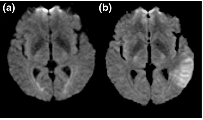
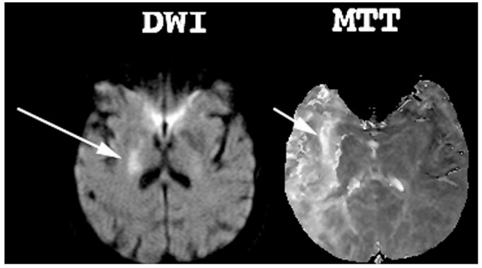
The appeal of MRI methods is that, whereas the standard CT examination of acute ischemic stroke will typically appear normal in the first hours after stroke onset, the methods of magnetic resonance angiography, perfusion weighted imaging (PWI), and diffusion weighted imaging (DWI) provide information on arterial patency, tissue blood flow, and parenchymal injury from the earliest times after onset of ischemic symptoms in a brief, non-invasive examination. DWI (Fig. 1) detects tissue injury within minutes of ischemia, has high sensitivity and specificity for the diagnosis of ischemic stroke, and permits measurement of lesion volumes that correlate with clinical severity and prognosis [6,7,8,9,10,11,12]. If untreated, the lesion seen with DWI typically enlarges over hours to days and will progress to infarction. PWI depicts focal cerebral ischemia. The volume of ischemic tissue seen with PWI, in the majority of cases, is greater than the region of parenchymal injury evident on DWI, and this diffusion-perfusion mismatch is considered to be a marker of the ischemic penumbra (Fig. 2), the tissue at greatest risk for infarct progression [13,14,15,16,17,18,19,20,21]. Furthermore, increasing theoretical, experimental, and clinical evidence suggests that MRI using magnetic susceptibility weighted pulse sequences may be sensitive to the early detection of hemorrhage [22,23,24]. Although prospective comparisons of MRI and CT for sensitivity to hemorrhage detection have yet to be reported, the proper acquisition and interpretation of MRI can eliminate the need for a screening CT scan and regain some of the time spent on adding a MRI examination to a screening evaluation. The target pathology revealed by MRI also represents the biological marker of the disease that can serve as a surrogate measure for assessing the effects of a therapy.
Figure 1.

The diffusion weighted imaging (DWI) of 3 h stroke. (a) The fluid-attenuated inversion recovery image without diffusion weighting shows no acute lesion. (b) DWI demonstrates the acute lesion as a region of hyperintensity (brightness) in the left temporal lobe.
Figure 2.

The diffusion-perfusion mismatch. A small lesion on the diffusion weighted image (DWI) in a 3 h stroke relative to the larger perfusion weighted image abnormality on a relative mean transit time (MTT) image.
Three potential uses of MRI in clinical trials have been proposed: patient selection, proof of pharmacologic principle, and as an outcome measure.
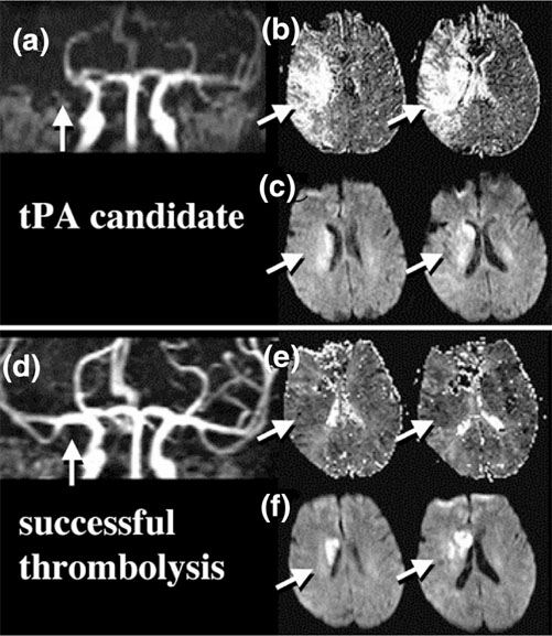
In using MRI as a selection criterion in patient selection (Table 1), the goal would be a sample based on a positive imaging diagnosis of a pathology rationally linked to the drug's mechanisms of action. Requiring a positive diagnosis of acute ischemic injury by DWI would ideally assure that no patients with diagnoses mimicking stroke are included in the sample, a desirable objective unachievable in trials using bedside impression and normal CT as the basis of inclusion. The goal of image-based patient selection is to narrow the range of patient characteristics, leading to a more homogeneous sample, reducing within-group variance, and increasing the statistical power of the experimental design to demonstrate efficacy. Optimal patient selection would be based on positive imaging evidence of the ischemic pathology that the therapy has been developed to treat. The simplest use as an inclusion criterion would include the presence of a lesion on DWI to increase the diagnostic certainty of ischemic stroke. The optimal target of therapy for reperfusion therapies would be patients with evidence of an arterial occlusion or hypoper-fusion (Fig. 3) [17,25]. Optimal selection of patients for neuroprotective drugs would be acute lesions involving the cerebral cortex and with a larger region of hypoperfusion - the diffusion-perfusion mismatch indicative of tissue at risk for infarction (Fig. 2 and Table 2). Patients might also be excluded from the trial at screening if subacute or chronic lesions are found that may confound measurements of lesion volumes or clinical severity as outcome variables. Because of a relatively large error of measurement associated with small lesions [26], lesions larger than a minimum volume (eg 5 cm3) may be desirable. Furthermore, an upper limit of lesion volume at enrollment would permit an opportunity for lesion growth and may better differentiate the effect on lesion size of an effective treatment from placebo. Selection of patients by DWI is also optimally suited for using the lesion volume change as a direct measure of the neuroprotective effect of the drug.
Table 1.
Proposed uses of magnetic resonance imaging (DWI, PWI, and MRA) as a selection tool in stroke trials
| Positive radiological diagnosis of ischemic lesion by DWI |
| Select by location (eg cortical, MCA territory, etc) |
| Select by size (DWI) |
| Select by perfusion defect (PWI, MRA) for reperfusion therapies |
| Select by diffusion/perfusion mismatch (DWI, PWI) for neuroprotective |
| drugs |
| Exclude if confounding subacute or chronic lesions |
DWI, diffusion weighted imaging; MCA, middle cerebral artery; MRA, magnetic resonance angiography; PWI, perfusion weighted imaging.
Figure 3.

Example of magnetic resonance imaging based selection for thrombolysis. (a) Magnetic resonance angiography demonstrates right middle cerebral artery occlusion (arrow). (b) Two representative perfusion weighted imaging slices demonstrate delayed relative mean transit time in the entire right middle cerebral artery (MCA) territory. (c) Two corresponding diffusion weighted imaging slices demonstrate parenchymal injury only in the deeper parts of the right MCA territory (arrows), a diffusion-perfusion mismatch. Following tissue-type plasminogen activation (tPA) therapy, recanalization of the right MCA is seen (d), with normalization of perfusion (e), and limitation of the parenchymal damage (f).
Table 2.
The diffusion-perfusion mismatch
| MRI marker of the ischemic penumbra |
| The strongest predictor of lesion growth from baseline |
| Present in approximately 80% of MCA territory strokes up to 6 h |
| poststroke |
| Distinction of benign hypoperfusion from true tissue at risk not yet |
| possible prospectively |
MRI, magnetic resonance imaging; MCA, middle cerebral artery.
The proof of pharmacological principle uses MRI as a marker of response to therapy, replicating the preclinical experiment in patients. Before an experimental stroke therapy is brought from the laboratory to clinical trial, it is necessary to demonstrate that the treatment causes reduction in lesion volume in experimental models. The fundamental premise of drug discovery and development in acute stroke is that treatments that reduce lesion size are those most likely to lead to clinical benefit. In clinical trial programs that depend solely on clinical endpoints as indices of benefit, drugs may be brought to phase III testing - costing several years and tens of millions of dollars - without the slightest evidence that the drug will have the therapeutic effect observed in the experimental model. Only a safe and acceptable dose must be demonstrated by the end of phase II. The question of whether the treatment causes reduction of lesion volume, however, may be answerable in the study of 100-200 patients in phase II, whereas 5-10 times as many patients are typically tested in phase III studies to evaluate the treatment with clinical endpoints. A phase II MRI endpoint trial to replicate the preclinical experiment in a patient population may thus be a rational and cost-effective basis of deciding whether to proceed with phase III testing. A positive lesion outcome study in late phase II would be supportive of the decision to proceed with phase III trials.
MRI measurements have proven to be a marker of clinical severity measured by stroke scales [11,15,27,28], and changes in lesion volume over time are associated with change in clinical severity (Table 3) [29]. The exact sample size that is required for detecting the effect of lesion volume change with MRI will depend on many factors in the design of a trial. The citicoline MRI trial [29], with approximately 40 evaluable patients per group, approached but did not reach significance. Estimates based on that study indicate that 58 patients per treatment arm would have been sufficient to demonstrate a neuroprotective effect in patients, a sample size compatible with typical phase II sample sizes. That study and natural history samples suggest that a sample size of 50-100 should be sufficient to demonstrate a neuro-protective effect on lesion volume in patients.
Table 3.
Magnetic resonance imaging as a biomarker of clinical status [29]
| Acute lesion volumes correlated with clinical scales and with final | |||
| lesion volumes | |||
| Strong association of reduction in lesion volume with clinical | |||
| improvement:* | |||
| % patients | Median | Mean | |
| Clinical | with lesion | change | change |
| improvement | decrease† | (cm3)‡ | (SE) (cm3)† |
| Yes | 74 | -2.8 | 3.8 (3.8) |
| No | 36 | 3.7 | 25.5 (6.8) |
*Week 12 minus baseline lesion volume change related to clinical improvement (n = 81, from [29]). SE, standard error. †P < 0.001; ‡P < 0.01.
It is proposed that a treatment emergent advantage on a measure of lesion volume is a surrogate of clinical benefit for stroke trials (Table 4). The rationale for the use of lesion volume as a surrogate measure in stroke trials may be summarized as follows. Lesion volume reduction in animal models is both necessary and sufficient evidence of neuroprotection. The clinical benefit for neuroprotective drugs is mediated through a reduction in cell death and brain tissue loss. Drugs that reduce infarct volume are those most likely to cause clinical benefit.
Table 4.
Magnetic resonance imaging as outcome measure
| Necessary but not sufficient evidence of protective effect |
| Protective effect may be attenuation of expected lesion growth or |
| partial DWI lesion reversal |
| Clinical benefit unlikely if no protective effect on lesion volume (go/no |
| go decision at phase II) |
| Smaller sample size requirements than for typical clinical endpoints |
| (∼ 50-100 per arm) |
| May be confirmatory evidence supporting positive clinical endpoint trial |
| for regulatory approval |
DWI, diffusion weighted imaging.
The factors required for validation of MRI as a surrogate marker are summarized in Table 5. The first four of these requirements have been met (see earlier discussion and cited references). Confirmation of the validity of many of these features of DWI and PWI in acute stroke has recently come from the first prospective multicenter stroke trial using MRI as an inclusion and primary outcome measure, the citicoline MRI stroke trial [29]. In that study, identical MRI hardware and software were used in 17 centers across the United States to study 100 patients with ischemic stroke within 24 h of onset. Patients were randomly assigned to 500 mg/day citicoline or placebo. Diffusion and perfusion MRI were obtained before treatment, and 1 and 12 weeks after treatment. Image data processing and volumetric analysis were performed at a single central laboratory using a single expert reader blinded to patient clinical severity and treatment assignment. The primary MRI inclusion criterion was a lesion of volume 1-120 cm3 in middle cerebral artery territory gray matter. The primary efficacy endpoint was a change in lesion volume from pretreatment to week 12. Although the primary efficacy endpoint of an effect of citicoline on lesion growth was numerically different (181% increase in lesion volume in placebo patients versus 34% increase for citicoline treated patients), it was not statistically significant. However, the study replicated the findings of other investigations regarding the relationship of MRI-derived lesion volumes to patients' clinical status. Acute lesion volumes by DWI in 100 patients correlated significantly with acute clinical severity on NIH stroke scale scores (r = 0.64) and with chronic lesion volume (r = 0.79); the chronic lesion volume by T2-weighted MRI significantly correlated with chronic NIH stroke scale score (r = 0.63). The strongest predictor of change in lesion size from baseline in the 81 patients who completed their week 12 assessment was the size of the perfusion abnormality (P < 0.0001 by co-variance analysis). The volume change over the 12 weeks of observation was significantly related to the patient's clinical improvement. Patients meeting the protocol specified criterion of clinical improvement (improvement on the NIH stroke scale of seven points or more) had a significantly more favorable response on the lesion volume change outcome variable than those who did not improve. The differentiation of improved from not improved was present whether the lesion volume change was assessed as an absolute decrease (74% versus 36%), median change (-2.8 cm3 versus 3.7 cm3), or mean (SE) change (3.8 [3.8] cm3 versus 25.5 [6.8] cm3) (Table 5). This prospective multicenter, centrally analyzed trial confirmed the value of MRI as a marker of disease severity and progression in stroke trials, and indicated that the change in MRI lesion size is likely to predict clinical improvement in clinical trials.
Table 5.
Requirements of a validated surrogate for DWI and PWI
| To fully establish diffusion and perfusion MRI as a useful tool and | |
| validated surrogate in stroke trials, several conditions need to be | |
| satisfied (the first four have been met; see text): | |
| 1. | DWI and PWI as biologic markers of the disease process in |
| ischemic stroke | |
| 2. | The tests are sensitive and specific for the diagnosis of stroke |
| in patients | |
| 3. | Lesion volumes correlate with clinical function as measured by |
| clinical rating scales, predict outcome, and co-vary over time | |
| with clinical severity | |
| 4. | Rational co-variates affecting lesion volumes identified |
| 5. | Utility in identifying effective treatments in trials proven |
DWI, diffusion weighted imaging; MRI, magnetic resonance imaging; PWI, perfusion weighted imaging.
The fifth criterion of validation, the concordance of effects on clinical outcomes and surrogate outcomes, remains to be demonstrated. Effective drugs will show benefit on both clinical and imaging outcome measures. The citicoline trials provide support for this, wherein trends on both clinical and imaging outcomes measures have been observed [29,30,31,32]. Ineffective drugs will show benefit on neither clinical nor imaging outcome measures. The latter has been found for the Glycine Antagonist in Neuroprotection (GAIN) trials, which showed no effect on clinical or MRI surrogate outcomes [33,34]. This comparison is only meaningful if studies are optimally designed and equally powered to show effect on their respective outcome measures; that is, the optimal sample size for imaging studies may be too small to show clinical effects. Possible explanations for discordant clinical versus surrogate marker results are presented in Table 6.
Table 6.
Validation of magnetic resonance imaging lesion volumes as a surrogate outcome: explanations of possible discordance between clinical and surrogate lesion volume measures
| If clinical endpoint shows a benefit but lesion volume does not: |
| Imaging methods are insensitive to neuroprotection |
| Clinical benefit not mediated by neuroprotection |
| If lesion volume shows a benefit but the clinical endpoint does not: |
| Trial design or clinical measures are insensitive to detecting a |
| clinical effect |
| Toxicity offsets neuroprotective effect |
The concept that improvement as a measure of brain lesion volume is a proper surrogate outcome for destructive central nervous system diseases has been already accepted by academic and regulatory communities alike. Approval of beta-interferon for the treatment of multiple sclerosis was based, in part, on lesion volume as a surrogate marker of disease activity, even though the surrogate was not considered fully validated. A surrogate outcome measure in clinical trials does not need to be fully validated as a condition of drug approval. Recent changes to the Federal Food Drug and Cosmetic Act, which regulates the Food and Drug Administration approval process, have specified a fast-track drug designation to expedite review for drugs that have "the potential to address unmet medical needs for serious and life-threatening conditions" [35]. Drugs for treatment of stroke have fallen under this designation. A drug must ordinarily have a beneficial effect on a clinical endpoint or on a validated surrogate endpoint to demonstrate effectiveness. The new regulations state that a drug "may be approved if it has an effect on a surrogate endpoint that is reasonably likely to predict clinical benefit. Such surrogate endpoints are considered not to be validated because, while suggestive of clinical benefit, their relationship to clinical outcomes, such as morbidity and mortality, is not proven" [35] (emphasis added). The issue with regard to MRI as a surrogate in stroke trials is whether it is 'reasonably likely to predict clinical benefit'. The hypothesis that neuroprotection, the restriction of infarct volume, is reasonably likely to be clinically beneficial to patients is the premise of virtually all acute stroke drugs being developed. The clinical data already discussed supports the value of measuring infarct value as a surrogate.
Strict validation must eventually be proven but, as we see from Food and Drug Administration regulations, it is no longer required to use lesion volume by MRI as a surrogate outcome in stroke trials. A benefit on the surrogate may be acceptable as an independent source of confirmatory data in support of a clinical benefit seen in a single trial. The question, therefore, is no longer whether MRI surrogates should be used in trials, but how they should be used.
The pharmaceutical industry has taken the initiative in investigating this final step in validation. The results of several industry-sponsored drug trials using MRI as a surrogate will be known over the next several years, and those studies should provide the most decisive information regarding the utility of MRI as a surrogate outcome measure in stroke trials. Three multicenter randomized clinical trials using MRI as a key selection and outcome variable have been completed and reported. Several other trials are in progress or being planned.
In conclusion, there have been concerns raised in the past that the use of MRI in stroke clinical trails is impractical for technical and logistical reasons (eg scan duration and availability). The practical limitations have disappeared with the widespread availability of ultrafast echoplanar imaging with diffusion and perfusion capability on commercial MRI scanners. A highly motivated, well-coordinated center can perform emergency diffusion and perfusion MRI with a latency to scan and scanning session duration comparable with that of emergency head CT. There are now over 100 centers worldwide capable of and experienced in performing these types of acute MRI examinations. Key design issues with regard to the use of diffusion and perfusion MRI in stroke trials are proposed in Table 7. MRI-based recruitment into trials with a time window of 6 h has proven feasible, as has specific selection based on lesion size, location, and the diffusion-perfusion mismatch. As the field of stroke clinical trials examines opportunities for improving trial design, positive imaging diagnoses in patient selection and use of imaging as treatment assessments is likely to assume an increasingly useful role. Patient selection and outcomes based exclusively on clinical assessment and non-hemorrhagic CT scans may no longer be appropriate for all trials.
Table 7.
Proposed features of clinical trials
| Imaging methods | DWI, PWI, MRA, T2/FLAIR |
| Selection criteria | Cortical DWI lesion > 5 cm3 |
| Diffusion-perfusion mismatch | |
| No pre-existing lesions in same vascular | |
| territory | |
| Outcome variable | Change in lesion volume, pretreatment to |
| chronic (3 months) | |
| Data analysis | Transformed lesion volume (percentage |
| change, log, cube root) | |
| Co-variance analysis on baseline variables: | |
| NIHSS, volume of hypoperfusion, initial | |
| lesion volume |
DWI, diffusion weighted imaging; PWI, perfusion weighted imaging; MRA, magnetic resonance angiography; FLAIR, fluid-attenuated inversion recovery; NIHSS, National Institutes of Health Stroke Scales.
References
- The National Institute of Neurological Disorders and Stroke rt-PA Stroke Study Group Tissue plasminogen activator for acute ischemic stroke. N Engl J Med. 1995;333:1581–1587. doi: 10.1056/NEJM199512143332401. [DOI] [PubMed] [Google Scholar]
- Furlan A, Higashida R, Wechsler L, Gent M, Rowley H, Kase C, Pessin M, Ahuja A, Callahan F, Clark WM, Silver F, Rivera F. Intra-arterial prourokinase for acute ischemic stroke. The PROACT II study: a randomized controlled trial. Prolyse in Acute Cerebral Thromboembolism. JAMA. 1999;282:2003–2011. doi: 10.1001/jama.282.21.2003. [DOI] [PubMed] [Google Scholar]
- Hacke W, Kaste M, Fieschi C, von Kummer R, Davalos A, Meier D, Larrue V, Bluhmki E, Davis S, Donnan G, Schneider D, Diez-Tejedor E, Trouillas P. Randomised double-blind placebo-controlled trial of thrombolytic therapy with intravenous alteplase in acute ischaemic stroke (ECASS II). Second European-Australasian Acute Stroke Study Investigators. Lancet. 1998;352:1245–1251. doi: 10.1016/S0140-6736(98)08020-9. [DOI] [PubMed] [Google Scholar]
- Hacke W, Kaste M, Fieschi C, Toni D, Lesaffre E, von Kummer R, Boysen G, Bluhmki E, Hoxter G, Mahagne MH. Intravenous thrombolysis with recombinant tissue plasminogen activator for acute hemispheric stroke. The European Cooperative Acute Stroke Study (ECASS). JAMA. 1995;274:1017–1025. doi: 10.1001/jama.274.13.1017. [DOI] [PubMed] [Google Scholar]
- Clark WM, Wissman S, Albers GW, Jhamandas JH, Madden KP, Hamilton S. Recombinant tissue-type plasminogen activator (Alteplase) for ischemic stroke 3 to 5 hours after symptom onset. The ATLANTIS Study: a randomized controlled trial. Alteplase Thrombolysis for Acute Noninterventional Therapy in Ischemic Stroke. JAMA. 1999;282:2019–2026. doi: 10.1001/jama.282.21.2019. [DOI] [PubMed] [Google Scholar]
- Moseley ME, Cohen Y, Mintorovitch J, Chileuitt L, Shimizu H, Kucharczyk J, Wendland MF, Weinstein PR. Early detection of regional cerebral ischemia in cats: comparison of diffusion-and T2-weighted MRI and spectroscopy. Magn Reson Med. 1990;14:330–346. doi: 10.1002/mrm.1910140218. [DOI] [PubMed] [Google Scholar]
- Lovblad KO, Laubach HJ, Baird AE, Curtin F, Schlaug G, Edelman RR, Warach S. Clinical experience with diffusion-weighted MR in patients with acute stroke. Am J Neuroradiol. 1998;19:1061–1066. [PMC free article] [PubMed] [Google Scholar]
- Warach S, Chien D, Li W, Ronthal M, Edelman RR. Fast magnetic resonance diffusion-weighted imaging of acute human stroke. Neurology. 1992;42:1717–1723. doi: 10.1212/wnl.42.9.1717. [DOI] [PubMed] [Google Scholar]
- Warach S, Gaa J, Siewert B, Wielopolski P, Edelman RR. Acute human stroke studied by whole brain echo planar diffusion-weighted magnetic resonance imaging. Ann Neurol. 1995;37:231–241. doi: 10.1002/ana.410370214. [DOI] [PubMed] [Google Scholar]
- Baird AE, Warach S. Magnetic resonance imaging of acute stroke. J Cereb Blood Flow Metab. 1998;18:583–609. doi: 10.1097/00004647-199806000-00001. [DOI] [PubMed] [Google Scholar]
- Lovblad KO, Baird AE, Schlaug G, Benfield A, Siewert B, Voetsch B, Connor A, Burzynski C, Edelman RR, Warach S. Ischemic lesion volumes in acute stroke by diffusion-weighted magnetic resonance imaging correlate with clinical outcome. Ann Neurol. 1997;42:164–170. doi: 10.1002/ana.410420206. [DOI] [PubMed] [Google Scholar]
- Warach S, Dashe JF, Edelman RR. Clinical outcome in ischemic stroke predicted by early diffusion-weighted and perfusion magnetic resonance imaging: a preliminary analysis. J Cereb Blood Flow Metab. 1996;16:53–59. doi: 10.1097/00004647-199601000-00006. [DOI] [PubMed] [Google Scholar]
- Barber PA, Darby DG, Desmond PM, Yang Q, Gerraty RP, Jolley D, Donnan GA, Tress BM, Davis SM. Prediction of stroke outcome with echoplanar perfusion- and diffusion-weighted MRI. Neurology. 1998;51:418–426. doi: 10.1212/wnl.51.2.418. [DOI] [PubMed] [Google Scholar]
- Barber PA, Davis SM, Darby DG, Desmond PM, Gerraty RP, Yang Q, Jolley D, Donnan GA, Tress BM. Absent middle cerebral artery flow predicts the presence and evolution of the ischemic penumbra. Neurology. 1999;52:1125–1132. doi: 10.1212/wnl.52.6.1125. [DOI] [PubMed] [Google Scholar]
- Beaulieu C, de Crespigny A, Tong DC, Moseley ME, Albers GW, Marks MP. Longitudinal magnetic resonance imaging study of perfusion and diffusion in stroke: evolution of lesion volume and correlation with clinical outcome. Ann Neurol. 1999;46:568–578. doi: 10.1002/1531-8249(199910)46:4<568::AID-ANA4>3.0.CO;2-R. [DOI] [PubMed] [Google Scholar]
- Darby DG, Barber PA, Gerraty RP, Desmond PM, Yang Q, Parsons M, Li T, Tress BM, Davis SM. Pathophysiological topography of acute ischemia by combined diffusion-weighted and perfusion MRI. Stroke. 1999;30:2043–2052. doi: 10.1161/01.str.30.10.2043. [DOI] [PubMed] [Google Scholar]
- Marks MP, Tong DC, Beaulieu C, Albers GW, de Crespigny A, Moseley ME. Evaluation of early reperfusion and i.v. tPA therapy using diffusion- and perfusion-weighted MRI. Neurology. 1999;52:1792–1798. doi: 10.1212/wnl.52.9.1792. [DOI] [PubMed] [Google Scholar]
- Warach S, Li W, Ronthal M, Edelman RR. Acute cerebral ischemia: evaluation with dynamic contrast-enhanced MR imaging and MR angiography. Radiology. 1992;182:41–47. doi: 10.1148/radiology.182.1.1727307. [DOI] [PubMed] [Google Scholar]
- Schlaug G, Benfield A, Baird AE, Siewert B, Lovblad KO, Parker RA, Edelman RR, Warach S. The ischemic penumbra: operationally defined by diffusion and perfusion MRI. Neurology. 1999;53:1528–1537. doi: 10.1212/wnl.53.7.1528. [DOI] [PubMed] [Google Scholar]
- Baird AE, Benfield A, Schlaug G, Siewert B, Lovblad KO, Edelman RR, Warach S. Enlargement of human cerebral ischemic lesion volumes measured by diffusion-weighted magnetic resonance imaging. Ann Neurol. 1997;41:581–589. doi: 10.1002/ana.410410506. [DOI] [PubMed] [Google Scholar]
- Tong DC, Yenari MA, Albers GW, O'Brien M, Marks MP, Moseley ME. Correlation of perfusion- and diffusion-weighted MRI with NIHSS score in acute (6.5 hour) ischemic stroke. Neurology. 1998;50:864–870. doi: 10.1212/wnl.50.4.864. [DOI] [PubMed] [Google Scholar]
- Linfante I, Llinas RH, Caplan LR, Warach S. MRI features of intracerebral hemorrhage within 2 hours from symptom onset. Stroke. 1999;30:2263–2267. doi: 10.1161/01.str.30.11.2263. [DOI] [PubMed] [Google Scholar]
- Schellinger PD, Jansen O, Fiebach JB, Hacke W, Sartor K. A standardized MRI stroke protocol: comparison with CT in hyperacute intracerebral hemorrhage. Stroke. 1999;30:765–768. doi: 10.1161/01.str.30.4.765. [DOI] [PubMed] [Google Scholar]
- Patel MR, Edelman RR, Warach S. Detection of hyperacute primary intraparenchymal hemorrhage by magnetic resonance imaging. Stroke. 1996;27:2321–2324. doi: 10.1161/01.str.27.12.2321. [DOI] [PubMed] [Google Scholar]
- Schellinger PD, Jansen O, Fiebach JB, Heiland S, Steiner T, Schwab S, Pohlers O, Ryssel H, Sartor K, Hacke W. Monitoring intravenous recombinant tissue plasminogen activator thrombolysis for acute ischemic stroke with diffusion and perfusion MRI. Stroke. 2000;31:1318–1328. doi: 10.1161/01.str.31.6.1318. [DOI] [PubMed] [Google Scholar]
- Laubach HJ, Jakob PM, Loevblad KO, Baird AE, Bovo MP, Edelman RR, Warach S. A phantom for diffusion-weighted imaging of acute stroke. J Magn Reson Imaging. 1998;8:1349–1354. doi: 10.1002/jmri.1880080627. [DOI] [PubMed] [Google Scholar]
- Baird AE, Lovblad KO, Dashe JF, Connor A, Burzynski C, Schlaug G, Straroselskaya I, Edelman RR, Warach S. Clinical correlations of diffusion and perfusion lesion volumes in acute ischemic stroke. Cerebrovasc Dis. 2000;10:441–448. doi: 10.1159/000016105. [DOI] [PubMed] [Google Scholar]
- van Everdingen KJ, van der Grond J, Kappelle LJ, Ramos LM, Mali WP. Diffusion-weighted magnetic resonance imaging in acute stroke [see comments]. Stroke. 1998;29:1783–1790. doi: 10.1161/01.str.29.9.1783. [DOI] [PubMed] [Google Scholar]
- Warach S, Pettigrew LC, Dashe JF, Pullicino P, Lefkowitz DM, Sabounjian L, Harnett K, Schwiderski U, Gammans R. Effect of citicoline on ischemic lesions as measured by diffusion-weighted magnetic resonance imaging. Ann Neurol. 2000;48:713–722. doi: 10.1002/1531-8249(200011)48:5<713::AID-ANA4>3.3.CO;2-R. [DOI] [PubMed] [Google Scholar]
- Clark WM, Williams BJ, Selzer KA, Zweifler RM, Sabounjian LA, Gammans RE. A randomized efficacy trial of citicoline in patients with acute ischemic stroke. Stroke. 1999;30:2592–2597. doi: 10.1161/01.str.30.12.2592. [DOI] [PubMed] [Google Scholar]
- Clark W, Gunion-Rinker L, Lessov N, Hazel K. Citicoline treatment for experimental intracerebral hemorrhage in mice. Stroke. 1998;29:2136–2140. doi: 10.1161/01.str.29.10.2136. [DOI] [PubMed] [Google Scholar]
- Warach S, Sabounjian LA. ECCO 2000 study of citicoline for treatment of acute ischemic stroke: effects on infarct volumes measured by MRI [abstract]. Stroke. 2000;31:42. [Google Scholar]
- Lees KR, Asplund K, Carolei A, Davis SM, Diener HC, Kaste M, Orgogozo JM, Whitehead J. Glycine antagonist (gavestinel) in neuroprotection (GAIN International) in patients with acute stroke: a randomised controlled trial. GAIN International Investigators [see comments]. Lancet. 2000;355:1949–1954. doi: 10.1016/S0140-6736(00)02326-6. [DOI] [PubMed] [Google Scholar]
- Warach S, Kaste M, Fisher M. The effect of GV150526 on ischemic lesion volume: the GAIN Americas and GAIN International MRI Substudy. Neurology. 2000;54 (suppl 3):A87–A88. [Google Scholar]
- House of Representatives Prescription Drug User Fee Reauthorization and Drug Regulatory Modernization Act of 1997 House of Representatives Report, 105th Congress, 1st session, Report 105-310, Section 4 Washington, DC: House of Representatives, 1997. pp. 54–56.


