SUMMARY
Purpose
The main aim of the present study is to present a case of mandibular transposition between lateral incisor and canine in a paediatric patient.
Materials and methods
A fixed multibracket orthodontic treatment was performed by means of a modified welded arch as to correct the transposition and obtaining a class I functional and symmetrical occlusion, also thanks to the early diagnosis of the eruption anomaly.
Results
Our case report shows that a satisfactory treatment of mandibular transpositions is obtained when detected at an early stage of the tooth development.
Conclusions
The main treatment options to be taken into consideration in case of a mandibular transposition are two: correcting the transposition or aligning it leaving the dental elements in their transposed order; in both cases, the follow-ups show a stable condition, maintained without relapses. Several factors, such as age of the patient, occlusion, aesthetics, patient’s collaboration, periodontal support and duration of treatment have to be considered as to prevent potential damage to dental elements and support appliances. The choice between the two treatment approaches for mandibular lateral incisor/canine transpositions mainly depends on the time the anomaly is detected.
Keywords: mandibular transposition, malocclusion, orthodontic treatment, tooth eruption
Introduction
Transposition is an eruption anomaly consisting in the exchange of position between two adjacent teeth (1). Mandibular transpositions are less frequent and show less variability than maxillary ones (2); five types of maxillary transpositions have been so far identified and described whereas only two maxillary transpositions are known: 1) mandibular lateral incisor/canine (Mn.I2. C); 2) transmigrated mandibular canine/erupted canine (Mn. C. trans-erupted) (3).
Both mandibular transpositions are very rare. The prevalence of Mn. L2. C transposition is 0.03%, that of Mn. C. trans-erupted transposition is 0.02% (3).
We also distinguish complete and incomplete forms depending on the involvement of both crown and root, or the crown only.
Depending on the transposition development stage, different characteristics of this dental anomaly emerge, first described in a study by Peck et al. (4):
Transposition initial phase: it is characterised by early distal tipping, coronal movement and severe mesio-lingual rotation (between 60 and 120 degrees) of the lateral incisor of the lower arch, thus affecting the physiological eruption path of the canine which develops transposed mesially to the lateral. The first deciduous molar is often damaged by the ectopic development of the lateral incisor thus inducing its premature exfoliation. At this stage, Mn. L2. C anomaly shows crown transposition of the mandibular lateral incisor and the canine; however, the roots are not in inverted position yet.
Mature transposition phase: this phase is characterised by an ectopic position of the mandibular canine between the lateral and central incisor. In several cases, at this stage, the crowns of the canine and the lateral are clearly and neatly transposed, the roots can be overlaid or in full transposition. At the age of 13, Mn. L2. C. transposition is usually complete for the roots as well. The aetiology of the transposition is multifactorial: in 1998, Peck (3) observed an association between mandibular transposition and dental anomalies, thus supporting the genetic hypothesis.
In mandibular lateral incisor/canine transposition cases, the development phase of the anomaly and the age of the patient are important factors to consider when choosing the most appropriate treatment option (2).
Case presentation
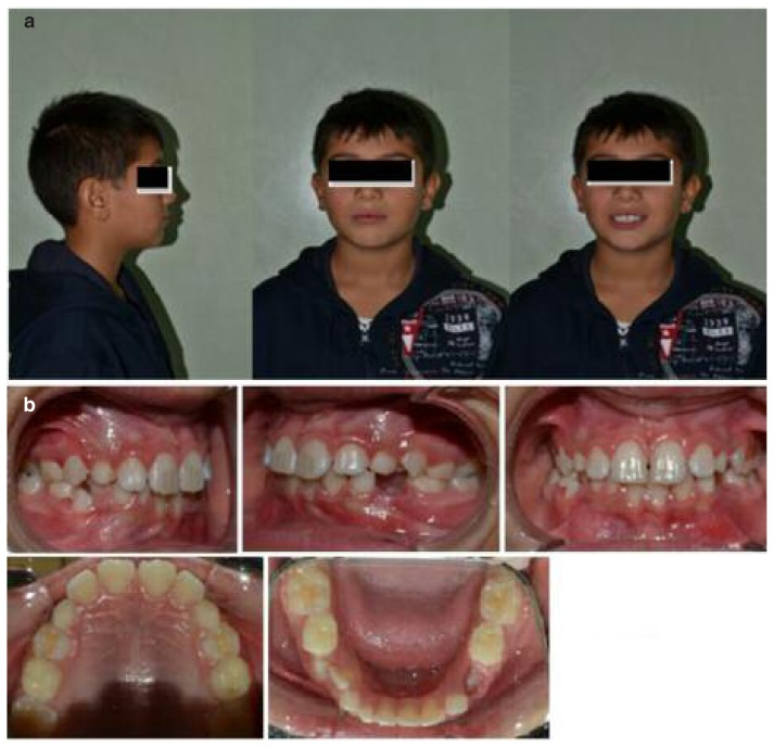
A 9-year-old male patient, nonsyndromic and free of other associated dental anomalies, under treatment at the orthodontics department of the Dental School of University of Bari for the presence of a mandibular lateral incisor/canine transposition. The patient shows a slightly convex profile, harmonious smile (Figure 1 a), cross bit occlusion and a tendency to molar face to face (class I occlusion tending to class II). The canine class cannot be defined because the lower canine has not erupted in arch yet (Figure 1 b).
Figure 1.
a) Pre-treatment facial pictures. b) Pre-treatment intraoral pictures.
The overjet is 3 millimetres, the overbite 3.5 millimetres (Figure 1 b).
The dental midline is not symmetrical because the mandibular line is deviated 1 millimetre right in relationship with the maxillary, dental and facial line (Figure 1 b).
There is evidence of cross of molars, cross of incisors is not detected. The right permanent lower lateral incisor is next to the permanent first premolar, in a phase of early eruption. The deciduous canine and lower lateral incisor persist in the arch. The permanent lateral incisor is located distally to the deciduous canine (Figure 1 b).
The oral hygiene is inadequate, with presence of mild marginal gingivitis and plaque build-up (Figure 1 b).
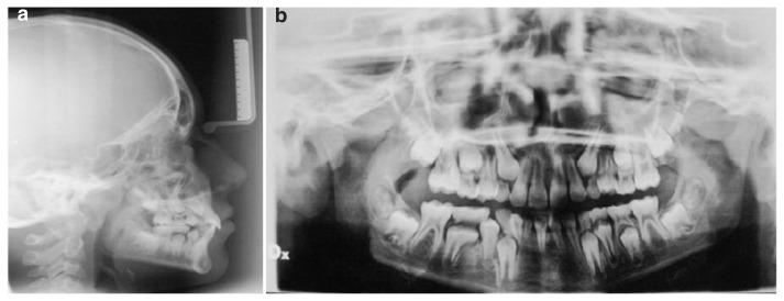
The radiographic examination highlights the position of the permanent canine cusp aligned with the permanent lateral incisor already erupted, whose root is hidden by the canine crown. The first is positioned lingually to the permanent first premolar, its apex is lingual to the erupting canine (Figure 2 a). All the other teeth are in tardive dentition phase; the tooth germs of the permanent third molars are present (Figure 2 b).
Figure 2.
a) Pre-treatment latero-lateral teleradiography. b) Pre-treatment orthopantomography.
On the basis of the orthodontic check-up, the following treatment plan is set:
establishment of a class I dental occlusion;
development of an ideal overjet and overbite and correction of the cross-bite;
correction of the mandibular lateral incisor/canine transposition;
correction of the root inclination and angulation;
establishment a skeletal class I.
Treatment phases
The first phase of the treatment consists in the extraction of the persistent deciduous lateral incisor and canine.
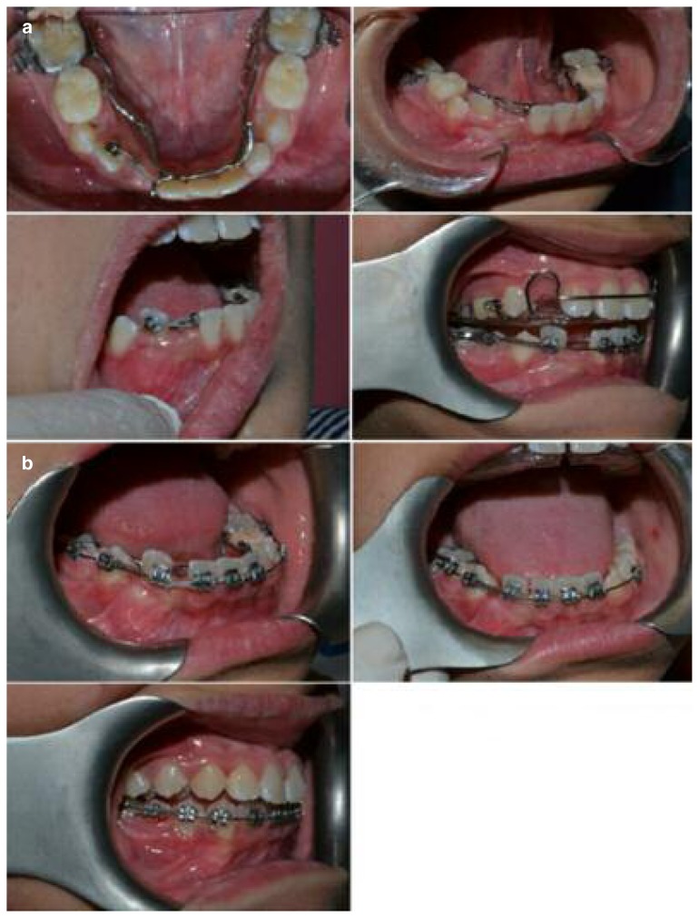
A lower welded arch was positioned with a distal stop at the permanent central incisor and with a lingual loop connecting the lateral incisor and the deciduous second molar as to allow the placing of a button on the lingual surface of the permanent lateral incisor. The same stop on the permanent central incisor enables the placing of an elastic module exerting a mesial traction on the permanent lateral incisor (Figure 3 a).
Figure 3.
a) Pictures of the different phases of the orthodontic treatment. b) Orthodontic treatment phases.
A plate with an anterior hitting plane is then inserted on the upper arch in order to avoid interferences during the movement of the tooth (Figure 3 a).
After about 3 months a button is then applied on the vestibular surface as well, in order to obtain a lingual traction and to detach the root of the lateral incisor from the crown of the canine thus avoiding any radicular damage (Figure 3 a).
Four months from the beginning of the treatment a fixed multibrackets retainer (022-028) is placed, with the insertion of a coil spring open between 42 and 45 to enable the mesialization of 42 without a further diversion of the mandibular line (Figure 3 a).
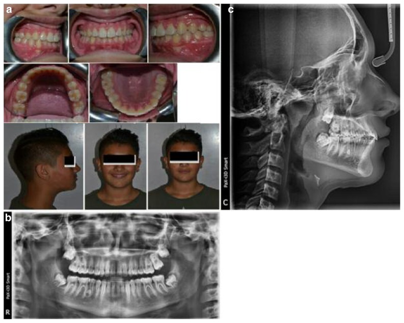
The use of elastic chains allowed us to accomplish the mesial movement of the lateral incisor (Figure 3 b). With a steady activation of the coil spring open we obtained the eruption of the canine on which, once erupted in the arch, a bracket is positioned for the alignment of the canine (Figure 3 b). The treatment provides for the sequence of round threads from 014 to 018 until the rotation is corrected and the complete levelling is achieved; after this phase, rectangular threads 017 and 025 were used as to achieve the correct inclination of the roots of all the dental elements. The patient undergoes regular checkups each 3–5 weeks during the active phases of the treatment (Figure 3 b). When the retainer is removed, a restraining plate is made in order to preserve the obtained results. At the end of treatment the patient is 11 years old (Figure 4 a) and he did an orthopantomography and a latero-lateral teleradiography (Figure 4 b, c).
Figure 4.
a) End of treatment. b) Orthopantomography post treatment. c) Latero-lateral teleradiography post treatment.
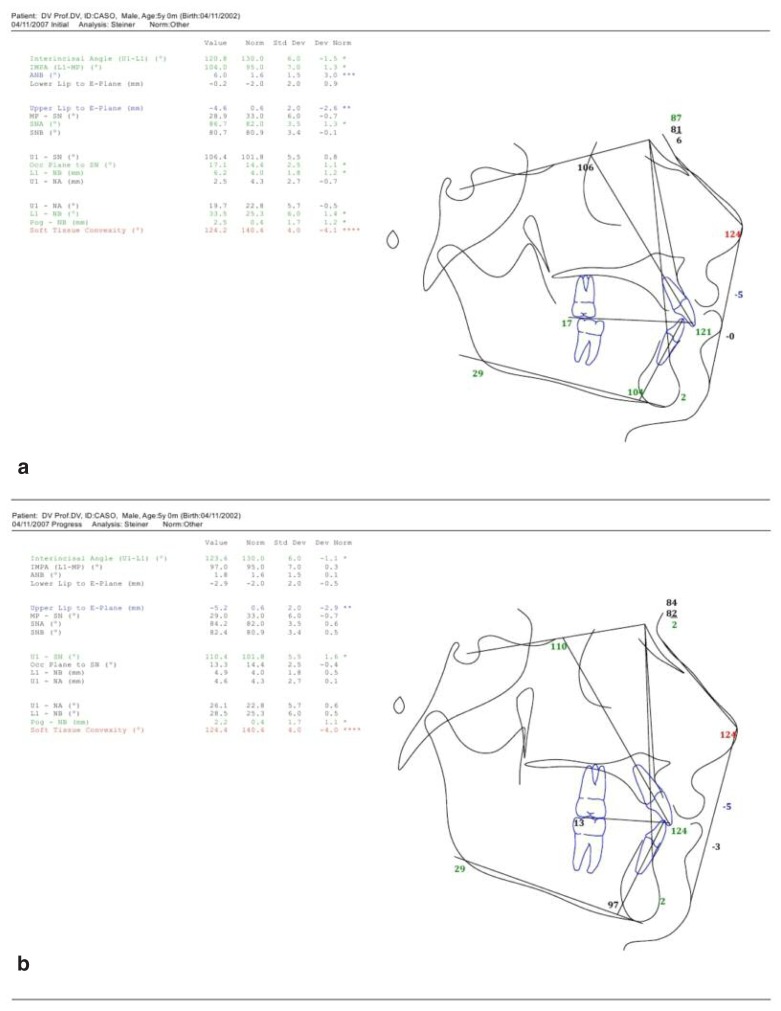
In Figure 5 a and b we can see cephalometric tracings pre and post treatment.
Figure 5.
a) Cephalometric tracing pre treatment. b) Cephalometric tracing post treatment.
Discussion
The main treatment options to be taken into consideration in case of a mandibular transposition are two (5–7): correcting the transposition or aligning it leaving the dental elements in their transposed order; in both cases, the follow-ups show a stable condition, maintained without relapses. Several factors, such as age of the patient, occlusion, aesthetics, patient’s collaboration, periodontal support and duration of treatment have to be considered as to prevent potential damage to dental elements and support appliances (8–31).
The choice between the two treatment approaches for mandibular lateral incisor/canine transpositions mainly depends on the time the anomaly is detected (5, 6):
if the transposition is diagnosed at an early stage, the position of the teeth can be corrected by means of a fixed orthodontic treatment; in the first stage of the treatment extractions are carried out as to reduce the duration of treatment.
-
if the transposition is diagnosed after the transposed teeth erupt in their anomalous position, it is preferable not to correct it and to align the dental elements in their transposed order. For this purpose, two options are to be considered depending on the length of the arch:
- if there is not enough room for the transposed teeth to be aligned in the arch, the extraction could be necessary. The posterior teeth are thus mesially aligned. It is however important to consider the occlusion and the condition of the teeth; if the other teeth are in good conditions, the lateral incisor can be extracted;
- if there is enough room to align the transposed teeth in the arch, the transposition is then kept and the shape of the transposed teeth can be modified using aesthetic restoration materials.
Peck and Peck (2) advise to correct only the pseudo-transpositions and to keep the transposed order of the dental elements in all the types of real transpositions. Weeks and Power (7) clarify the necessary interventions to obtain an acceptable compromise without correcting the transposition. The correction of teeth in their normal position is reported by several other Authors (6–8).
Correction is a complex process, potentially detrimental for the teeth and the support appliances; they also present the advantages and disadvantages of alignment and the correction. Our case report shows that a satisfactory treatment of mandibular transpositions is obtained when detected at an early stage of the tooth development.
References
- 1.Peck S, Peck L. Classification of maxillary toothtranspositions. Am J Orthod Dentofac Orthop. 1995;107:505–17. doi: 10.1016/s0889-5406(95)70118-4. [DOI] [PubMed] [Google Scholar]
- 2.Peck S, Peck L, Kataja M. Mandibular lateral incisorcanine transposition, concomitant dental anomalies, and genetic control. Angle Orthod. 1998 Oct;68(5):455–66. doi: 10.1043/0003-3219(1998)068<0455:MLICTC>2.3.CO;2. [DOI] [PubMed] [Google Scholar]
- 3.Peck S. On the phenomenon of intraosseous migration of nonerupting teeth. Am J Orthod Dentofacial Orthop. 1998 May;113(5):515–7. doi: 10.1016/s0889-5406(98)70262-8. [DOI] [PubMed] [Google Scholar]
- 4.Peck L, Peck S, Attia Y. Maxillary canine-first premolar transposition, associated dental anomalies and genetic basis. Angle Orthod. 1993 Summer;63(2):99–109. doi: 10.1043/0003-3219(1993)063<0099:MCFPTA>2.0.CO;2. discussion 110. [DOI] [PubMed] [Google Scholar]
- 5.Shapira Y, Kuftinec MM, Stom D. A unique treatment approach for maxillary canine-lateral incisor transposition. Am J Orthod Dentofac Orthop. 2001;119:540–5. doi: 10.1067/mod.2001.111221. [DOI] [PubMed] [Google Scholar]
- 6.Laptook T, Silling G. Canine transposition-approaches to treatment. J Am Dent Assoc. 1983 Nov;107(5):746–8. doi: 10.14219/jada.archive.1983.0330. [DOI] [PubMed] [Google Scholar]
- 7.Weeks EC, Power SM. The presentations and management of transposed teeth. Br Dent J. 1996;18i:421–4. doi: 10.1038/sj.bdj.4809280. [DOI] [PubMed] [Google Scholar]
- 8.Bocchieri A, Braga G. Correction of a bilateral maxillary canine-first premolar transposition in the late mixed dentition. Am J Orthod Dentofacial Orthop. 2002 Feb;121(2):120–8. doi: 10.1067/mod.2002.120755. [DOI] [PubMed] [Google Scholar]
- 9.Di Venere D, Corsalini M, Stefanachi G, Tafuri S, De Tommaso M, Cervinara F, Re A, Pettini F. Quality of life in fibromyalgia patients with craniomandibular disorders. Open Dent J. 2015 Jan 30;9:9–14. doi: 10.2174/1874210601509010009. [DOI] [PMC free article] [PubMed] [Google Scholar]
- 10.Favia G, Corsalini M, Di Venere D, Pettini F, Favia G, Capodiferro S, Maiorano E. Immunohistochemical evaluation of neuroreceptors in healthy and pathological temporo-mandibular joint. Int J Med Sci. 2013 Sep 25;10(12):1698–701. doi: 10.7150/ijms.6315. [DOI] [PMC free article] [PubMed] [Google Scholar]
- 11.Scarano A, Nardi G, Murmura G, Rapani M, Mortellaro C. Evaluation of the Removal Bacteria on Failed Titanium Implants After Irradiation With Erbium-Doped Yttrium Aluminium Garnet Laser. J Craniofac Surg. 2016 Jul;27(5):1202–4. doi: 10.1097/SCS.0000000000002735. [DOI] [PubMed] [Google Scholar]
- 12.Di Venere D, Gaudio RM, Laforgia A, Stefanachi G, Tafuri S, Pettini F, Silvestre F, Petruzzi M, Corsalini M. Correlation between dento-skeletal characteristics and craniomandibular disorders in growing children and adolescent orthodontic patients: retrospective case-control study. Oral Implantol (Rome) 2016 Nov 16;9(4):175–184. doi: 10.11138/orl/2016.9.4.175. [DOI] [PMC free article] [PubMed] [Google Scholar]
- 13.Grassi FR, Pappalettere C, Di Comite M, Corsalini M, Mori G, Ballini A, Crincoli V, Pettini F, Rapone B, Boccaccio A. Effect of different irrigating solutions and endodontic sealers on bond strength of the dentin-post interface with and without defects. Int J Med Sci. 2012;9(8):642–54. doi: 10.7150/ijms.4998. [DOI] [PMC free article] [PubMed] [Google Scholar]
- 14.Di Comite M, Crincoli V, Fatone L, Ballini A, Mori G, Rapone B, Boccaccio A, Pappalettere C, Grassi FR, Favia A. Quantitative analysis of defects at the dentin-post space in endodontically treated teeth. Materials. 2015;8:3268–3283. [Google Scholar]
- 15.Corsalini M, Pettini F, Di Venere D, Ballini A, Chiatante G, Lamberti L, Pappalettere C, Fiorentino M, Uva AE, Monno G, Boccaccio A. An Optical System to Monitor the Displacement Field of Glass-fibre Posts Subjected to Thermal Loading. Open Dent J. 2016 Nov16;10:610–618. doi: 10.2174/1874210601610010610. [DOI] [PMC free article] [PubMed] [Google Scholar]
- 16.Corsalini M, Di Venere D, Pettini F, Stefanachi G, Catapano S, Boccaccio A, Lamberti L, Pappalettere C, Carossa S. A comparison of shear bond strength of ceramic and resin denture teeth on different acrylic resin bases. Open Dent J. 2014 Dec 29;8:241–50. doi: 10.2174/1874210601408010241. [DOI] [PMC free article] [PubMed] [Google Scholar]
- 17.Pettini F, Savino M, Corsalini M, Cantore S, Ballini A. Cytogenetic genotoxic investigation in peripheral blood lymphocytes of subjects with dental composite restorative filling materials. J Biol Regul Homeost Agents. 2015 Jan-Mar;29(1):229. [PubMed] [Google Scholar]
- 18.Pettini F, Corsalini M, Savino MG, Stefanachi G, Venere DD, Pappalettere C, Monno G, Boccaccio A. Roughness Analysis on Composite Materials (Micro-filled, Nanofilled and Silorane) After Different Finishing and Polishing Procedures. Open Dent J. 2015 Oct 26;9:357–67. doi: 10.2174/1874210601509010357. [DOI] [PMC free article] [PubMed] [Google Scholar]
- 19.Romeo U, Nardi GM, Libotte F, Sabatini S, Palaia G, Grassi FR. The Antimicrobial Photodynamic Therapy in the Treatment of Peri-Implantitis. Int J Dent. 2016;2016:7692387. doi: 10.1155/2016/7692387. [DOI] [PMC free article] [PubMed] [Google Scholar]
- 20.Lauritano D, Petruzzi M, Nardi GM, Carinci F, Minervini G, Di Stasio D, Lucchese A. Single application of a dessicating agent in the treatment of recurrent aphtous stomatitis. J Biol Regul Homeost Agents. 2015 Jul-Sep;29(3 Suppl 1):59–66. [PubMed] [Google Scholar]
- 21.Kalemaj Z, Scarano A, Valbonetti L, Rapone B, Grassi FR. Bone response to four dental implants with different surface topography: a histologic and histometric study in minipigs. Int J Periodontics Restorative Dent. 2016 Sep-Oct;36(5):745–54. doi: 10.11607/prd.2719. [DOI] [PubMed] [Google Scholar]
- 22.Laforgia A, Corsalini M, Stefanachi G, Pettini F, Di Venere D. Assessment of Psychopatologic Traits in a Group of Patients with Adult Chronic Periodontitis: Study on 108 Cases and Analysis of Compliance during and after Periodontal Treatment. Int J Med Sci. 2015 Oct 4;12(10):832–9. doi: 10.7150/ijms.12317. [DOI] [PMC free article] [PubMed] [Google Scholar]
- 23.Rapone B, Nardi GM, Di Venere D, Pettini F, Grassi FR, Corsalini M. Oral hygiene in patients with oral cancer undergoing chemotherapy and/or radiotherapy after prosthesis rehabilitation: protocol proposal. Oral Implantol (Rome) 2016 Dic;9(01 suppl):90–97. doi: 10.11138/orl/2016.9.1S.090. [DOI] [PMC free article] [PubMed] [Google Scholar]
- 24.Nardi GM, Sabatini S, Lauritano D, Silvestre F, Petruzzi M. Effectiveness of two different desensitizing varnishes in reducing tooth sensitivity: a randomized double-blind clinical trial. Oral Implantol (Rome) 2016 Nov 16;9(4):185–189. doi: 10.11138/orl/2016.9.4.185. [DOI] [PMC free article] [PubMed] [Google Scholar]
- 25.Corsalini M, Rapone B, Grassi FR, Di Venere D. A study on oral rehabilitation in stroke patients: analysis of a group of 33 patients. Gerodontology. 2010 Sep;27(3):178–82. doi: 10.1111/j.1741-2358.2009.00322.x. [DOI] [PubMed] [Google Scholar]
- 26.Di Venere D, Pettini F, Nardi GM, Laforgia A, Stefanachi G, Notaro V, Rapone B, Grassi FR, Corsalini M. Correlation between parodontal indexes and orthodontic retainers: prospective study in a group of 16 patients. Oral Implantology. 2017 Apr 10;10(1):78–86. doi: 10.11138/orl/2017.10.1.078. eCollection 2017 Jan–Mar. [DOI] [PMC free article] [PubMed] [Google Scholar]
- 27.Mori G, Ballini A, Carbone C, Oranger A, Brunetti G, Di Benedetto A, Rapone B, Cantore S, Di Comite M, Colucci S, Grano M, Grassi FR. Osteogenic differentiation of dental follicle stem cells. Int J Med Sci. 2012;9(6):480–487. doi: 10.7150/ijms.4583. Epub 2012 Aug 13. [DOI] [PMC free article] [PubMed] [Google Scholar]
- 28.Ballini A, Cantore S, Fatone L, Montenegro V, De Vito D, Pettini F, Crincoli V, Antelmi A, Romita P, Rapone B, Miniello G, Perillo L, Grassi FR, Foti C. Transmission of non-viral sexually transmitted infections and oral sex. Journal of Sexual Medicine. 2012 Feb;9(2):372–384. doi: 10.1111/j.1743-6109.2011.02515.x. Epub 2011 Oct 24. [DOI] [PubMed] [Google Scholar]
- 29.Mussano F, Genova T, Corsalini M, Schierano G, Pettini F, Di Venere D, Carossa S. Cytokine, Chemokine, and Growth Factor Profile Characterization of Undifferentiated and Osteoinduced Human Adipose-Derived Stem Cells. Stem Cells Int. 2017;2017:6202783. doi: 10.1155/2017/6202783. Epub 2017 May 10. [DOI] [PMC free article] [PubMed] [Google Scholar]
- 30.Bruno V, O’Sullivan DB, Badino M, Catapano S. Preserving soft tissue after placing implants in fresh extraction sockets in the maxillary esthetic zone and a prosthetic template for interim crown fabrication: A prospective study. Journal of Prosthetic Dentistry. 2014 Mar;111(3):195–202. doi: 10.1016/j.prosdent.2013.09.008. [DOI] [PubMed] [Google Scholar]
- 31.Bruno V, Badino M, Sacco R, Catapano S. The use of a prosthetic template to maintain the papilla in the esthetic zone for immediate implant placement by means of a radiographic procedure. Journal of Prosthetic Dentistry. 2012 Dec;108(6):394–397. doi: 10.1016/S0022-3913(12)60199-1. [DOI] [PubMed] [Google Scholar]


