CASE PRESENTATION
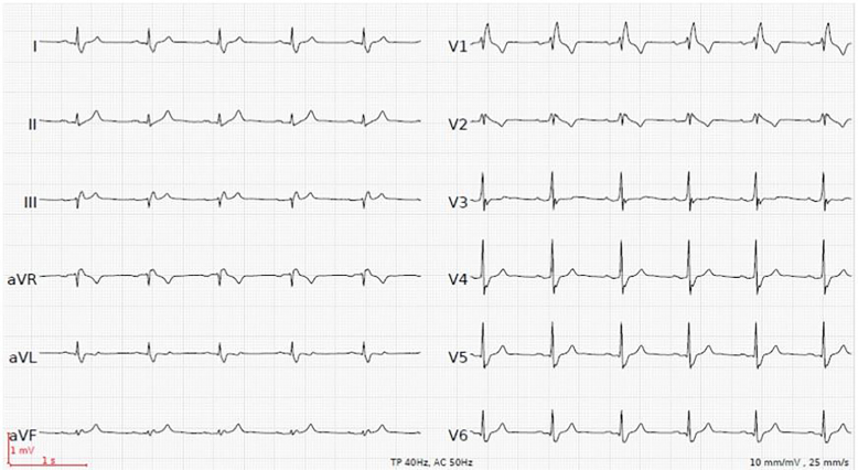
A 49-year-old man presented to our emergency department complaining of progressive muscle weakness in his legs for three days. He had no past history of significant health issues, and denied any illicit or recreational drug use. At presentation, vital signs were normal. Physical exam revealed reduced strength (3/5) in his lower extremities but no focal deficits. Laboratory studies showed severe hyperkalemia of 8.6 mmol/l (3.6–4.8 mmol/l), hyponatremia of 130 mmol/l (135–145 mmol/l) and mild hyperchloremic metabolic acidosis. Kidney function was moderately impaired (serum creatinine 120 μmol/l (50–100 μmol/l)). The electrocardiogram (ECG) demonstrated a sinus rhythm with normal heart rate, prolonged PR- and QRS-intervals, tall peaked T-waves and type I Brugada-like pattern in leads V1 and V2 (Image 1).
Image 1.
Initial electrocardiogram with a heart rate of 54 beats per minute, PR-interval 270 milliseconds, QRS-interval 122 milliseconds. Potassium level of 8.6 mmol/l.
Four hours after treatment with calcium gluconate, insulin with glucose, and nebulized beta-2 agonist, the ECG returned to baseline with preexisting right bundle branch block at a potassium level of 6.6 mmol/l (Image 2). Hyponatremia persisted with 130 mmol/l. The patient’s symptoms completely resolved.
Image 2.
Post-treatment electrocardiogram with a heart rate of 66 beats per minute, PR-interval 170 milliseconds, QRS-interval 120 milliseconds. Return to pre-existing right bundle branch block pattern. Potassium level of 6.6 mmol/l.
Autoimmune primary adrenal insufficiency (Addison’s disease) was found as the underlying cause of this severe hyperkalemia.
DIAGNOSIS
Hyperkalemia is found in up to 40% of patients with primary adrenal insufficiency due to mineralocorticoid deficiency.1 Severe hyperkalemia typically causes muscle weakness and rhythm disturbances. Muscle weakness typically begins in the legs with progression to the trunk and arms. Sphincter tone and cranial nerve function are typically not affected. Cardiac manifestations include electrocardiographic changes, conduction abnormalities and cardiac arrhythmias (Table 1).2,3 Of note, the progression and severity of ECG changes do not correlate well with the serum potassium level.4
Table 1.
Cardiac manifestations in hyperkalemia.
| Electrocardiographic changes | Conduction abnormalities | Cardiac arrhythmias |
|---|---|---|
| tall peaked T-waves with shortened QT-interval prolonged PR- and QRS-intervals with small or disappearing P-waves further prolongation of the QRS-interval to a sine wave pattern |
right or left bundle branch block bifascicular block advanced atrioventricular block type I Brugada pattern (characterized by high takeoff ≥2 mm coved ST-segment elevation followed by a negative T-wave in at least two precordial leads) |
sinus bradycardia, sinus arrest slow idioventricular rhythms ventricular tachycardia, ventricular fibrillation asystole |
This case highlights that hyperkalemia-induced muscle weakness with associated electrocardiographic changes can be the major presenting symptom in primary adrenal insufficiency. The treatment consists of lowering potassium levels and replacement of hydrocortisone and fludrocortisone.1
CPC-EM Capsule.
What do we already know about this clinical entity?
Patients with primary adrenal insufficiency can present with nonspecific symptoms such as weakness. Hyperkalemia is found in up to 40% of patients with primary adrenal insufficiency.
What is the major impact of the image(s)?
The images show reversible hyperkalemia-induced electrocardiographic manifestations (including type I Brugada-like pattern) as a main finding in primary adrenal insufficiency.
How might this improve emergency medicine practice?
This case highlights that hyperkalemia-induced muscle weakness with associated electrocardiographic changes may be the only presenting symptom in primary adrenal insufficiency.
Footnotes
Section Editor: Rick A. McPheeters, DO
Full text available through open access at http://escholarship.org/uc/uciem_cpcem
Conflicts of Interest: By the CPC-EM article submission agreement, all authors are required to disclose all affiliations, funding sources and financial or management relationships that could be perceived as potential sources of bias. The authors disclosed none.
REFERENCES
- 1.Bancos I, Hahner S, Tomlinson J, et al. Diagnosis and management of adrenal insufficiency. Lancet Diabetes Endocrinol. 2015;3(3):216–26. doi: 10.1016/S2213-8587(14)70142-1. [DOI] [PubMed] [Google Scholar]
- 2.Alfonzo AV, Isles C, Geddes C, et al. Potassium disorders--clinical spectrum and emergency management. Resuscitation. 2006;70(1):10–25. doi: 10.1016/j.resuscitation.2005.11.002. [DOI] [PubMed] [Google Scholar]
- 3.Littmann L, Monroe MH, Taylor L, 3rd, et al. The hyperkalemic Brugada sign. J Electrocardiol. 2007;40(1):53–9. doi: 10.1016/j.jelectrocard.2006.10.057. [DOI] [PubMed] [Google Scholar]
- 4.Montague BT, Ouellette JR, Buller GK. Retrospective review of the frequency of ECG changes in hyperkalemia. Clin J Am Soc Nephrol. 2008;3(2):324–30. doi: 10.2215/CJN.04611007. [DOI] [PMC free article] [PubMed] [Google Scholar]


