Abstract
Objective:
To evaluate the image quality improvement and noise reduction in routine dose, non-enhanced chest CT imaging by using a new generation adaptive statistical iterative reconstruction (ASIR-V) in comparison with ASIR algorithm.
Methods:
30 patients who underwent routine dose, non-enhanced chest CT using GE Discovery CT750HU (GE Healthcare, Waukesha, WI) were included. The scan parameters included tube voltage of 120 kVp, automatic tube current modulation to obtain a noise index of 14HU, rotation speed of 0.6 s, pitch of 1.375:1 and slice thickness of 5 mm. After scanning, all scans were reconstructed with the recommended level of 40%ASIR for comparison purpose and different percentages of ASIR-V from 10% to 100% in a 10% increment. The CT attenuation values and SD of the subcutaneous fat, back muscle and descending aorta were measured at the level of tracheal carina of all reconstructed images. The signal-to-noise ratio (SNR) was calculated with SD representing image noise. The subjective image quality was independently evaluated by two experienced radiologists.
Results:
For all ASIR-V images, the objective image noise (SD) of fat, muscle and aorta decreased and SNR increased along with increasing ASIR-V percentage. The SD of 30% ASIR-V to 100% ASIR-V was significantly lower than that of 40% ASIR (p < 0.05). In terms of subjective image evaluation, all ASIR-V reconstructions had good diagnostic acceptability. However, the 50% ASIR-V to 70% ASIR-V series showed significantly superior visibility of small structures when compared with the 40% ASIR and ASIR-V of other percentages (p < 0.05), and 60% ASIR-V was the best series of all ASIR-V images, with a highest subjective image quality. The image sharpness was significantly decreased in images reconstructed by 80% ASIR-V and higher.
Conclusion:
In routine dose, non-enhanced chest CT, ASIR-V shows greater potential in reducing image noise and artefacts and maintaining image sharpness when compared to the recommended level of 40%ASIR algorithm. Combining both the objective and subjective evaluation of images, non-enhanced chest CT images reconstructed with 60% ASIR-V have the highest image quality.
Advances in knowledge:
This is the first clinical study to evaluate the clinical value of ASIR-V in the same patients using the same CT scanner in the non-enhanced chest CT scans. It suggests that ASIR-V provides the better image quality and higher diagnostic confidence in comparison with ASIR algorithm.
Keywords: Computed tomography, Adaptive statistical iterative reconstruction, Image noise, Image quality
Introduction
CT is being widely used in clinical practice, owing to its advantages of high-density resolution, faster imaging speed and convenience. However, related data shows the incidence of radiation-induced carcinogenesis from CT is increasing.1–3 More and more people begin to pay attention to the risk of radiation in routine dose CT scan. Since reducing image noise and improving image quality have a significant potential for reducing radiation dose, many imaging equipment manufacturers and clinical researchers have been focusing their efforts to develop and evaluate different image reconstruction algorithms to effectively reduce image noise and improve image quality.
As the first-generation iterative reconstruction technique, the adaptive statistical iterative reconstruction (ASIR) algorithm has been known to have considerable potential for reducing image noise in many studies.4–7 However, indicating that high percentage ASIR may produce waxy artefacts in certain applications, and reduce the sharpness of anatomical structures, resulting in the relatively poor image quality.8 ASIR percentages can be selected in a spectrum from 0 to 100%. The 40%ASIR means that 40% of the ASIR image is blended with 60% of filtered back projection image, which is a recommended level for ASIR by several articles9–11 and based on our own experiences to balance image noise and sharpness. ASIR-V is a new generation iterative reconstruction algorithm that contains more advanced noise modelling and object modelling than the previous version ASIR. ASIR-V has also added some physics modelling to be more robust in terms of balancing image noise and spatial resolution. ASIR-V can also adjust the image noise reduction power through changing the ASIR-V percentage weight.12,13 However, there are a relatively small number of studies on ASIR-V currently, and it is still not coming to consensus on the optimal percentage ranges of ASIR-V in many clinical applications.
The purpose of this study was to evaluate the clinical value of ASIR-V on image noise reduction and diagnostic confidence improvement compared with ASIR in the non-enhanced chest CT imaging, and to provide the optimal ASIR-V percentage in such application. To the best of our knowledge, this was the first clinical study to evaluate image quality in the same patients using the same CT scanner in routine dose chest CT, which avoided the effect of hardware and software differences on image quality due to the different models of CT scanners.
Methods and Materials
Patient population
From March 12 to June 5, 2016, 34 participants who came for routine non-enhanced chest CT examination were initially recruited to our study. The inclusion criteria were as follows: (1) the participants who could walk autonomously and had clear state of consciousness; (2) the participants with complete clinical data and (3) the participants aged 20 to 80 years old. Our clinical exclusion criteria were as follows: (1) the participants with severe respiratory symptoms that interfered with their CT examination; (2) the participants could not lie flat on the scanning bed and lift their arms over the head and (3) the image had severe artefacts that affected the data measurement.
Finally, 30 patients were enrolled (4 patients were excluded because of the large image artefacts), including 14 males and 16 females (age range, 27–78 years; mean age, 59.70 years; body mass index, 22.31 ± 3.11 kg m−2).
Scanning techniques
All patients underwent non-enhanced chest routine CT on a GE Discovery CT750HD (GE Healthcare, Waukesha, WI). The scan parameters were as follows: tube voltage of 120 kVp and automatic tube current modulation (Auto mA; GE Healthcare) to obtain a noise index of 14HU at 5 mm image slice thickness, rotation speed of 0.6 s and pitch of 1.375:1. The volumetric CT dose index produced using this scan protocol was 5.61 ± 2.48 mGy for the patient population in our study.
Image reconstruction
All the images were reconstructed using the data from the same scanner. First, the images were reconstructed with the adaptive statistical iterative reconstruction (ASIR; GE Healthcare) for daily clinical practice. Specifically, the reconstructed images were based on a 40% ASIR. Then, all the images were reconstructed again with the new generation iterative reconstruction algorithm ASIR-V (GE Healthcare) with percentage weights varying from 10 to 100% in a 10% increment. All reconstructed images were axial, with a layer thickness of 0.625 mm. Finally, we obtained a total of 11 series images, including 40% ASIR, 10% ASIR-V, 20% ASIR-V, 30% ASIR-V, 40% ASIR-V, 50% ASIR-V, 60% ASIR-V, 70% ASIR-V, 80% ASIR-V, 90% ASIR-V and 100% ASIR-V.
Objective image quality analysis
All images were transferred to and reviewed on a GE AW4.6 CT workstation (GE Healthcare, Waukesha, WI) with all patient information removed. The window width of 350HU and window level of 40HU was used for the mediastinal window, and window width of 1500HU and window level of −800HU was used for the lung window. The objective parameters of all images were measured at the level of tracheal carina in the mediastinal window. The CT attenuation values and SD of subcutaneous fat, back muscle and descending aorta were measured using a region of interest (ROI). The ROI covered a half of descending aorta in area, and adjusted for the subcutaneous fat or muscle. The SD value represented the objective image noise. The signal-to-noise ratio (SNR) of subcutaneous fat, back muscle and descending aorta were calculated using the following formula: SNR = CT value/SD value. All the data were measured three times, and the average value used as the final statistical result.
Subjective image quality analysis
Two experienced radiologists (one with 9 years of experience in the diagnosis of chest CT, and the other with 11 years of experience in the diagnosis of X-ray and chest CT) blindly and independently scored the image quality first and the final results were obtained by consensus. Specifically, the subjective image noise, visibility of small structures (for bronchus, mediastinal lymph nodes, blood vessels, pericardium, chest wall and other structures), image artefacts and diagnostic acceptability were all graded on a scale from one to five (Table 1), ignoring patient information and reconstruction algorithms. The sharpness of bronchus was observed in the lung window, while, the lymph nodes, blood vessels, pericardium and chest wall were observed in the mediastinal window. When the evaluation was inconsistent, the final score was decided by two radiologists after consulting each other.
Table 1.
Subjective scoring of image quality analysis
| Grading score | Qualitative image analysis | |||
|---|---|---|---|---|
| Subjective noise | Visibility | Artefacts | Diagnostic acceptability | |
| 1 | Severe image noise | Unacceptable visibility, cannot distinguish small structures | Severe artefacts, not diagnostic | Without diagnostic confidence |
| 2 | Heavy image noise | Small structures are not displayed very well, seriously impact diagnosis | Substantial artefacts affecting diagnostic | Insufficient confidence and could not establish the diagnosis |
| 3 | Mderate (acceptable) image noise | Small structures can be displayed, and enough for diagnosis | Moderate artefacts, but images are diagnostic | Low confidence but diagnosis possible |
| 4 | Mild image noise | Small structures can be clearly displayed with good contrast | Minor artefacts not affecting diagnosis | Good diagnostic acceptability |
| 5 | Little image noise | Small structures can be clearly displayed with excellent contrast | No artefacts | Excellent diagnostic acceptability |
Statistical analysis
All measurements were analysed by using SPSS® v. 19.0 (IBM Corp, New York, NY; formerly SPSS Inc, Chicago, IL). For continuous data, the results were showed as mean ±SD. The results of objective image noise and SNR were compared using the one-way ANOVA. Differences in scores of the subjective image noise, visibility of small structures, image artefacts and diagnostic acceptability were analysed using the Wilcoxon signed-rank test. A p-value of less than 0.05 was considered statistically significant. Interobserver agreements were evaluated using Cohen’s kappa test to determine the consistency of image quality scores between the two radiologists. Agreements were analysed as follows: (1) κ value of 0–0.20, poor agreement; (2) κ value of 0.21–0.40, fair agreement; (3) κ value of 0.41–0.60, moderate agreement; (4) κ value of 0.61–0.80, good agreement and (5) κ value of 0.81–1.00, almost excellent agreement.
Results
Objective measurement
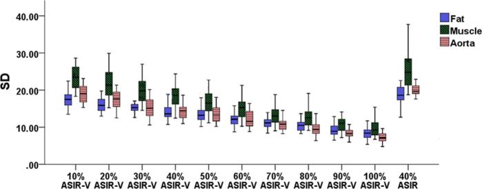
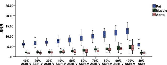
The image noise (SD) and SNR values of fat, muscle and aorta among different ASIR-V series (from 10 to 100% in a 10% increment), and 40% ASIR are listed in Table 2. For all ASIR-V images, the SD values of fat, muscle and aorta decreased and SNR values increased along with increasing ASIR-V percentage (Figures 1 and 2).
Table 2.
Comparison of image noise and SNR in different percentages of ASIR-V and 40% ASIR
| Noise (SD) | SNR | |||||
|---|---|---|---|---|---|---|
| Fat | Muscle | Aorta | Fat | Muscle | Aorta | |
| ASIR-V | ||||||
| 10% | 17.71 ± 2.84a | 23.45 ± 3.09a | 19.06 ± 2.32 | 6.13 ± 1.06 | 2.03 ± 0.42 | 1.70 ± 0.40 |
| 20% | 16.35 ± 2.26a | 21.61 ± 3.62a | 17.50 ± 2.54a | 6.69 ± 1.13a | 2.13 ± 0.42 | 1.85 ± 0.47 |
| 30% | 15.35 ± 1.72a | 19.92 ± 3.01a | 15.35 ± 2.48a | 7.03 ± 0.93a | 2.26 ± 0.49 | 2.06 ± 0.63 |
| 40% | 14.26 ± 2.17a | 18.51 ± 2.80a | 14.36 ± 2.10a | 7.63 ± 1.47a | 2.50 ± 0.67a | 2.27 ± 0.68a |
| 50% | 13.49 ± 2.06a | 16.74 ± 3.01a | 13.58 ± 2.22a | 8.08 ± 1.43a | 2.78 ± 0.76a | 2.37 ± 0.65a |
| 60% | 12.09 ± 1.75a | 15.04 ± 2.73a | 12.14 ± 2.40a | 9.15 ± 1.70a | 3.03 ± 0.77a | 2.67 ± 0.87a |
| 70% | 11.14 ± 1.31a | 13.12 ± 2.30a | 10.90 ± 1.73a | 9.71 ± 1.28a | 3.59 ± 0.97a | 2.84 ± 0.84a |
| 80% | 10.42 ± 1.47a | 12.62 ± 2.35a | 9.67 ± 1.82a | 10.48 ± 1.86a | 3.77 ± 0.88a | 3.52 ± 0.97a |
| 90% | 9.21 ± 1.62a | 10.92 ± 2.42a | 8.38 ± 1.10a | 11.92 ± 2.30a | 4.37 ± 1.42a | 3.84 ± 1.13a |
| 100% | 8.36 ± 1.48a | 10.07 ± 3.55a | 7.33 ± 1.35a | 13.19 ± 2.99a | 4.82 ± 1.51a | 4.40 ± 1.56a |
| 40% ASIR | 19.02 ± 3.50 | 25.45 ± 4.77 | 20.01 ± 1.50 | 5.81 ± 1.30 | 1.94 ± 0.56 | 1.72 ± 0.46 |
| p valueb | <0.05 | <0.05 | <0.05 | <0.05 | <0.05 | <0.05 |
ASIR, adaptive statistical iterative reconstruction; ASIR-V, adaptive statistical iterative reconstruction V; SNR, signal-to-noise ratio.
ap < 0.05 for paired comparison, ASIR-V series vs 40% ASIR.
bp values for the one-way ANOVA test for all groups.
Figure 1.
Comparison of objective image noise (SD) among different percentages of ASIR-V and 40% ASIR images. The graph showed that the SD values of fat, muscle and aorta on all ASIR-V series significantly decreased along with the increasing ASIR-V percentage when compared to the images reconstructed using 40% ASIR. ASIR, adaptive statistical iterative reconstruction; ASIR-V, adaptive statistical iterative reconstruction V.
Figure 2.
Comparison of the SNR among different percentages of ASIR-V and 40% ASIR images. The graph showed that the SNR values of fat, muscle and aorta on all ASIR-V series significantly increased along with the increasing ASIR-V percentage in comparison with the images reconstructed using 40% ASIR. ASIR, adaptive statistical iterative reconstruction; ASIR-V, adaptive statistical iterative reconstruction V; SNR, signal-to-noise ratio.
When comparing the 40% ASIR images to different ASIR-V series, the SD values of fat and muscle on all ASIR-V series were significantly lower than those with 40% ASIR. The SD values of aorta on the 20% ASIR-V to 100% ASIR-V images were lower in comparison with 40% ASIR images with statistically significant differences (p < 0.05) (Table 2).
In terms of SNR, the SNR values of fat on the 20% ASIR-V to 100% ASIR-V images were significantly higher than those on 40% ASIR. The SNR values of muscle and aorta on the 40% ASIR-V to 100% ASIR-V images were significantly improved when compared to 40% ASIR (p < 0.05) (Table 2).
Subjective measurement
The two radiologists had good to excellent consistencies for the subjective evaluation of the subjective image noise (κ = 0.73, p < 0.001), visibility of small structures (Kappa = 0.79, p < 0.001), image artefacts (Kappa = 0.84, p < 0.001) and diagnosis acceptability (Kappa = 0.83, p < 0.001). The subjective image quality scores of all images are listed in Table 3. The subjective image noise on ASIR-V series was significantly decreased with the increasing ASIR-V percentage and the 100% ASIR-V had the lowest image noise. Besides, scores of the subjective image noise on the 40% ASIR-V to 100% ASIR-V images were significantly better than those on the 40% ASIR images (p < 0.05). In addition, the 50% ASIR-V to 70% ASIR-V series showed significantly superior visibility of small structures, lower image artefacts and better diagnostic acceptability when compared with 40% ASIR (p < 0.05) (Figures 3 and 4). The 60% ASIR-V algorithm yielded the numerically highest overall image quality score. On the other hand, images reconstructed with ASIR-V of higher percentages (80%ASIR-V or higher) were more likely to present relatively poor image quality, due to the worsened sharpness of anatomical structures and diagnostic acceptability in comparison with other ASIR-V series.
Table 3.
Comparison of subjective image data in different percentages of ASIR-V and 40% ASIR
| Score | ||||
|---|---|---|---|---|
| Subjective noise | Visibility | Artefacts | Diagnosis acceptability | |
| ASIR-V | ||||
| 10% | 3.60 ± 0.50 | 3.77 ± 0.50 | 3.70 ± 0.47 | 4.37 ± 0.67 |
| 20% | 3.63 ± 0.49 | 3.83 ± 0.59 | 3.77 ± 0.43 | 4.47 ± 0.57 |
| 30% | 3.83 ± 0.59 | 4.07 ± 0.64a | 3.93 ± 0.74 | 4.50 ± 0.51 |
| 40% | 4.10 ± 0.61a | 4.30 ± 0.54a | 4.19 ± 0.48a | 4.57 ± 0.50 |
| 50% | 4.43 ± 0.57a | 4.47 ± 0.57a | 4.33 ± 0.48a | 4.73 ± 0.45a |
| 60% | 4.63 ± 0.56a | 4.63 ± 0.49a | 4.50 ± 0.51a | 4.87 ± 0.35a |
| 70% | 4.73 ± 0.45a | 4.60 ± 0.50a | 4.47 ± 0.51a | 4.83 ± 0.38a |
| 80% | 4.80 ± 0.41a | 3.93 ± 0.58 | 4.24 ± 0.45a | 4.54 ± 0.49 |
| 90% | 4.87 ± 0.35a | 3.60 ± 0.62 | 3.97 ± 0.56 | 4.50 ± 0.51 |
| 100% | 4.93 ± 0.25a | 3.37 ± 0.56 | 3.90 ± 0.48 | 4.47 ± 0.51 |
| 40% ASIR | 3.70 ± 0.47 | 3.73 ± 0.52 | 3.83 ± 0.38 | 4.33 ± 0.66 |
| p valueb | <0.05 | <0.05 | <0.05 | <0.05 |
ASIR, adaptive statistical iterative reconstruction; ASIR-V, adaptive statistical iterative reconstruction V.
ap < 0.05 for paired comparison, ASIR-V series vs 40% ASIR.
bp values for the one-way ANOVA test for all groups.
Figure 3.
Transverse non-enhanced chest CT images of a 62year-old male with a body mass index of 24.07 kg m−2. Images reconstructed included 40% ASIR (A1), 50% ASIR-V (A2), 70% ASIR-V (A3) in mediastinal window from the same patient. The SD of ROI (circle) for fat, muscle and aorta of 50% ASIR-V (A2) and 70% ASIR-V (A3) images were significantly lower than those with 40% ASIR (A1). ASIR, adaptive statistical iterative reconstruction; ASIR-V, adaptive statistical iterative reconstruction V; ROI, region of interest.
Figure 4.
Transverse non-enhanced chest CT images of a 58-year-old female with lesions in the lower lobe of the lungs. The edges of lesions and bronchus (arrows) of images reconstructed with 50% ASIR-V (B2) and 70% ASIR-V (B3) in lung window were clearly in comparison with 40% ASIR images (B1). The scores of the visibility of small structures of 40% ASIR (B1), 50% ASIR-V (B2) and 70% ASIR-V (B3) were 3, 4 and 4, respectively. ASIR, adaptive statistical iterative reconstruction; ASIR-V, adaptive statistical iterative reconstruction V.
Discussion
In recent years, iterative reconstruction algorithms have been used and studied by many scholars for improving image quality. The biggest advantage of iterative reconstruction algorithm is that even if the original data were obtained at low signals, it is still possible to reconstruct a higher quality image with lower noise and less artefacts. As the first generation iterative reconstruction algorithm, ASIR has been widely used in clinical practice. ASIR technique takes full account of the statistical noise model of data and through the iterative reactions with the raw data to quickly reduce the image noise without severely affecting the image spatial resolution compared with the simple image smoothing operations.14–16
However, ASIR technique also has its own limitations. ASIR only includes the system noise model and object model, and lacking the more complexed physics model and optical model. Therefore, it may be more likely to cause changes in noise frequency, resulting in over-smoothness in correcting image noise. On the other hand, the full model-based iterative reconstruction (MBIR) algorithm includes not only the system noise model and the object model, but also the physics model and optical model, which can reduce the image noise more effectively than the ASIR and improve image spatial resolution at the same time.17–20 However, due to its longer operation time, MBIR is yet to be used routinely in the clinical work.
As a newly developed iterative reconstruction algorithm, ASIR-V is a new reconstruction algorithm between ASIR and MBIR algorithms, and has its own characteristics. Unlike MBIR that incorporates the four models, ASIR-V discards the system optical model, which is the most computational expensive model, to make it a real-time reconstruction algorithm.21 Therefore, the operation speed of ASIR-V is much faster than that of MBIR. At the same time, ASIR-V retains the upgraded system noise model, the object model and the physics model, which in theory can help to reduce image noise more effectively, and improve the density resolution and suppress image artefacts, thus providing images with superior anatomical details and higher diagnosis acceptability and producing a greater clinical value than ASIR.
In our study with 30 patients, we compared the value between ASIR-V and ASIR in image noise reduction and diagnostic performance. Since our previous clinical experience and many other researchers have demonstrated that the 40% is the “optimal” percentage for ASIR algorithm in the non-contrast enhanced CT scans,9–11 we have selected the recommended level of 40%ASIR for comparison purpose. From the results of the subjective and objective evaluation, we demonstrated that ASIR-V in a wide percentage range (30–80%) could significantly reduce image noise and improve image quality compared to 40% ASIR.
When the images from the same patients were reconstructed using ASIR-V, it was proved that ASIR-V had a more significant noise reduction capability than ASIR. In the assessments of objective image quality, the image noise on 40% ASIR-V was significantly reduced in comparison with 40% ASIR. In addition, in this study, we also analysed the subjective image scores; it was clearly demonstrated that the subjective image noise decreased with increased ASIR-V initially, and the image contrast resolution increased accordingly. However, when ASIR-V increased to a certain extent (80% ASIR-V and above this level), the images started to produce a “waxy” texture or blotchy appearance, unfamiliar to radiologists accustomed to the 40% ASIR images in clinical work. And we observed that ASIR-V percentages were not always positively correlated with subjective scores; the scores of a higher percentage of ASIR-V started to decline because of the changed noise structure, affecting the diagnosis acceptability.22 The image quality fall-offs of ASIR-V at high percentages was similar to ASIR algorithm, but at a much higher level.
There were several limitations in our present study. First, this study mainly evaluated the overall image quality, and did not classify the different lung diseases. However, different diseases might have an effect on the display of the image details, resulting in the deviation of the subjective image evaluation. Second, our study focused on the non-enhanced chest CT scans with different imaging characteristics from the contrast enhanced CT scans and CT angiography. Therefore, the “optimal” percentage of ASIR-V may be different for the contrast-enhanced CT scans. Third, although qualitative assessment was made by two experienced radiologists who blindly scored the subjective image noise and visibility of anatomical structure without patient basic information on the images, there still existed too much observer bias due to the differences in the appearances of the natural data characteristics between the ASIR and ASIR-V series. Fourth, the sample size was not large enough, we just studied a small number of patients, therefore, a large sample and more detailed study is still expected. Finally, in our study, we only evaluated the noise reduction potential of ASIR-V, but not the radiation dose saving potential since the scan protocol was fixed. Although the noise reduction potential and the optimal strength for ASIR-V are of interest for people in the field looking for optimal ASIR-V protocols, it might be more interesting to do research on the dose reduction potential. However, since there is a known relationship between noise and radiation dose, our results about the noise reduction potential can easily be converted into dose reduction potential, but further research is needed to verify this potential.
Conclusion
In conclusion, ASIR-V shows greater potential in reducing image noise and artefacts when compared to the recommended level of 40% ASIR in the non-enhanced chest CT scans. The 50% ASIR-V to 70% ASIR-V series showed significantly superior image quality among all the image series, and the reconstructions with 60% ASIR-V yield the best image quality and highest diagnostic confidence.
Consent
This prospective clinical study was approved by the Ethics Committees of our hospital, and written informed consent was signed by all participants.
Contributor Information
Nan Yu, Email: 349863320@qq.com.
Yongjun Jia, Email: jiayongjun1985@163.com.
Yong Yu, Email: 22434158@qq.com.
Haifeng Duan, Email: dhf98@163.com.
Dong Han, Email: 147690660@qq.com.
Guangming Ma, Email: 416725386@qq.com.
Chenglong Ren, Email: 442981552@qq.com.
Taiping He, Email: htpkeyan@163.com.
References
- 1.Bischoff B, Hein F, Meyer T, Hadamitzky M, Martinoff S, Schömig A, , et al. Trends in radiation protection in CT: present and future status. J Cardiovasc Comput Tomogr 2009; 3(Suppl 2): S65–S73. [DOI] [PubMed] [Google Scholar]
- 2.Alkhorayef M, Babikir E, Alrushoud A, Al-Mohammed H, Sulieman A, . Patient radiation biological risk in computed tomography angiography procedure. Saudi J Biol Sci 2017; 24: 235–40. [DOI] [PMC free article] [PubMed] [Google Scholar]
- 3.Yerramasu A, Venuraju S, Atwal S, Goodman D, Lipkin D, Lahiri A, . Radiation dose of CT coronary angiography in clinical practice: objective evaluation of strategies for dose optimization. Eur J Radiol 2012; 81: 1555–61. [DOI] [PubMed] [Google Scholar]
- 4.Chen JH, Jin EH, He W, Zhao LQ, . Combining automatic tube current modulation with adaptive statistical iterative reconstruction for low-dose chest CT screening. PLoS One 2014; 9: e92414. [DOI] [PMC free article] [PubMed] [Google Scholar]
- 5.Prezzi D, Goh V, Virdi S, Mallett S, Grierson C, Breen DJ, , et al. Adaptive statistical iterative reconstruction improves image quality without affecting perfusion CT quantitation in primary colorectal cancer. Eur J Radiol Open 2017; 4: 69–74. [DOI] [PMC free article] [PubMed] [Google Scholar]
- 6.Rapalino O, Kamalian S, Kamalian S, Payabvash S, Souza LC, Zhang D, , et al. Cranial CT with adaptive statistical iterative reconstruction: improved image quality with concomitant radiation dose reduction. AJNR Am J Neuroradiol 2012; 33: 609–15. [DOI] [PMC free article] [PubMed] [Google Scholar]
- 7.Kaul D, Grupp U, Kahn J, Ghadjar P, Wiener E, Hamm B, , et al. Reducing radiation dose in the diagnosis of pulmonary embolism using adaptive statistical iterative reconstruction and lower tube potential in computed tomography. Eur Radiol 2014; 24: 2685–91. [DOI] [PubMed] [Google Scholar]
- 8.Qi LP, Li Y, Tang L, Li YL, Li XT, Cui Y, , et al. Evaluation of dose reduction and image quality in chest CT using adaptive statistical iterative reconstruction with the same group of patients. Br J Radiol 2012; 85: e906–11. [DOI] [PMC free article] [PubMed] [Google Scholar]
- 9.Kim HG, Chung YE, Lee YH, Choi JY, Park MS, Kim MJ, , et al. Quantitative analysis of the effect of iterative reconstruction using a phantom: determining the appropriate blending percentage. Yonsei Med J 2015; 56: 253–61. [DOI] [PMC free article] [PubMed] [Google Scholar]
- 10.Brady SL, Moore BM, Yee BS, Kaufman RA, . Pediatric CT: implementation of ASIR for substantial radiation dose reduction while maintaining Pre-ASIR image noise. Radiology 2014; 270: 223–31. [DOI] [PMC free article] [PubMed] [Google Scholar]
- 11.Vorona GA, Ceschin RC, Clayton BL, Sutcavage T, Tadros SS, Panigrahy A, . Reducing abdominal CT radiation dose with the adaptive statistical iterative reconstruction technique in children: a feasibility study. Pediatr Radiol 2011; 41: 1174–82. [DOI] [PubMed] [Google Scholar]
- 12.Benz DC, Gräni C, Mikulicic F, Vontobel J, Fuchs TA, Possner M, , et al. Adaptive statistical iterative reconstruction-V: impact on image quality in ultralow-dose coronary computed tomography angiography. J Comput Assist Tomogr 2016; 40: 958–63. [DOI] [PubMed] [Google Scholar]
- 13.Lim K, Kwon H, Cho J, Oh J, Yoon S, Kang M, , et al. Initial phantom study comparing image quality in computed tomography using adaptive statistical iterative reconstruction and new adaptive statistical iterative reconstruction v. J Comput Assist Tomogr 2015; 39: 1–8. [DOI] [PubMed] [Google Scholar]
- 14.Sagara Y, Hara AK, Pavlicek W, Silva AC, Paden RG, Wu Q, . Abdominal CT: comparison of low-dose CT with adaptive statistical iterative reconstruction and routine-dose CT with filtered back projection in 53 patients. AJR Am J Roentgenol 2010; 195: 713–9. [DOI] [PubMed] [Google Scholar]
- 15.Hussain FA, Mail N, Shamy AM, Suliman A, Saoudi A, . A qualitative and quantitative analysis of radiation dose and image quality of computed tomography images using adaptive statistical iterative reconstruction. J Appl Clin Med Phys 2016; 17: 419–32. [DOI] [PMC free article] [PubMed] [Google Scholar]
- 16.Brady SL, Moore BM, Yee BS, Kaufman RA, . Pediatric CT: implementation of ASIR for substantial radiation dose reduction while maintaining pre-ASIR image noise. Radiology 2014; 270: 223–31. [DOI] [PMC free article] [PubMed] [Google Scholar]
- 17.Annoni AD, Andreini D, Pontone G, Formenti A, Petullà M, Consiglio E, , et al. Ultra-low-dose CT for left atrium and pulmonary veins imaging using new model-based iterative reconstruction algorithm. Eur Heart J Cardiovasc Imaging 2015; 16: 1366–73. [DOI] [PubMed] [Google Scholar]
- 18.Smith EA, Dillman JR, Goodsitt MM, Christodoulou EG, Keshavarzi N, Strouse PJ, . Model-based iterative reconstruction: effect on patient radiation dose and image quality in pediatric body CT. Radiology 2014; 270: 526–34. [DOI] [PMC free article] [PubMed] [Google Scholar]
- 19.Yasaka K, Katsura M, Hanaoka S, Sato J, Ohtomo K, . High-resolution CT with new model-based iterative reconstruction with resolution preference algorithm in evaluations of lung nodules: Comparison with conventional model-based iterative reconstruction and adaptive statistical iterative reconstruction. Eur J Radiol 2016; 85: 599–606. [DOI] [PubMed] [Google Scholar]
- 20.Deák Z, Grimm JM, Treitl M, Geyer LL, Linsenmaier U, Körner M, , et al. Filtered back projection, adaptive statistical iterative reconstruction, and a model-based iterative reconstruction in abdominal CT: an experimental clinical study. Radiology 2013; 266: 197–206. [DOI] [PubMed] [Google Scholar]
- 21.Kwon H, Cho J, Oh J, Kim D, Cho J, Kim S, , et al. The adaptive statistical iterative reconstruction-V technique for radiation dose reduction in abdominal CT: comparison with the adaptive statistical iterative reconstruction technique. Br J Radiol 2015; 88: 20150463. [DOI] [PMC free article] [PubMed] [Google Scholar]
- 22.Lee S, Kwon H, Cho J, . The detection of focal liver lesions using abdominal CT: a comparison of image quality between adaptive statistical iterative reconstruction V and adaptive statistical iterative reconstruction. Acad Radiol 2016; 23: 1532–8. [DOI] [PubMed] [Google Scholar]


