Abstract
Mucous-mesh grazers (pelagic tunicates and thecosome pteropods) are common in oceanic waters and efficiently capture, consume and repackage particles many orders of magnitude smaller than themselves. They feed using an adhesive mucous mesh to capture prey particles from ambient seawater. Historically, their grazing process has been characterized as non-selective, depending only on the size of the prey particle and the pore dimensions of the mesh. The purpose of this review is to reverse this assumption by reviewing recent evidence that shows mucous-mesh feeding can be selective. We focus on large planktonic microphages as a model of selective mucus feeding because of their important roles in the ocean food web: as bacterivores, prey for higher trophic levels, and exporters of carbon via mucous aggregates, faecal pellets and jelly-falls. We identify important functional variations in the filter mechanics and hydrodynamics of different taxa. We review evidence that shows this feeding strategy depends not only on the particle size and dimensions of the mesh pores, but also on particle shape and surface properties, filter mechanics, hydrodynamics and grazer behaviour. As many of these organisms remain critically understudied, we conclude by suggesting priorities for future research.
Keywords: bentho-pelagic coupling, selective feeding, grazers, tunicates, pteropods
1. Introduction
Particles in the ocean exhibit striking diversity in size, shape and chemistry [1,2]. If and how grazers select particles from this mixed assemblage influences food web structure and carbon flux [3,4]. Some of the more abundant marine grazers use mucous meshes to capture prey—a process considered to have minimal potential for dietary selection [5–7]. Here, we review recent developments in the literature that collectively portray mucous-mesh feeding as a more selective process than historically assumed.
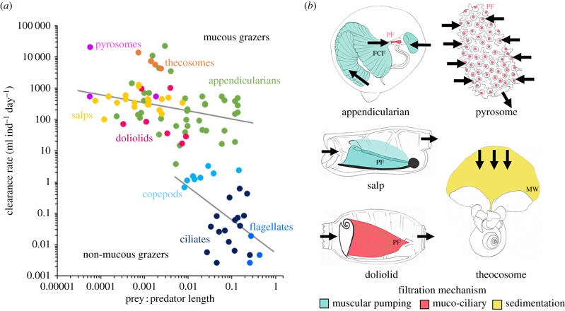
Although mucus-based feeding mechanisms have independently evolved in multiple animal classes, this review is restricted to ‘large planktonic microphages' [8] (pelagic tunicates and thecosome pteropods) because of their important, yet understudied, roles as benthic–pelagic links and key species in the planktonic food web [4,8,9]. All of these animals use mucous filters with large surface areas to maximize particle capture rates (figure 1a). We use the term ‘mucous-mesh grazers’ throughout, although the term ‘grazers’ is used loosely, because some taxa are omnivorous rather than strictly herbivorous [10–15].
Figure 1.
Mucous-mesh grazing: (a) clearance rates of mucous-mesh grazers and other common, non-mucus microphagous grazers versus prey-to-predator length (electronic supplementary material, table S1). (b) Schematic showing location of the mucous mesh for different grazers. The mucous mesh is highlighted according to the mechanism used to drive flow through or across the mesh. (b) IF, inlet filter; FCF, food-concentrating filter; PF, pharyngeal filter; MW, mucous web. Drawings by Caitlyn Webster. (Online version in colour.)
Although mucous-mesh grazers tend to be overlooked—with the majority of species distributed far offshore—their ecological impacts are pronounced. Not only does their feeding process typify a mechanistic interaction between small particles and mucus, they also occupy a unique role in the ocean food web. While the consumer–prey size ratio for heterotrophic filter-feeding plankton ranges from approximately 5 : 1 to 100 : 1 [16], mucous-mesh grazers can achieve ratios greater than 10 000 : 1 (figure 1a). This makes them uniquely capable of short-circuiting the microbial loop and providing a more efficient linkage in the trophic chain [8,17,18].
Below, we highlight important differences in the hydrodynamics and filtration mechanics of each of the three groups of mucous-mesh grazers. Next, we summarize the different ways these organisms impact ocean biogeochemical cycling. Then we review the passive and behavioural mechanisms by which mucous-mesh grazers can selectively feed. Equal weight is not given to all taxa because some are less studied than others, owing to patchy or episodic distribution, difficulties with handling, laboratory maintenance or observations of feeding [11,18,19]. We conclude by suggesting future research directions to help remedy these gaps.
2. Mucous-mesh grazers
(a). Appendicularians
Tunicates in the class Appendicularia feed using an external cellulose and mucus filtration apparatus (the house) and an internal mucus filter (the pharyngeal filter) (figure 1b). Sinusoidal beating of the muscular tail drives flow into the house and through the food-concentrating filter, which, for Oikopleura dioica, concentrates particles through serial adhesion and detachment in coordination with the tail beating [20]. After being conveyed through the food-concentrating filter, fluid and suspended particles move through the buccal tube and into the mouth, where they are captured on the pharyngeal filter for ingestion (figure 1b). Appendicularians discard and build new houses at species-specific rates, ranging from two to 40 houses per individual per day [21].
(b). Thaliaceans
The tunicate class Thaliacea includes three orders—Salpida, Doliolida and Pyrosomida—that all feed by secreting a mucous mesh that moves posteriorly towards the oesophagus, where it is rolled into a mucus string by cilia and ingested [22]. Salps and doliolids are barrel-shaped zooids that pass water from an afferent siphon out through an efferent siphon (figure 1b). Salps achieve high filtration rates [23] and produce swimming wakes through muscular contraction (figures 1b and 2) [28]. The feeding current of doliolids is achieved through ciliary beating (figure 1b) [29,30]. Pyrosomes are permanently colonial, with zooids held side by size in a gelatinous tunic [31]. Like salps and doliolids, the individual zooids have an afferent and efferent siphon. The tubular colonies move slowly by the continuous expulsion of fluid through the individual efferent siphons and out of a common aperture (figure 1b) [32].
Figure 2.
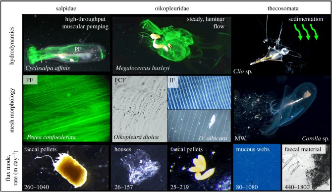
Hydrodynamics, mesh morphologies, and flux of mucous-mesh grazers. PF: pharyngeal filter; FCF: food-concentrating filter; IF: inlet filter prior- (top) and post-inflation of the house (bottom); MW: mucous web. Photographs courtesy of: Linda Ianniello for Clio sp. and Corolla sp., S. Bush for Peraclidae mucous web, © 2008 MBARI, Ron Gilmer for Cavolinia uncinata faecal material [10]. Salpidae flux rates based on the faecal pellets of Pegea confoederata [24]; Oikopleuridae flux rates based on Oikopleura dioica faecal pellets [25] and houses [26]; Thecosomata flux rates based on the mucous webs of Limacinia retroversa [27] and faecal material of Corolla spectabilis [9]. (Online version in colour.)
(c). Thecosome pteropods
Thecosomes are a holoplanktonic order of gastropods that feed using a large external mucous web suspended above the animal (figure 1b) [12,33]. Their molluscan foot has been modified into a wing-like appendage. Unlike tunicates, thecosomes are not true filter-feeders because they do not generate feeding currents; instead, they cease swimming and attain near or complete neutral buoyancy [12], passively entrapping suspended particles via ‘flux feeding,’ a variant of passive ambush feeding (figure 1b) [33,34]. Motile organisms also can be trapped by swimming into the web [10–12]. After prey capture, thecosomes ingest the web by pulling it into the pharynx [19,35].
3. Ecological impacts
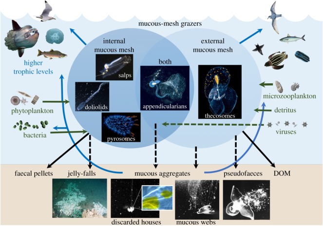
Mucous-mesh grazers are uniquely capable of capturing a wide range of particle sizes, and filtering water at high rates (figure 1a). Because mucus is adhesive, the mesh can capture particles smaller than its pores through hydrosol filtration mechanisms [9,27,33,36,37]. Although mucous-mesh grazers were once considered a trophic ‘dead end’ because of their high water content and consequently low to moderate caloric value per unit volume [38], they can have relatively high carbon and protein per unit dry mass; as such, they may represent a valuable food source, particularly at times of prey scarcity [39,40]. Mucous-mesh grazers are increasingly recognized as prey for higher trophic levels (figure 3). Thaliaceans [18,41–44], appendicularians [45,46] and pteropods [11,47,48] contribute a significant proportion of the diet for several fish species and as such can provide a more expedited trophic link to fisheries production [8].
Figure 3.
Contributions of mucous-mesh grazers to the ocean food web. Arrows show common flux pathways (solid line) and pathways unique to a specific group (dashed line), including jelly-falls (Pyrosoma atlanticum, courtesy of S. Marion), appendicularian houses (inset shows fluorescent inclusions in the house rudiment of Oikopleura albicans), mucous webs and pseudofaeces (Corella calceola from Gilmer [12], courtesy of R. Gilmer). Pyrosome and thecosome photographs courtesy of Mike Bartick; appendicularian photograph courtesy of Linda Ianniello. (Online version in colour.)
Mucous-mesh grazers also influence the marine particle field through their production of mucous aggregates that contribute to the downward flux of organic matter, sinking at rates ranging from 80 to 800 m day−1 (figure 2) [27,49]. Discarded appendicularian houses and thecosome webs contain accumulated pico- and nano-plankton [50–52]. They can also serve as microhabitats with elevated levels of heterotrophic bacterial growth and remineralization [53–56]. Because many of these mucous aggregates can fluoresce and luminesce [57], both visual feeders and non-visual flux-feeders consume them [15,52,58–62] (figure 3).
All mucous-mesh grazers sequester biogenic carbon through production of faecal pellets that sink at high rates (figure 2) [63], except those of doliolids, which are not as compact [64]. Faecal pellets tend to contain more refractory carbon than discarded feeding structures, but can also be a food source for other zooplankton [65]. Only pteropods produce pseudofaeces, composed of mucus and rejected food particles (figures 2 and 3) [58].
Abundance of some mucous-mesh grazers can be pulsed. As these episodic populations die, the carcasses contribute to ‘jelly-falls,’ which provide particulate organic matter to the seabed [66]. Pyrosomes and salps are important contributors to jelly-falls, and doliolids and pteropods may also contribute to a lesser-known extent [67–69] (figure 3).
4. Selective grazing using mucous meshes
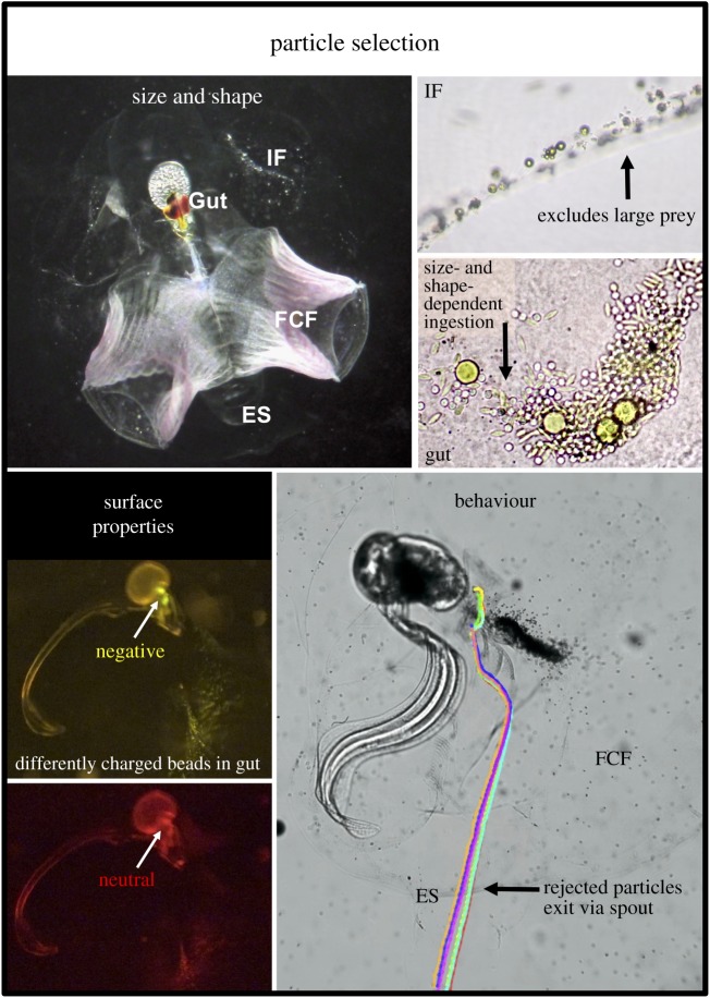
We define selective feeding as ‘an imbalance between the proportion of prey types in a predator's diet and the proportion in the environment’ [70]. Defining selectivity for appendicularians is complicated by the distinction between the preferential ingestion of certain particles by the animal and the differential retention of particles by the house (figure 4), although both processes can affect ambient particle size spectra and composition [71]. We review physical selection mechanisms that depend on the properties of the particles and mechanisms that depend on grazer behaviour (figure 4). Although this framework suggests these mechanisms are discrete, in many cases the selection process depends on the interaction between particle properties and a behavioural response.
Figure 4.
Physical and behavioural particle selection mechanisms of mucous-mesh grazers using the appendicularian Oikopleura dioica as a model. The inlet filters (IF) exclude large and spinous prey from entering the house. Small particles, such as the red carmine dye, are more likely to adhere to the food-concentrating filter (FCF). Both of these filtration processes determine what reaches the pharyngeal filter and gut. Surface properties, such as charge, influence particle interactions with the mucous filters (courtesy of A. Karim). Particles rejected by behavioural mechanisms (shown by coloured tracks) can exit the house via the exit spout (ES). (Online version in colour.)
5. Physical selection mechanisms
(a). Size
Mucous-mesh grazers feed in a low Reynolds number environment with thick viscous boundary layers because of the fine mucous-mesh fibres [34]. Within this viscous regime, all mucous-mesh grazers exhibit mechanical, size-dependent selection (figure 1) with a lower limit of particle retention (set in part by the dimensions of the filter pores), and an upper limit that is set by the diameter of the mucous mesh or the animal's mouth (electronic supplementary material, table S2). The upper and lower limits of particle retention vary considerably by species (figure 1), but all appear to capture submicron particles with imperfect (less than 100%) efficiency [9,27,36,72–76]. Despite this, cells in the picoplankton size range (0.2–2 µm) can still constitute an important contribution to the energetic demands of these organisms [36].
The effective pore size of the mesh is inconstant, depending upon environmental conditions and animal behaviour—for example, through mesh clogging, which can depend upon the ambient particle size and concentrations, or the mucus translational speed, which may affect mesh stretching (electronic supplementary material, table S2). While the mucous filters of tunicates are usually arranged in a rectangular mesh pattern (figure 2) [36,77], the pores of pteropod webs are more irregular [51]. We evaluate the differential size retention patterns by each taxon.
Although historically appendicularians have been assumed to feed non-selectively [5,6,78], we argue that this is a misrepresentation, because the house necessarily causes size-dependent selection by preventing some particles from being ingested [71,79]. For most species of appendicularians, size-dependent selection first occurs at the inlet filter, which excludes large particles (approx. 15–54 µm, depending on the species) from entering the house [80,81]. Spinous particles, such as Trichodesmium or foraminifera, as well as large dinoflagellates and diatoms, detritus and metazoans are often excluded [50,80,81]. These particles may or may not remain associated with the house when it is discarded, depending on how strongly they adhere to the filter and whether or not they are dislodged during tail arrests and associated back-flushing of the filters [82–84]. Some appendicularians lack inlet filters and thus the dimensions of the incurrent openings are the only limitation on the size of particles that may enter the house [80]. In addition to inlet filters, Fritillaria borealis can exclude large particles (greater than 30 µm) by arresting them against the anterior wall of the tail chamber and ejecting them from the house [83].
In Oikopleurids, size-dependent selection then occurs at the food-concentrating filters on which smaller particles are more likely to remain stuck [5,20,85,86]. After conveyance through the food-concentrating filter, particles reach the pharyngeal filter, which has a left-skewed retention efficiency curve that declines below approximately 3 µm for larger species (O. vanhoeffeni) [87,88] and approximately 1–2 µm for smaller species (O. dioica and F. borealis) [89]. However, gut content analysis and experiments with live prey indicate that appendicularians can consume small bacteria and large viruses (less than 0.3 µm) [90–92]. Oikopleura dioica can even filter viruses (160–180 nm) at rates comparable to those of larger algae (2–50 ml−1 ind−1 day−1) [92].
The pharyngeal filter of thaliaceans appears less efficient at retaining small particles than does that of appendicularians. Experimental evidence shows that many species of salps retain particles less than 2–4 µm with less than 100% efficiency [93,94], with only approximately 15% efficiency of 1.0 µm particles [73], although mathematical models predict higher retention of smaller particles through hydrosol mechanisms [36]. Experimental results show Pegea confoederata can ingest particles down to 0.5 µm and mathematical modelling suggests it may capture particles as small as 0.05 µm through hydrosol mechanisms, but only at less than 2% efficiency [36].
The size-retention patterns of doliolids and pyrosomes remain less clear [77]. Evidence from chemostats [74], mesocosms [75], incubations [95] and faecal pellet analysis [96] indicates doliolids can capture submicron free-living bacteria (0.2 µm) with unknown efficiency. Measurements of the mesh of the pyrosome Pyrosoma atlanticum [97] suggest submicron particle capture is likely (electronic supplementary material, table S2). The only study to date on size selectivity of pyrosomes showed favourable selection of particles greater than 10 µm [76]. The smallest cells identified in P. atlanticum faecal pellets were 3–5 µm phytoplankton [72], but a recent study hypothesized that a swarm of P. spinosum was sustained by high densities of Synechococcus and flagellates approximately 1–3 µm [98].
Thecosomes capture a wide range of particles, including small copepods, diatoms, dinoflagellates, coccolithophores, protozoans, detritus and bacteria [35,51,99]. The upper size limit for particle capture is set by the maximum dimensions of the tract used to transport the web to the mouth, which is in the range of 200–800 µm [51]. Clearance of small particles (less than 1 µm) may be facilitated by particle aggregation in mucus produced during spawning, or by direct capture through adhesion to the mucous web [27,51].
(b). Shape
Filter-feeding is defined as ‘feeding by passing the surrounding water through structures that retain particles mainly according to size and shape’ [100]. Despite this, only two studies have explicitly examined the effect of shape on selectivity by mucous-mesh grazers; both focused on appendicularians. In one, the minimum diameter of ellipsoidal particles was the key variable for determining how cells were grazed by O. dioica [71]. In another, retention by O. dioica depended on algal cell shape, algal concentration, and whether the animals were fed a monoculture or a mixed algal suspension [101]. A smaller alga with projecting spines was often retained on the inlet filters and blocked the entry of the larger particles into the house [101]. A few other studies have suggested that appendicularians may exhibit reduced ingestion of spinous prey [50,85,102]. Otherwise, the effects of particle shape on selection remain largely overlooked in spite of the abundance of non-spherical particles in the ocean.
(c). Surface properties
A growing body of work calls for a re-evaluation of the role of particle surface properties in dietary selection in the ocean. Surface properties, including charge [103–105], biochemical coatings [106] and hydrophobicity [103,105,107–109] affect particle retention by suspension-feeders. Understanding how surface properties affect selection by mucous-mesh grazers requires a biochemical characterization of both the grazer's mucus and the prey particles. Although gastropod mucus has been characterized—consisting of protein–polysaccharides, often with negatively charged acidic carbohydrates—the mucus of pteropod webs has not [110]. Benthic tunicate mucous meshes also contain acidic mucopolysaccharides and mucoproteins [77,111], but the molecular compositions of pelagic tunicate meshes appear to be quite distinct [112].
Despite the high likelihood that particle surface properties play a role in dietary selection, only one study to date has examined this in mucous-mesh grazers. Oikopleura albicans removed cyanobacteria, but had null or low retention of the similarly sized SAR 11 heterotrophic bacterial clade [108]. Cell surface hydrophobicity was invoked as a probable mechanism for the observed retention patterns because the SAR 11 clade had a lower hydrophobic interaction chromatography index than other bacterial phylotypes measured [108].
6. Behavioural selection mechanisms
Among mucous-mesh grazers, appendicularians exhibit the greatest array of behavioural mechanisms for selection of particles (figure 4). At least three particle rejection mechanisms exist: (i) secretion of the pharyngeal filter may cease, causing particles to exit via the spiracles [113]; (ii) the spiracles can create a flow reversal when undesirable particles contact sensory hairs on the lips, rejecting individual particles out of the mouth or buccal tube [20,82,114–116]; (iii) the lower lip can cover the mouth, causing bulk particles to be rejected non-selectively via ‘pipe-smoking’ [113,114,117], possibly in response to satiation at high particle concentrations (greater than 20 000 cells ml−1) [118].
Thaliaceans have a limited array of behavioural selection mechanisms [119]. The different classes have a sensory structure—some shared and some unique—to respond primarily to mechanical and chemical stimuli [120]. Doliolids and salps can perform a ‘crossed reflex’ to reject food, swimming backwards to prevent large objects from entering the pharynx [29,121,122]. Doliolids can also arrest the cilia of the gill bars when large or noxious particles contact the mouth, crushing spinous cells into smaller pieces by cyclically reversing the mucus cord prior to ingestion [29]. Pyrosomes can also arrest the gill cilia in response to large particles [120].
Only a few field observations have been made of thecosomes feeding [12,51,58]. Like thaliaceans, thecosomes have a behavioural response that allows them to dislodge excessive food particles through vigorous beating of the wings [51], but production of pseudofaeces is their primary mechanism for behavioural selection of particles. The ciliary pathways on the mantle lining, footlobes and wings sort and reject prey particles prior to ingestion [35]. Rejected material mixes with mucus (pseudofaeces) and is transported away from the mouth and web by cilia on the footlobes [35].
Swimming is an additional mechanism for behavioural selection by appendicularians, pteropods and some thaliaceans. After ingesting a web, thecosomes can either swim to a new location to secrete a new web, or may set sequential webs in the same location [12]. By regulating the speed of the tail beat, cultured O. dioica may move through different particle environments and select favourable patches to dwell [82]. Oikopleura dioica swim more at low particle concentrations and reduce tail beat frequency at high particle concentrations—a behavioural mechanism that reduces the negative effects of high food concentrations [118]. House abandonment may be an additional response to an undesirable particle field [5].
7. Future directions
Mounting evidence overturns the paradigm that mucus-mesh grazers are non-selective and instead shows that mesh morphology, behaviour, hydrodynamics and particle properties play important roles in determining particle selection. Further advances will yield a more informed understanding of selective processes and the consequences for food-web dynamics and particle export.
Culture advancements have been made for the pteropod Limacina helicina [123] and the appendicularians O. dioica [124] and F. borealis (JM Bouquet 2017, personal communication). Earlier efforts to culture salps [125] and doliolids [126] have not been replicated; still, continued developments in culture techniques could allow for more detailed observations at the level of feeding structures and controlled feeding incubations.
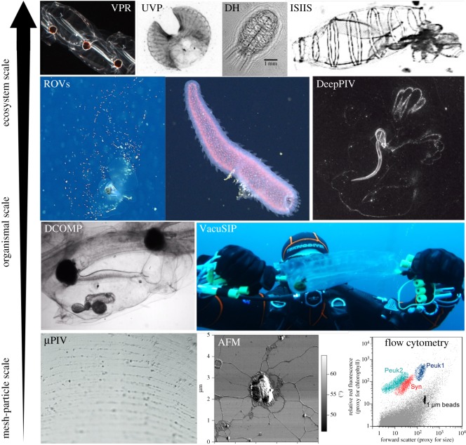
The fragile nature of mucous-mesh grazers has hampered previous efforts to study them. The most promising tools allow for undisturbed observations in the field, including diver-operated, towed and remotely operated systems (figure 5). Most recently, the filtration rates [133] and size selectivity [134] of giant appendicularians were quantified with DeepPIV, and efforts are underway to investigate the selectivity of salps using the VacuSIP technique [135] coupled with Next Generation Sequencing (A Dadon-Pilosof 2017, personal communication) (figure 5). Many of these systems allow for quantification of non-uniform selection on natural prey assemblages at the same time that they allow for visualizations of the particular mechanisms driving selection.
Figure 5.
Future directions for investigating feeding by mucous-mesh grazers at different spatial scales. Mesh-particle scale techniques: micro-scale particle image velocimetry (μPIV) [127]; atomic force microscopy (AFM) topographic image of two conjoint Synechococcus cells surrounded by a gel matrix (courtesy of F. Malfatti [128]); flow cytometry cytogram showing prey particles (Syn: Synechoccocus, Peuks: picoeukaryotes) from pyrosome gut contents (courtesy of A. Thompson). Diver-operated methods: Diver Controlled Observation and Measurement of Plankton (DCOMP) (Weelia cylindrica); VacuSIP (courtesy of A. Dadon-Pilosof). Remotely operated systems: remotely operated vehicles (ROVs) (Pteropod © 2008 MBARI, courtesy of S. Bush; Pyrosoma atlanticum © 2014 MBARI); DeepPIV (Bathochordaeus stygius, courtesy of K. Katija). Towed systems: video plankton recorder (VPR) (Salpa aspera, courtesy of C. Davis [129]); underwater vision profiler (UVP) (appendicularian house, courtesy of L. Stemmann [130]); digital holography (DH) (Thalia democratica, courtesy of N. Loomis [131]); In situ Ichthyoplankton Imaging System (ISIIS) (budding doliolid, courtesy of M. Schmid [132]). (Online version in colour.)
Understanding the mechanisms of selection by mucous-mesh grazers is particularly important in the context of changing ocean conditions. Climate change may impact mucous-mesh grazers through various means, including ocean temperature, density gradients, pH, nutrient distributions, and changes in primary production, cell size or morphology [136–138]. A better understanding of the selectivity of mucous-mesh grazers is a prerequisite to predicting how their grazing impact may shift under changing ocean regimes. For example, if ambient particle size spectra shift, measurements of size-driven selection will inform how particles will be differentially grazed. Ultimately, such interactions can have significant ramifications for ocean food-web dynamics and biogochemical cycling.
Supplementary Material
Acknowledgements
We thank Larry Madin and Ron Gilmer for their comments on a draft of the manuscript; Ferdinando Boero and one anonymous reviewer; and all those who provided photographs.
Data accessibility
This article has no additional data.
Authors' contributions
K.R.S. and K.R.C. conceived of the review. K.R.C. led the writing of the manuscript. K.R.S. and F.L. helped in the drafting and revising of the article. All the authors gave their final approval for publication.
Competing interests
We declare we have no competing interests.
Funding
This work was funded by the National Science Foundation (OCE-1537201 to K.R.S.), the Alfred P. Sloan Foundation (K.R.S.), the United States–Israel Binational Science Foundation (grant no. 2012089 to K.R.S.) and a Robert E. Malouf Marine Studies Scholarship from Oregon Sea Grant (K.R.C.).
References
- 1.Karp-Boss L, Boss E. 2016. The elongated, the squat and the spherical: selective pressures for phytoplankton shape. In Aquatic microbial ecology and biogeochemistry: a dual perspective (eds Gilbert PM, Kana T), pp. 25–34. Amsterdam, the Netherlands: Elsevier. [Google Scholar]
- 2.Menden-Deuer S, Lessard EJ. 2000. Carbon to volume relationships for dinoflagellates, diatoms, and other protist plankton. Limnol. Oceanogr. 45, 569–579. ( 10.4319/lo.2000.45.3.0569) [DOI] [Google Scholar]
- 3.Stamieszkin K, Poulton N, Pershing A. 2017. Zooplankton grazing and egestion shifts particle size distribution in natural communities. Mar. Ecol. Prog. Ser. 575, 43–56. ( 10.3354/meps12212) [DOI] [Google Scholar]
- 4.D'Alelio D, Libralato S, Wyatt T, Ribera D'Alcalà M. 2016. Ecological-network models link diversity, structure and function in the plankton food-web. Sci. Rep. 6, 137 ( 10.1038/srep21806) [DOI] [PMC free article] [PubMed] [Google Scholar]
- 5.Bedo AW, Acuna JL, Robins D, Harris RP. 1993. Grazing in the micron and the sub-micron particle size range: the case of Oikopleura dioica (Appendicularia). Bull. Mar. Sci. 53, 2–14. [Google Scholar]
- 6.Gorsky G, Chrétiennot-Dinet MJ, Blanchot J, Palazzoli I. 1999. Picoplankton and nanoplankton aggregation by appendicularians: fecal pellet contents of Megalocercus huxleyi in the equatorial Pacific. J. Geophys. Res. 104, 3381–3390. ( 10.1029/98jc01850) [DOI] [Google Scholar]
- 7.Bochdansky AB, Deibel D. 1999. Functional feeding response and behavioral ecology of Oikopleura vanhoeffeni (Appendicularia, Tunicata). J. Exp. Mar. Bio. Ecol. 233, 181–211. ( 10.1016/S0022-0981(98)00109-9) [DOI] [Google Scholar]
- 8.Fortier L, Le Fèvre J, Legendre L. 1994. Export of biogenic carbon to fish and to the deep ocean: the role of large planktonic microphages. J. Plankton Res. 16, 809–839. ( 10.1093/plankt/16.7.809) [DOI] [Google Scholar]
- 9.Bruland KW, Silver MW. 1981. Sinking rates of fecal pellets from gelatinous zooplankton (salps, pteropods, doliolids). Mar. Biol. 63, 295–300. ( 10.1007/BF00395999) [DOI] [Google Scholar]
- 10.Gilmer RW, Harbison GR. 1991. Diet of Limacina helicina (Gastropoda: Thecosomata) in Arctic waters in midsummer. Mar. Ecol. Prog. Ser. 77, 125–134. ( 10.3354/meps077125) [DOI] [Google Scholar]
- 11.Hunt BPV, Pakhomov EA, Hosie GW, Siegel V, Ward P, Bernard K. 2008. Pteropods in Southern Ocean ecosystems. Prog. Oceanogr. 78, 193–221. ( 10.1016/j.pocean.2008.06.001) [DOI] [Google Scholar]
- 12.Gilmer RW. 1990. In situ observations of feeding behavior of thecosome pteropod molluscs. Am. Malacol. Bull. 8, 53–59. [Google Scholar]
- 13.Gannefors C, Böer M, Kattner G, Graeve M, Eiane K, Gulliksen B, Hop H, Falk-Petersen S. 2005. The Arctic sea butterfly Limacina helicina: lipids and life strategy. Mar. Biol. 147, 169–177. ( 10.1007/s00227-004-1544-y) [DOI] [Google Scholar]
- 14.Lancraft TM, Hopkins TL, Torres JJ, Donnelly J. 1991. Oceanic micronektonic/macrozooplanktonic community structure and feeding in ice covered Antarctic waters during the winter (AMERIEZ 1988). Polar Biol. 11, 157–167. ( 10.1007/BF00240204) [DOI] [Google Scholar]
- 15.Lombard F, Eloire D, Gobet A, Stemmann L, Dolan JR, Sciandra A, Gorsky G. 2010. Experimental and modeling evidence of appendicularian–ciliate interactions. Limnol. Oceanogr. 55, 77–90. ( 10.4319/lo.2010.55.1.0077) [DOI] [Google Scholar]
- 16.Hansen B, Bjornsen PK, Hansen PJ. 1994. The size ratio between planktonic predators and their prey. Limnol. Oceanogr. 39, 395–403. ( 10.4319/lo.1994.39.2.0395) [DOI] [Google Scholar]
- 17.Boero F, Bouillon J, Gravili C, Miglietta MP, Parsons T, Piraino S. 2008. Gelatinous plankton: irregularities rule the world (sometimes). Mar. Ecol. Prog. Ser. 356, 299–310. ( 10.3354/meps07368) [DOI] [Google Scholar]
- 18.Henschke N, Everett JD, Richardson AJ, Suthers IM. 2016. Rethinking the role of salps in the ocean. Trends Ecol. Evol. 31, 720–733. ( 10.1016/j.tree.2016.06.007) [DOI] [PubMed] [Google Scholar]
- 19.Hamner WM, Madin LP, Alldredge AL, Gilmer RW, Hamner PP. 1975. Underwater observations of gelatinous zooplankton: sampling problems, feeding biology, and behavior. Limnol. Oceanogr. 20, 907–917. ( 10.4319/lo.1975.20.6.0907) [DOI] [Google Scholar]
- 20.Conley KR, Gemmell BJ, Bouquet J-M, Thompson EM, Sutherland KR. 2017. A self-cleaning biological filter: how appendicularians mechanically control particle adhesion and removal. Limnol. Oceanogr. 63, 927–938. ( 10.1002/lno.10680) [DOI] [Google Scholar]
- 21.Sato R, Tanaka Y, Ishimaru T. 2003. Species-specific house productivity of appendicularians. Mar. Ecol. Prog. Ser. 259, 163–172. ( 10.3354/meps259163) [DOI] [Google Scholar]
- 22.Madin LP. 1974. Field observations on the feeding behavior of salps (Tunicata: Thaliacea). Mar. Biol. 25, 143–147. ( 10.1007/BF00389262) [DOI] [Google Scholar]
- 23.Sutherland KR, Madin LP. 2010. A comparison of filtration rates among pelagic tunicates using kinematic measurements. Mar. Biol. 157, 755–764. ( 10.1007/s00227-009-1359-y) [DOI] [Google Scholar]
- 24.Caron DA, Madin LP, Cole JJ. 1989. Composition and degradation of salp fecal pellets: Implications for vertical flux in oceanic environments. J. Mar. Res. 47, 829–850. ( 10.1357/002224089785076118) [DOI] [Google Scholar]
- 25.Dagg MJ, Brown SL. 2005. The potential contribution of fecal pellets from the larvacean Oikopleura dioica to vertical flux of carbon in a river dominated coastal margin. GB Scientific Publisher pp. [Google Scholar]
- 26.Gorsky G, Fisher NS, Fowler SW. 1984. Biogenic debris from the pelagic tunicate, Oikopleura dioica, and its role in the vertical transport of a transuranium element. Estuar. Coast. Shelf Sci. 18, 13–23. ( 10.1016/0272-7714(84)90003-9) [DOI] [Google Scholar]
- 27.Noji TT, et al. 1997. Clearance of picoplankton-sized particles and formation of rapidly sinking aggregates by the pteropod, Limacina retroversa. J. Plankton Res. 19, 863–875. ( 10.1093/plankt/19.7.863) [DOI] [Google Scholar]
- 28.Sutherland KR, Madin LP. 2010. Comparative jet wake structure and swimming performance of salps. J. Exp. Biol. 213, 2967–2975. ( 10.1242/jeb.041962) [DOI] [PubMed] [Google Scholar]
- 29.Deibel D, Paffenhofer GA. 1988. Cinematographic analysis of the feeding mechanism of the pelagic tunicate Doliolum nationalis. Bull. Mar. Sci. 43, 404–412. [Google Scholar]
- 30.Bone Q, Braconnot JC, Carré C, Ryan KP. 1997. On the filter-feeding of Doliolum (Tunicata: Thaliacea). J. Exp. Mar. Bio. Ecol. 214, 179–193. ( 10.1016/S0022-0981(97)00001-4) [DOI] [Google Scholar]
- 31.Godeaux J, Bone Q, Braconnot JC. 1998. Anatomy of Thaliacea. In The biology of pelagic tunicates (ed. Bone Q.), pp. 1–24. Oxford, UK: Oxford University Press. [Google Scholar]
- 32.Bone Q. 1998. Locomotion, locomotor muscles, and buoyancy. In The biology of pelagic tunicates (ed. Bone Q.), pp. 35–53. Oxford, UK: Oxford University Press. [Google Scholar]
- 33.Gilmer RW. 1972. Free-floating mucus webs: a novel feeding adaptation for the open ocean. Science 176, 1239–1240. ( 10.1126/science.176.4040.1239) [DOI] [PubMed] [Google Scholar]
- 34.Kiørboe T. 2011. How zooplankton feed: mechanisms, traits and trade-offs. Biol. Rev. 86, 311–339. ( 10.1111/j.1469-185X.2010.00148.x) [DOI] [PubMed] [Google Scholar]
- 35.Lalli CM, Gilmer RW. 1989. Pelagic snails: the biology of holoplanktonic gastropod mollusks. Stanford, CA: Stanford University Press. [Google Scholar]
- 36.Sutherland KR, Madin LP, Stocker R. 2010. Filtration of submicrometer particles by pelagic tunicates. Proc. Natl Acad. Sci. USA 107, 15 129–15 134. ( 10.1073/pnas.1003599107) [DOI] [PMC free article] [PubMed] [Google Scholar]
- 37.Rubenstein DI, Koehl MAR. 1977. The mechanisms of filter feeding: some theoretical considerations. Am. Nat. 111, 981–994. ( 10.2307/2460393) [DOI] [Google Scholar]
- 38.Verity PG, Smetacek V. 1996. Organism life cycles, predation, and the structure of marine pelagic ecosystems. Mar. Ecol. Prog. Ser. 130, 277–293. ( 10.3354/meps130277) [DOI] [Google Scholar]
- 39.Dubischar CD, Pakhomov EA, von Harbou L, Hunt BPV, Bathmann UV. 2012. Salps in the Lazarev Sea, Southern Ocean: II. Biochemical composition and potential prey value. Mar. Biol. 159, 15–24. ( 10.1007/s00227-011-1785-5) [DOI] [Google Scholar]
- 40.Davis ND, Myers KW, Ishida Y. 1998. Caloric value of high-seas salmon prey organisms and simulated salmon ocean growth and prey consumption. North Pacific Anadromous Fish Comm. Bull. 1, 146–162. [Google Scholar]
- 41.Mianzan H, Pájaro M, Alvarez Colombo G, Madirolas A. 2001. Feeding on survival-food: gelatinous plankton as a source of food for anchovies. Hydrobiologia, 451, 45–53. ( 10.1023/A:1011836022232) [DOI] [Google Scholar]
- 42.Harbison GR. 1998. The parasites and predators of Thaliacea. In The biology of pelagic tunicates (ed. Bone Q.), pp. 187–214. Oxford, UK: Oxford University Press. [Google Scholar]
- 43.Bulman C, Althaus F, He X, Bax NJ, Williams A. 2001. Diets and trophic guilds of demersal fishes of the south-eastern Australian shelf. Mar. Freshw. Res. 52, 537–548. ( 10.1071/MF99152) [DOI] [Google Scholar]
- 44.D'Alelio D, Luongo G, Di Capua I. 2017. Plankton food for benthic fish: de visu evidence of trophic interaction between rainbow wrasse (Coris julis) and pelagic tunicates (Pegea confoederata). Adv. Oceanogr. Limnol. 8, 235–241. ( 10.4081/aiol.2017.6973) [DOI] [Google Scholar]
- 45.Purcell JE, Sturdevant MV, Galt CP. 2005. A review of appendicularians as prey of invertebrate and fish predators. In Response of marine ecosystems to global changes: ecological impact of appendicularians (eds Gorsky G, Youngbluth MJ, Deibel D), pp. 359–435. Paris, France: GB Scientific Publisher. [Google Scholar]
- 46.Llopiz JK, Richardson DE, Shiroza A, Smith SL, Cowen RK. 2010. Distinctions in the diets and distributions of larval tunas and the important role of appendicularians. Limnol. Oceanogr. 55, 983–996. ( 10.4319/lo.2010.55.3.0983) [DOI] [Google Scholar]
- 47.Armstrong JL, Myers KW, Beauchamp DA, Davis ND, Walker RV, Boldt JL, Piccolo JJ, Haldorson LJ, Moss JH. 2008. Interannual and spatial feeding patterns of hatchery and wild juvenile pink salmon in the gulf of alaska in years of low and high survival. Trans. Am. Fish. Soc. 137, 1299–1316. ( 10.1577/T07-196.1) [DOI] [Google Scholar]
- 48.Karnovsky NJ, Hobson KA, Iverson S, Hunt GL. 2008. Seasonal changes in diets of seabirds in the North Water Polynya: a multiple-indicator approach. Mar. Ecol. Prog. Ser. 357, 291–299. ( 10.3354/meps07295) [DOI] [Google Scholar]
- 49.Robison BH. 2005. Giant larvacean houses: rapid carbon transport to the deep sea floor. Science 308, 1609–1611. ( 10.1126/science.1109104) [DOI] [PubMed] [Google Scholar]
- 50.Alldredge AL. 1976. Discarded appendicularian houses as sources of food, surface habitats, and particulate organic matter in planktonic environments. Limnol. Oceanogr. 21, 14–24. ( 10.4319/lo.1976.21.1.0014) [DOI] [Google Scholar]
- 51.Gilmer RW. 1974. Some aspects of feeding in thecosomatous pteropod molluscs. J. Exp. Mar. Bio. Ecol. 15, 127–144. ( 10.1016/0022-0981(74)90039-2) [DOI] [Google Scholar]
- 52.Alldredge AL. 1972. Abandoned larvacean houses: a unique food source in the pelagic environment. Science 177, 885–887. ( 10.1126/science.177.4052.885) [DOI] [PubMed] [Google Scholar]
- 53.Alldredge AL, Gotschalk CC. 1990. The relative contribution of marine snow of different origins to biological processes in coastal waters. Cont. Shelf Res. 10, 41–58. ( 10.1016/0278-4343(90)90034-J) [DOI] [Google Scholar]
- 54.Alldredge AL, Silver MW. 1988. Characteristics, dynamics and significance of marine snow. Prog. Oceanogr. 20, 41–82. ( 10.1016/0079-6611(88)90053-5) [DOI] [Google Scholar]
- 55.Caron DA, Davis PG, Madin LP, Sieburth JM. 1986. Enrichment of microbial populations in macroaggregates (marine snow) from surface waters of the North Atlantic. J. Mar. Res. 44, 543–565. ( 10.1357/002224086788403042) [DOI] [Google Scholar]
- 56.Caron DA, Davis PG, Madin LP, Sieburth JM. 1982. Heterotrophic bacteria and bacterivorous protozoa in oceanic macroaggregates. Science 218, 795–797. ( 10.1126/science.218.4574.795) [DOI] [PubMed] [Google Scholar]
- 57.Martini S, Haddock SHD. 2017. Quantification of bioluminescence from the surface to the deep sea demonstrates its predominance as an ecological trait. Sci. Rep. 7, 45750 ( 10.1038/srep45750) [DOI] [PMC free article] [PubMed] [Google Scholar]
- 58.Gilmer RW, Harbison GR. 1986. Morphology and field behavior of pteropod molluscs: feeding methods in the families Cavoliniidae, Limacinidae and Peraclididae (Gastropoda: Thecosomata). Mar. Biol. 91, 47–57. ( 10.1007/BF00397570) [DOI] [Google Scholar]
- 59.Ohtsuka S, Kubo N, Okada M, Gushima K. 1993. Attachment and feeding of pelagic copepods on larvacean houses. J. Oceanogr. 49, 115–120. ( 10.1007/BF02234012) [DOI] [Google Scholar]
- 60.Gorsky G, Fenaux R. 1998. The role of appendicularians in marine food webs. In The biology of pelagic tunicates (ed. Bone Q.), pp. 161–169. Oxford, UK: Oxford University Press. [Google Scholar]
- 61.Lombard F, Koski M, Kiørboe T. 2013. Copepods use chemical trails to find sinking marine snow aggregates. Limnol. Oceanogr. 58, 185–192. ( 10.4319/lo.2013.58.1.0185) [DOI] [Google Scholar]
- 62.Miller MJ, Chikaraishi Y, Ogawa NO, Yamada Y, Tsukamoto K, Ohkouchi N. 2013. A low trophic position of Japanese eel larvae indicates feeding on marine snow. Biol. Lett. 9, 1–5. ( 10.1098/rsbl.2012.0826) [DOI] [PMC free article] [PubMed] [Google Scholar]
- 63.Turner JT. 2015. Zooplankton fecal pellets, marine snow, phytodetritus and the ocean's biological pump. Prog. Oceanogr. 130, 205–248. ( 10.1016/j.pocean.2014.08.005) [DOI] [Google Scholar]
- 64.Paffenhöfer GA, Köster M. 2005. Digestion of diatoms by planktonic copepods and doliolids. Mar. Ecol. Prog. Ser. 297, 303–310. ( 10.3354/meps297303) [DOI] [Google Scholar]
- 65.Ishimaru T, Nishida S, Marumo R. 1988. Food size selectivity of zooplankton evaluated from the occurrence of coccolethophorids in the guts. Bull. Plankt. Soc. Japan 35, 101–114. [Google Scholar]
- 66.Lebrato M, et al. 2012. Jelly-falls historic and recent observations: a review to drive future research directions. Hydrobiologia. 690, 227–245. ( 10.1007/s10750-012-1046-8) [DOI] [Google Scholar]
- 67.Lebrato M, Mendes PdeJ, Steinberg DK, Cartes JE, Jones BM, Birsa LM, Benavides R, Oschlies A. 2013. Jelly biomass sinking speed reveals a fast carbon export mechanism. Limnol. Oceanogr. 58, 1113–1122. ( 10.4319/lo.2013.58.3.1113) [DOI] [Google Scholar]
- 68.Martin B, Koppelmann R, Kassatov P. 2017. Ecological relevance of salps and doliolids in the northern Benguela upwelling system. J. Plankton Res. 39, 290–304. ( 10.1093/plankt/fbw095) [DOI] [Google Scholar]
- 69.Paffenhöfer GA. 2013. A hypothesis on the fate of blooms of doliolids (Tunicata, Thaliacea). J. Plankton Res. 35, 919–924. ( 10.1093/plankt/fbt048) [DOI] [Google Scholar]
- 70.Strom SL, Loukos H. 1998. Selective feeding by protozoa: model and experimental behaviours and their consequences for population stability. J. Plankton Res. 20, 831–846. ( 10.1093/plankt/20.5.831) [DOI] [Google Scholar]
- 71.Conley KR, Sutherland KR. 2017. Particle shape impacts export and fate in the ocean through interactions with the globally abundant appendicularian Oikopleura dioica. PLoS ONE 12, e0183105 ( 10.1371/journal.pone.0183105) [DOI] [PMC free article] [PubMed] [Google Scholar]
- 72.Drits AV, Arashkevich EG, Semenova TN. 1992. Pyrosoma atlanticum (Tunicata, Thaliacea): grazing impact on phytoplankton standing stock and role in organic carbon flux. J. Plankton Res. 14, 799–809. ( 10.1093/plankt/14.6.799) [DOI] [Google Scholar]
- 73.Kremer P, Madin LP. 1992. Particle retention efficiency of salps. J. Plankton Res. 14, 1009–1015. ( 10.1093/plankt/14.7.1009) [DOI] [Google Scholar]
- 74.Katechakis A, Stibor H, Sommer U, Hansen T. 2002. Changes in the phytoplankton community and microbial food web of Blanes Bay (Catalan Sea, NW Mediterranean) under prolonged grazing pressure by doliolids (Tunicata), cladocerans or copepods (Crustacea). Mar. Ecol. Prog. Ser. 234, 55–69. ( 10.3354/meps234055) [DOI] [Google Scholar]
- 75.Katechakis A, Stibor H, Sommer U, Hansen T. 2004. Feeding selectivities and food niche separation of Acartia clausi, Penilia avirostris (Crustacea) and Doliolum denticulatum (Thaliacea) in Blanes Bay (Catalan Sea, NW Mediterranean). J. Plankton Res. 26, 589–603. ( 10.1093/plankt/fbh062) [DOI] [Google Scholar]
- 76.Perissinotto R, Mayzaud P, Nichols PD, Labat JP. 2007. Grazing by Pyrosoma atlanticum (Tunicata, Thaliacea) in the south Indian Ocean. Mar. Ecol. Prog. Ser. 330, 1–11. ( 10.3354/meps330001) [DOI] [Google Scholar]
- 77.Bone Q, Carre C, Chang P. 2003. Tunicate feeding filters. J. Mar. Biol. Assoc. United Kingdom 83, 907–919. ( 10.1017/S002531540300804Xh) [DOI] [Google Scholar]
- 78.Dagg MJ, Green EP, McKee BA, Ortner PB. 1996. Biological removal of fine-grained lithogenic particles from a large river plume. J. Mar. Res. 54, 149–160. ( 10.1357/0022240963213466) [DOI] [Google Scholar]
- 79.Vaugeois M, Diaz F, Carlotti F. 2013. A mechanistic individual-based model of the feeding processes for Oikopleura dioica. PLoS ONE 8, e78255 ( 10.1371/journal.pone.0078255) [DOI] [PMC free article] [PubMed] [Google Scholar]
- 80.Alldredge AL. 1977. House morphology and mechanisms of feeding in the Oikopleuridae (Tunicata, Appendicularia). J. Zool. 181, 175–188. ( 10.1111/j.1469-7998.1977.tb03236.x) [DOI] [Google Scholar]
- 81.Sherlock RE, Walz KR, Robison BH. 2016. The first definitive record of the giant larvacean, Bathochordaeus charon, since its original description in 1900 and a range extension to the northeast Pacific Ocean. Mar. Biodivers. Rec. 9, 79 ( 10.1186/s41200-016-0075-9) [DOI] [Google Scholar]
- 82.Fenaux R. 1986. The house of Oikopleura dioica (Tunicata, Appendicularia): structure and functions. Zoomorphology 106, 224–231. ( 10.1007/BF00312043) [DOI] [Google Scholar]
- 83.Flood PR. 2003. House formation and feeding behaviour of Fritillaria borealis (Appendicularia: Tunicata). Mar. Biol. 143, 467–475. ( 10.1007/s00227-003-1075-y) [DOI] [Google Scholar]
- 84.Alldredge A. 1976. Appendicularians. Sci. Am. 235, 94–102. ( 10.1038/scientificamerican0776-94) [DOI] [Google Scholar]
- 85.Tiselius P, et al. 2003. Functional response of Oikopleura dioica to house clogging due to exposure to algae of different sizes. Mar. Biol. 142, 253–261. ( 10.1007/s00227-002-0961-z) [DOI] [Google Scholar]
- 86.Deibel D. 1986. Feeding mechanism and house of the appendicularian Oikopleura vanhoeffeni. Mar. Biol. 93, 429–436. ( 10.1007/BF00401110) [DOI] [Google Scholar]
- 87.Acuña JL, Deibel D, Morris CC. 1996. Particle capture mechanism of the pelagic tunicate Oikopleura vanhoeffeni. Limnol. Oceanogr. 41, 1800–1814. ( 10.4319/lo.1996.41.8.1800) [DOI] [Google Scholar]
- 88.Deibel D, Lee SH. 1992. Retention efficiency of sub-micrometer particles by the pharyngeal filter of the pelagic tunicate Oikopleura vanhoeffeni. Mar. Ecol. Prog. Ser. 81, 25–30. ( 10.3354/meps081025) [DOI] [Google Scholar]
- 89.Fernández D, López-Urrutia Á, Fernández A, Acuña JL, Harris R. 2004. Retention efficiency of 0.2 to 6 µm particles by the appendicularians Oikopleura dioica and Fritillaria borealis. Mar. Ecol. Prog. Ser. 266, 89–101. ( 10.3354/meps266089) [DOI] [Google Scholar]
- 90.Zubkov MV, López-Urrutia A. 2003. Effect of appendicularians and copepods on bacterioplankton composition and growth in the English Channel. Aquat. Microb. Ecol. 32, 39–46. ( 10.3354/ame032039) [DOI] [Google Scholar]
- 91.Gowing MM. 1993. Large virus-like particles from vacuoles of Phaeodarian radiolarians and from other marine samples. Mar. Ecol. Prog. Ser. 101, 33–44. ( 10.3354/meps101033) [DOI] [Google Scholar]
- 92.Lawrence J, Töpper J, Petelenz-Kurdziel E, Bratbak G, Larsen A, Thompson E, Troedsson C, Ray JL. 2017. Viruses on the menu: the appendicularian Oikopleura dioica efficiently removes viruses from seawater. Limnol. Oceanogr. 63, S244–S253. ( 10.1002/lno.10734) [DOI] [Google Scholar]
- 93.Harbison GR, Gilmer RW. 1976. The feeding rates of the pelagic tunicate, Pegea confoederata and two other salps. Limnol. Oceanogr. 21, 517–528. ( 10.4319/lo.1976.21.4.0517) [DOI] [Google Scholar]
- 94.Harbison GR, Mcalister VL. 1979. The filter-feeding rates and particle retention efficiencies of three species of Cyclosalpa (Tunicata, Thaliacea). Limnol. Oceanogr. 24, 875–892. ( 10.4319/lo.1979.24.5.0875) [DOI] [Google Scholar]
- 95.Crocker KM, Alldredge AL, Steinberg DK. 1991. Feeding rates of the doliolid, Dolioletta gegenbauri, on diatoms and bacteria. J. Plankton Res. 13, 77–82. ( 10.1093/plankt/13.1.77) [DOI] [Google Scholar]
- 96.Köster M, Sietmann R, Meuche A, Paffenhöfer GA. 2011. The ultrastructure of a doliolid and a copepod fecal pellet. J. Plankton Res. 33, 1538–1549. ( 10.1093/plankt/fbr053) [DOI] [Google Scholar]
- 97.Bone Q, Carre C, Ryan K. 2000. The endostyle and the feeding filter in salps (Tunicata). Mar. Biol. Assoc. 80, 523–534. ( 10.1017/S0025315400002228) [DOI] [Google Scholar]
- 98.Gauns M, Mochemadkar S, Pratihary A, Roy R, Naqvi SWA. 2015. Biogeochemistry and ecology of Pyrosoma spinosum from the central Arabian Sea. Zool. Stud. 54, 3 ( 10.1186/s40555-014-0075-6) [DOI] [PMC free article] [PubMed] [Google Scholar]
- 99.Lalli CM. 1970. Structure and function of the buccal apparatus of Clione limacina (Phipps) with a review of feeding in gymnosomatous pteropods. J. Exp. Mar. Bio. Ecol. 4, 101–118. ( 10.1016/0022-0981(70)90018-3) [DOI] [Google Scholar]
- 100.Jørgensen CB. 1966. Biology of suspension feeding. New York, NY: Pergamon Press. [Google Scholar]
- 101.Troedsson C, Frischer ME, Nejstgaard JC, Thompson EM. 2007. Molecular quantification of differential ingestion and particle trapping rates by the appendicularian Oikopleura dioica as a function of prey size and shape. Limnol. Oceanogr. 52, 416–427. ( 10.4319/lo.2007.52.1.0416) [DOI] [Google Scholar]
- 102.Scheinberg RD, Landry MR, Calbet A. 2005. Grazing of two common appendicularians on the natural prey assemblage of a tropical coastal ecosystem. Mar. Ecol. Prog. Ser. 294, 201–212. ( 10.3354/meps294201) [DOI] [Google Scholar]
- 103.Gerritsen J, Porter KG. 1982. The role of surface chemistry in filter feeding by zooplankton. Science 216, 1225–1227. ( 10.1126/science.216.4551.1225) [DOI] [PubMed] [Google Scholar]
- 104.Sanders RW. 1988. Feeding by Cyclidium sp. (Ciliophora, Scuticociliatida) on particles of different sizes and surface properties. Bull. Mar. Sci. 43, 446–457. [Google Scholar]
- 105.Rosa M, Ward JE, Holohan BA, Shumway SE, Wikfors GH. 2017. Physicochemical surface properties of microalgae and their combined effects on particle selection by suspension-feeding bivalve molluscs. J. Exp. Mar. Bio. Ecol. 486, 59–68. ( 10.1016/j.jembe.2016.09.007) [DOI] [Google Scholar]
- 106.Matz C, Boenigk J, Arndt H, Jürgens K. 2002. Role of bacterial phenotypic traits in selective feeding of the heterotrophic nanoflagellate Spumella sp. Aquat. Microb. Ecol. 27, 137–148. ( 10.3354/ame027137) [DOI] [Google Scholar]
- 107.Monger BC, Landry MR, Brown SL. 1999. Feeding selection of heterotrophic marine nanoflagellates based on the surface hydrophobicity of their picoplankton prey. Limnol. Oceanogr. 44, 1917–1927. ( 10.4319/lo.1999.44.8.1917) [DOI] [Google Scholar]
- 108.Dadon-Pilosof A, et al. 2017. Surface properties of SAR11 bacteria facilitate grazing avoidance. Nat. Microbiol. 2, 1608–1615. ( 10.1038/s41564-017-0030-5) [DOI] [PubMed] [Google Scholar]
- 109.Rosa M, Ward JE, Shumway SE, Wikfors GH, Pales-Espinosa E, Allam B. 2013. Effects of particle surface properties on feeding selectivity in the eastern oyster Crassostrea virginica and the blue mussel Mytilus edulis. J. Exp. Mar. Bio. Ecol. 446, 320–327. ( 10.1016/j.jembe.2013.05.011) [DOI] [Google Scholar]
- 110.Smith AM. 2010. Gastropod secretory glands and adhesive gels. In (eds von Byern J, Grunwald I) Biological adhesive systems. Vienna, Austria: Springer; ( 10.1007/978-3-7091-0286-2_4) [DOI] [Google Scholar]
- 111.Flood PR, Fiala-Medioni A. 1981. Ultrastructure and histochemistry of the food trapping mucous film in benthic filter-feeders (Ascidians). Acta Zool. 62, 53–65. ( 10.1111/j.1463-6395.1981.tb00616.x) [DOI] [Google Scholar]
- 112.Hosp J, Sagane Y, Danks G, Thompson EM. 2012. The evolving proteome of a complex extracellular matrix, the Oikopleura house. PLoS ONE 7, e40172 ( 10.1371/journal.pone.0040172) [DOI] [PMC free article] [PubMed] [Google Scholar]
- 113.Flood PR. 1991. Architecture of, and water circulation and flow rate in, the house of the planktonic tunicate Oikopleura labradoriensis. Mar. Biol. 111, 95–111. ( 10.1007/BF01986351) [DOI] [Google Scholar]
- 114.Lombard F, Selander E, Kiørboe T. 2011. Active prey rejection in the filter-feeding appendicularian Oikopleura dioica. Limnol. Oceanogr. 56, 1504–1512. ( 10.4319/lo.2011.56.4.1504) [DOI] [Google Scholar]
- 115.Galt CP, Mackie GO. 1971. Electrical correlates of ciliary reversal in Oikopleura. J. Exp. Biol. 55, 205–212. [Google Scholar]
- 116.Bone Q, Gorski G, Pulsford AL. 1979. On the structure and behaviour of Fritillaria (Tunicata: Larvacea). J. Mar. Biol. Assoc. United Kingdom 59, 399–411. ( 10.1017/S0025315400042715) [DOI] [Google Scholar]
- 117.Acuña JL, Kiefer M. 2000. Functional response of the appendicularian Oikopleura dioica. Limnol. Oceanogr. 45, 608–618. ( 10.4319/lo.2000.45.3.0608) [DOI] [Google Scholar]
- 118.Selander E, Tiselius P. 2003. Effects of food concentration on the behaviour of Oikopleura dioica. Mar. Biol. 142, 263–270. ( 10.1007/s00227-002-0949-8) [DOI] [Google Scholar]
- 119.Madin LP. 1995. Sensory ecology of salps (Tunicata, Thaliacea): more questions than answers. Mar. Freshw. Behav. Physiol. 26, 175–195. ( 10.1080/10236249509378938) [DOI] [Google Scholar]
- 120.Caicci F, Gasparini F, Rigon F, Zaniolo G, Burighel P, Manni L. 2013. The oral sensory structures of Thaliacea (Tunicata) and consideration of the evolution of hair cells in chordata. J. Comp. Neurol. 521, 2756–2771. ( 10.1002/cne.23313) [DOI] [PubMed] [Google Scholar]
- 121.Mackie GO, Bone Q. 1977. Locomotion and propagated skin impulses in salps (Tunicata: Thaliacea). Biol. Bull. 153, 180–197. ( 10.2307/1540700) [DOI] [PubMed] [Google Scholar]
- 122.Bone Q, Mackie GO. 1982. Urochordata. In Electrical conduction and behaviour in ‘simple’ invertebrates (ed. GAB Shelton.), pp. 473–535. New York, NY: Oxford University Press. [Google Scholar]
- 123.Howes EL, et al. 2014. Sink and swim: a status review of thecosome pteropod culture techniques. J. Plankton Res. 36, 299–315. ( 10.1093/plankt/fbu002) [DOI] [Google Scholar]
- 124.Bouquet JM, Spriet E, Troedsson C, Otter H, Chourrout D, Thompson EM. 2009. Culture optimization for the emergent zooplanktonic model organism Oikopleura dioica. J. Plankton Res. 31, 359–370. ( 10.1093/plankt/fbn132) [DOI] [PMC free article] [PubMed] [Google Scholar]
- 125.Heron AC. 1972. Population ecology of a colonizing species: the pelagic tunicate Thalia democratica. Oecologia 10, 269–293. ( 10.1007/BF00345733) [DOI] [PubMed] [Google Scholar]
- 126.Paffenhöfer G-A, Gibson DM.. 1999. Determination of generation time and asexual fecundity of doliolids (Tunicata, Thaliacea). J. Plankton Res. 21, 1183–1189. ( 10.1093/plankt/21.6.1183) [DOI] [Google Scholar]
- 127.Gemmell BJ, Jiang H, Buskey EJ. 2014. A new approach to micro-scale particle image velocimetry (μPIV) for quantifying flows around free-swimming zooplankton. J. Plankton Res. 36, 1396–1401. ( 10.1093/plankt/fbu067) [DOI] [Google Scholar]
- 128.Malfatti F, Azam F. 2010. Atomic force microscopy reveals microscale networks and possible symbioses among pelagic marine bacteria. Aquat. Microb. Ecol. 58, 1–14. ( 10.3354/ame01355) [DOI] [Google Scholar]
- 129.Davis CS, Gallager SM, Solow A. 1992. Microaggregations of oceanic plankton observed by towed video microscopy. Science 257, 230–232. ( 10.1126/science.257.5067.230) [DOI] [PubMed] [Google Scholar]
- 130.Forest A, Stemmann L, Picheral M, Burdorf L, Robert D, Fortier L, Babin M. 2012. Size distribution of particles and zooplankton across the shelf-basin system in southeast Beaufort Sea: combined results from an underwater vision profiler and vertical net tows. Biogeosciences 9, 1301–1320. ( 10.5194/bg-9-1301-2012) [DOI] [Google Scholar]
- 131.Loomis NC. 2011. Computational imaging and automated identification for aqueous environments. Cambridge, MA: Massachusetts Institute of Technology. [Google Scholar]
- 132.Cowen RK, Guigand CM. 2008. In situ ichthyoplankton imaging system (ISIIS): system design and preliminary results. Limnol. Oceanogr. Methods 6, 126–132. ( 10.4319/lom.2008.6.126) [DOI] [Google Scholar]
- 133.Katija K, Sherlock RE, Sherman AD, Robison BH. 2017. New technology reveals the role of giant larvaceans in oceanic carbon cycling. Sci. Adv. 3, e1602374 ( 10.1126/sciadv.1602374) [DOI] [PMC free article] [PubMed] [Google Scholar]
- 134.Katija K, Choy CA, Sherlock RE, Sherman AD, Robison BH. 2017. From the surface to the seafloor: how giant larvaceans transport microplastics into the deep sea. Sci. Adv. 3, e1700715 ( 10.1126/sciadv.1700715) [DOI] [PMC free article] [PubMed] [Google Scholar]
- 135.Morganti T, Yahel G, Ribes M, Coma R. 2016. VacuSIP, an improved InEx method for in situ measurement of particulate and dissolved compounds processed by active suspension feeders. J. Vis. Exp. 114, e54221. ( 10.3791/54221) [DOI] [PMC free article] [PubMed] [Google Scholar]
- 136.Troedsson C, et al. 2013. Effects of ocean acidification, temperature and nutrient regimes on the appendicularian Oikopleura dioica: a mesocosm study. Mar. Biol. 160, 2175–2187. ( 10.1007/s00227-012-2137-9) [DOI] [Google Scholar]
- 137.Bouquet J-M, et al. 2018. Increased fitness of a key appendicularian zooplankton species under warmer, acidified seawater conditions. PLoS ONE 13, e0190625 ( 10.1371/journal.pone.0190625) [DOI] [PMC free article] [PubMed] [Google Scholar]
- 138.Atkinson A, Siegel V, Pakhomov E, Rothery P. 2004. Long-term decline in krill stock and increase in salps within the Southern Ocean. Nature 432, 100–103. ( 10.1038/nature02996) [DOI] [PubMed] [Google Scholar]
Associated Data
This section collects any data citations, data availability statements, or supplementary materials included in this article.
Supplementary Materials
Data Availability Statement
This article has no additional data.


