Abstract
In metazoans that undergo sexual reproduction, genomic inheritance is ensured by two distinct types of cell cycle, mitosis and meiosis. Mitosis maintains the genomic ploidy in somatic cells reproducing within a generation, whereas meiosis reduces by half the ploidy in germ cells to prepare for successive generations. The meiotic cell cycle is believed to be a derived form of the mitotic cell cycle; however, the molecular mechanisms underlying both of these processes remain elusive. My laboratory has long studied the meiotic cell cycle in starfish oocytes, particularly the control of meiotic M-phase by maturation- or M phase-promoting factor (MPF) and the kinase cyclin B-associated Cdk1 (cyclin B-Cdk1). Using this system, we have unraveled the molecular principles conserved in metazoans that modify M-phase progression from the mitotic type to the meiotic type needed to produce a haploid genome. Furthermore, we have solved a long-standing enigma concerning the molecular identity of MPF, a universal inducer of M-phase both in mitosis and meiosis of eukaryotic cells.
Keywords: cell cycle, meiosis, M-phase, MPF, cyclin B-Cdk1, oocyte
1. Introduction
All living organisms are comprised of cells,1,2) and all cells only arise from pre-existing cells.3) Since these principles were discovered in the mid-19th century, cell reproduction has been a subject of intense investigation.4) Nearly a century later, notably in the same year (1953) that the DNA double-helix model was proposed by Watson and Crick, the process of cell reproduction in eukaryotes, designated the cell cycle, was first described to consist of four discrete phases — G1, S, G2, and M — by Howard and Pelc.5) Namely, a period of chromosomal DNA replication (S-phase) is separated from the preceding and succeeding periods of chromosomal DNA segregation (M-phase) by two gap periods, G1 and G2, respectively. The cell cycle ensures accurate genomic inheritance by maintaining genomic ploidy (chromosome number) throughout cell and organismal reproduction.
In eukaryotes that undergo sexual reproduction, genomic ploidy is maintained by two distinct types of cell cycle, the mitotic cell cycle (“mitosis” by Flemming6)) in somatic cells and the meiotic cell cycle (“meiosis” by Farmer and Moore7)) in germ cells (i.e., female oocytes and male spermatocytes).8) During mitosis, the chromosome number is first doubled and the chromosomes are subsequently divided equally into two daughter cells, thereby maintaining the ploidy within a generation. In contrast, meiosis halves the chromosome number, leading to the production of haploid gametes. Zygote formation (i.e., the conjugation of female and male gametes) at fertilization restores the chromosome number in the subsequent generation.
The reduction of the chromosome number to half during meiosis results from a meiosis-specific cell cycle pattern that consists of two consecutive M-phases (meiosis I and meiosis II) without an intervening S-phase. Because eukaryotes that do not utilize sexual reproduction usually only undergo mitosis, germ cell meiosis is believed to be a subtype of somatic cell mitosis. Clarifying the way in which the mitotic cell cycle can be modified to the meiotic cell cycle is key to understanding how meiosis can halve the chromosome number. However, despite intensive investigation, the molecular mechanisms underlying both of these cell division processes remain elusive.
In addition to the absence of S-phase during the meiosis I to II transition, the meiotic cell cycle in the oocytes of most metazoans has two unique features: the meiotic cell cycle arrests at two different times.9,10) The first arrest occurs at the prophase of meiosis I (prophase I, which is equivalent to the late mitotic G2-phase in somatic cells) during oogenesis. Oocytes undergoing the first arrest are defined as immature. The release of immature oocytes from this arrest is known as meiotic resumption or the meiotic G2/M-phase transition, which is followed by meiotic maturation or oocyte maturation. The second arrest occurs subsequently as the maturing/mature oocytes await fertilization. The second arrest occurs at different cell cycle stages in the oocytes of different species, but it is invariably released by sperm-oocyte interactions at fertilization. All of the meiotic resumption, the meiosis I to II transition, and the second meiotic arrest are aspects of M-phase progression, each of which is critical for the half-reduction of the chromosome number in oocyte meiosis. Our goal has been to understand the molecular controls that govern M-phase progression, and their crucial modifications that underlie the half-reduction of the chromosome number during egg formation.
The study of meiotic resumption and meiotic maturation in oocytes actually began even prior to the identification of the four cell cycle phases because of the importance of these processes for mammalian reproductive endocrinology. Release from the first arrest in oocytes usually occurs at ovulation and is under the control of gonadotropic hormones (see reviews Refs. 11, 12). Over the years, the merging of reproductive endocrinology and cell cycle approaches to the study of oocyte-specific meiotic M-phase progression has proven to be highly rewarding.
My laboratory has long studied the mechanisms of meiotic cell cycle control in starfish oocytes. Here, I would like to retrace from a personal perspective how our studies developed through interactions with related research areas. Our research was originally based on work that was performed half a century ago in Japan on the reproductive endocrinology of oocyte maturation in starfish. Our findings over the years have contributed to the clarification of a key molecular principle that alters the mitotic cell cycle into the meiotic cell cycle, and we have further been able to determine the molecular identity of the universal M-phase inducer that is conserved in both mitosis and meiosis (for reviews Refs. 10, 13, 14).
2. The beginnings of invertebrate reproductive endocrinology
2.1. Starfish gonadotropin.
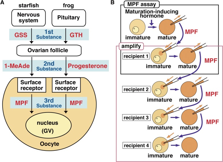
Reproductive endocrinology in the mid-20th century established that luteinizing hormone (LH), a gonadotropin secreted by the pituitary gland, induces ovulation and oocyte maturation in vertebrates including humans, mice, rats, and frogs. It was not believed at the time, however, that neural hormones similar to vertebrate gonadotropins could control reproduction in invertebrates. In 1959, Chaet and McConnaughy nonetheless reported that gamete shedding, which is analogous to ovulation, can be induced in ripe starfish by injecting a hot-water extract of the starfish radial nerve into the coelomic cavity.15) This “unexpected finding”15) opened the window on to the reproductive endocrinology in starfish (for reviews Refs. 16, 17). The active factor(s) in the radial nerve extract was later designated alternatively as gamete-shedding substance (GSS; subsequently renamed as gonad-stimulating substance) or radial nerve factor. Because GSS was only detectable in the coelomic fluid when starfish are naturally spawning, GSS was considered to be a gonadotropin-like hormone similar to LH in vertebrates (Fig. 1A).
Figure 1.
Hormonal control of oocyte maturation and demonstration of MPF. (A) In the endocrine control of oocyte maturation, starfish GSS (released from the nervous system) or frog GTH (released from the pituitary) functions as the first substance acting on ovarian follicles. The second substance, maturation-inducing hormone (starfish 1-MeAde or frog progesterone), is produced by and released from follicles, and acts on the oocyte surface. Based on these, a third substance that is responsible for oocyte maturation was hypothesized in the oocyte cytoplasm, and subsequently designated as maturation-promoting factor (MPF) upon its demonstration as shown in B. (B) MPF, the third substance, was demonstrated by cytoplasmic transfer from maturation-inducing hormone-treated maturing oocytes into untreated immature oocytes, which in turn undergo maturation (upper box). At the same time, the MPF activity was shown not to decrease through multiple successive transfers into immature oocytes in which de novo protein synthesis was prevented (lower box). This was called the “amplification” of MPF, implying that the inactive form of MPF is present in immature oocytes and that it can be autocatalytically activated by the active form of MPF. GV, germinal vesicle (oocyte nucleus).
GSS, the first gonadotropin-like hormone demonstrated in invertebrates, was characterized preliminarily as a single peptide with a molecular weight either of ∼4.8 kDa (42 amino acid residues) (by Chaet; see Ref. 16) or of ∼2.1 kDa (22 amino acid residues18)). Much more recently, GSS was finally purified from starfish radial nerves and characterized as a heterodimeric peptide with a molecular weight of 4,737 kDa (chains of 24 and 19 amino acid residues, which are cross-linked by three disulfide bonds). The molecule was phylogenetically classified as a member of the insulin/insulin-like growth factor (IGF)/relaxin superfamily.19)
2.2. Maturation-inducing hormone.
Even though the molecular identity of GSS remained unclear in the 1960s, Haruo Kanatani and his colleagues spearheaded important advances during that decade into the reproductive endocrinology of starfish. They found that GSS induces not only gamete shedding, but also simultaneously meiotic resumption in oocytes,20) and they established that the action of GSS on these processes is indirect (Fig. 1A). Namely, they found that GSS acts on ovarian follicles surrounding each oocyte to induce the synthesis of a second hormone, meiosis-inducing substance (MIS; subsequently renamed as maturation-inducing substance; also known as maturation-inducing hormone, MIH), which in turn induces both oocyte maturation and oocyte spawning21–23) (for a review Ref. 16). Soon thereafter, starfish MIS was purified and identified as 1-methyladenine (1-MeAde) by Kanatani and colleagues.24) Indeed, 1-MeAde acts at the oocyte surface to induce the in vitro maturation of immature starfish oocytes cultured in seawater.25)
1-MeAde was thus the first chemically identified bona fide MIH in metazoans.26) This finding in starfish introduced to the field of reproductive endocrinology the novel concepts that gonadotropins indirectly regulate ovulation and oocyte maturation, and that ovarian follicles directly control these processes (Fig. 1A).
In the late 1960s, the hormone progesterone was also found to induce oocyte maturation in frogs (Fig. 1A).27–29) However, because various steroids produced downstream of LH shows MIS-like effects in vitro,30) it was only four decades later that the physiological MIS of frogs was likely settled to be progesterone itself.31) In any event, the discoveries of 1-MeAde and progesterone at the latter half of the 1960s opened the way towards in vitro studies on oocyte maturation using immature oocytes isolated from non-mammalian, invertebrate starfish and vertebrate frogs.
2.3. Maturation-promoting factor (MPF).
How then does 1-MeAde induce maturation in starfish oocytes? Because GSS from nervous systems and MIS/1-MeAde from ovarian follicles were regarded as the first and second substances, respectively, for the hormonal induction of oocyte maturation (Fig. 1A), an emerging idea was that the cytoplasm of 1-MeAde-treated oocytes might contain a third key maturation-inducing molecule.16) The necessity for an additional substance was dictated by the finding that microinjection of 1-MeAde into immature starfish oocytes failed to induce maturation.25) The existence of the putative third substance, designated by Yoshio Masui as maturation-promoting factor (MPF), was first demonstrated in progesterone-treated frog oocytes (Fig. 1A).32,33)
My first successful research project in the Kanatani laboratory established that 1-MeAde-treated starfish oocytes also contain MPF as a transferable cytoplasmic activity.34) That is, cytoplasm taken from 1-MeAde-treated donor oocytes induces maturation upon its microinjection into untreated immature recipient starfish oocytes (Fig. 1B, upper box). The finding of MPF in both invertebrates and vertebrates brought into the field of reproductive endocrinology a new perspective that hormonal control of oocyte maturation is a cascade consisting of three successive substances: gonadotropins (first), MIS/MIH (second), and MPF (third) (Fig. 1A). It should be noted, however, that in mammals the concept of MIS/MIH is replaced by a somewhat more complex system.12,35)
3. The cell biology of M-phase control
3.1. MPF is a universal inducer of M-phase.
In the early 1970s, it appeared that the maturation induction systems in starfish and frogs might be different. For example, although progesterone was detectable in the starfish ovary, it was unable to induce oocyte maturation in this organism (see a review Ref. 17). Furthermore, it was already clear that the molecular nature of MIS/MIH in the two species was quite different. Of greatest importance here, it could not be assumed at the time that starfish MPF and frog MPF were related molecules, or even that MPF from one species would be effective in oocytes in the other species. However, in early 1978, we found that frog MPF can induce maturation in recipient starfish oocytes.36) Thus, in contrast to the first and second substances (gonadotropins and MIS/MIH; see Fig. 1A), cytoplasmic MPF activity in oocytes was shown to be cross-reactive between invertebrates and vertebrates.
This finding represented a turning point in studies on MPF. Shortly thereafter, MPF was detected in cleaving blastomeres (i.e., mitotic M-phase) of frog37,38) and starfish;36) in extracts of mammalian cultured somatic cells synchronized at M-phase;36,39) in extracts of budding yeast cdc mutants that were arrested at M-phase;40,41) and as a transferable cytoplasmic activity from mouse oocytes.42) In every case, MPF was assayed as an activity that can induce the meiotic G2/M-phase transition upon microinjection into immature oocytes of frog or starfish. Taken collectively, these observations clarified that MPF is conserved across different species. Furthermore, these experiments established that MPF activity varies with respect to the cell cycle, being detectable only at M-phase during oocyte meiosis and somatic cell mitosis (for a review Ref. 43).
In all of the above studies, MPF was demonstrated operationally by microinjection into immature oocytes, and to the best of my knowledge, even to the current date (2017) no study has reported the use of somatic cells as recipients of MPF injection. Nonetheless, earlier fusion experiments between mammalian somatic cells in M-phase and in other cell cycle phases implied that an activity equivalent to MPF must exist.44) Furthermore, the microinjection of partially purified frog MPF induced nuclear envelope breakdown (NEBD; a hallmark of M-phase entry) in frog embryos arrested in a G2 phase-like state.45) These observations supported the supposition that MPF can induce M-phase in somatic cell mitosis as well.
Based on these findings, the concept emerged in the early 1980s that MPF is a universal inducer of M-phase in all eukaryotic cells, whether they are undergoing mitosis or meiosis. Hence, MPF was renamed from “maturation-promoting factor” to “M-phase promoting factor” with the same abbreviation.46) Although its existence was first established in the field of reproductive endocrinology, MPF was soon recognized as a player key to M-phase control in the biology of all cells (for a review Ref. 43).
3.2. Cyclin B-Cdc2 kinase as an essential component of MPF.
The initial demonstrations of MPF in oocytes of frog and starfish immediately raised the issue of the molecular identity of MPF. Wasserman and Masui (1976) first succeeded in extracting MPF from crushed frog oocytes by centrifugation, because homogenization alone failed to release MPF from oocytes.47) Based on this observation, many laboratories tried to purify MPF from oocytes or somatic cells using microinjection into immature oocytes as a bioassy, but these efforts were unsuccessful for more than a decade because the material rapidly lost activity (e.g., Refs. 48–50). Finally, Manfred Lohka in Jim Maller’s laboratory successfully purified MPF biochemically from mature frog eggs by assaying fractions from conventional column chromatographies with frog egg cell-free extracts.51) Purified frog MPF was comprised of two major proteins (32 and 45 kDa) that together exhibited kinase activity phosphorylating histone H1. Within two years, these 32 kDa and 45 kDa components were identified as a homolog of the fission yeast cdc2 gene product and cyclin B, respectively, leading to the conclusion that MPF is a histone H1 kinase that consists of the cyclin B-Cdc2 complex (see original papers Refs. 52–58 as representatives of many outstanding papers; for reviews Refs. 59–61).
Great achievements in four separate research fields converged to generate the powerful paradigm that the evolutionarily conserved complex of kinase enzyme cyclin B-Cdc2 is MPF. These research fields were: (1) cell division cycle (cdc) mutant genes in the yeasts,62) particularly fission yeast cdc2 and its human homolog;63–65) (2) cyclin protein, which was first found as a particular protein band on SDS-PAGE whose abundance cycled as a function of time after fertilization of sea urchin eggs,66) the cDNA of which was first isolated from surf clam eggs;67) (3) M phase-specific histone H1 kinase (growth-associated histone H1 kinase, or Ca2+- and cyclic nucleotide-independent histone H1 kinase), derived from cultured mammalian cells68) and starfish oocytes;69) and (4) MPF, assayed in the microinjection studies described above.
Although the convergence of these ideas in 1988 was a monumental achievement in the history of cell reproduction research, Bill Dunphy and John Newport pointed out at that time that it was not yet clear “whether this single enzymatic activity is sufficient for fully competent MPF”.59) Certainly, the cyclin B-Cdc2 complex is the molecular identity of M phase-specific histone H1 kinase, and it is an indispensable component of MPF. However, because all the assays for MPF activity at the time of the convergence relied on the frog egg system so far as experimental data were presented, no data could prove the simple equation of the cyclin B-Cdc2 complex with the totality of MPF. Indeed, as will be discussed in a later section, studies performed in our laboratory on the intracellular regulation of cyclin B-Cdc2 activation at meiotic resumption in starfish oocytes established that despite its unquestioned importance, this crucial kinase is only one of the components of MPF (see Section 5: Reconsideration of MPF).
3.3. Regulation of cyclin B-Cdc2 activity.
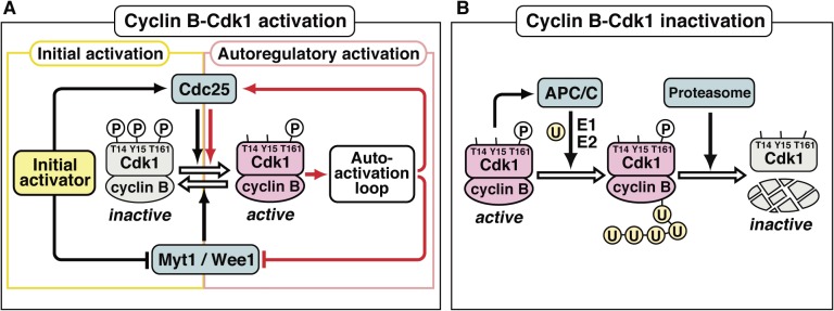
Within 5 years after the identification of cyclin B-Cdc2, the outline of the regulatory mechanisms controlling its activity was clarified. This effort was led first by yeast genetics and then progressed largely through biochemical analyses in frog egg extracts. Core elements for the regulation at mitotic entry are the kinase Wee1/Myt1, which directly phosphorylates cyclin B-associated Cdc2, leading to its inhibition; and the phosphatase Cdc25, which directly dephosphorylates the sites on cyclin B-associated Cdc2 that were phosphorylated by Wee1/Myt1, leading to the activation of cyclin B-associated Cdc2 (Fig. 2A) (for a review Ref. 70). At the G2/M-phase border, the balance between the activities of Wee1/Myt1 and Cdc25 is inclined to the inhibitory phosphorylation of Cdc2, thereby maintaining cyclin B-Cdc2 in an inactive state until before the cell is ready to enter M-phase.
Figure 2.
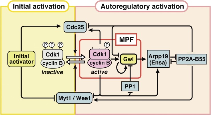
Activation of the cyclin B-Cdk1 complex at entry into M-phase and its subsequent inactivation at exit from M-phase. Cdc2 was renamed as cyclin-dependent kinase 1 (Cdk1) in the designation of the Cdk family in 1991.78) (A) At the G2/M-phase border, Myt1/Wee1 (which phosphorylates cyclin B-associated Cdk1 on Thr14 and Tyr15 for inhibition) surpasses Cdc25 (which dephosphorylates cyclin B-associated Cdk1 on the sites phosphorylated by Myt1/Wee1 for activation), and accordingly, cyclin B-Cdk1 remains in an inactive form, in which all three sites on Cdk1 are phosphorylated (indicated by the circled P). At the transition into M-phase, the initial activator (which is different depending on the cell type) first tips the balance between Myt1/Wee1 and Cdc25 activities to trigger the initial activation of cyclin B-Cdk1. Subsequently, active cyclin B-Cdk1 directly phosphorylates Cdc25 and Myt1/Wee1 for activation and inhibition, respectively, and hence the Myt1/Wee1-Cdc25 balance is further reversed via an autoactivation loop, leading to the robust and full autoregulatory activation of cyclin B-Cdk1. Details on the autoactivation loop are shown in Fig. 9. (B) At exit from M-phase, active cyclin B-Cdk1 activates an E3 ligase, the anaphase-promoting complex/cyclosome (APC/C), leading to poly-ubiquitination of Cdk1-associated cyclin B. Subsequently, the proteasome recognizes the poly-ubiquitin chain, dissociates poly-ubiquitinated cyclin B from Cdk1, and then proteolyses cyclin B, resulting in inactivation of cyclin B-Cdk1. U, ubiquitin; E1, ubiquitin-activating enzyme; E2, ubiquitin-conjugating enzyme.
At the onset of M-phase, the balance of activities between Wee1/Myt1 and Cdc25 is reversed in two ways (Fig. 2A). First, cyclin B-Cdc2-independent upstream signaling (designated here as the “initial activator”) reverses the balance just enough to trigger activation of a small population of cyclin B-Cdc2. Subsequently, this small amount of active cyclin B-Cdc2 starts an autoregulatory activation loop, in which active cyclin B-Cdc2 directly phosphorylates and thus activates Cdc25, while the cyclin B-Cdc2 kinase simultaneously phosphorylates and thus inactivates Wee1/Myt1. This autoregulatory loop swiftly and robustly leads to the activation of a much larger population of cyclin B-Cdc2 (for reviews Refs. 70–73). Although the molecular identity of the initial activator is different in different circumstances (discussed in Section 4.2.1 below), the mechanism for the autoregulatory activation loop is well conserved (see Section 5.2).
At the exit from M-phase, ubiquitin-dependent degradation of cyclin B inactivates cyclin B-Cdc274) (Fig. 2B) (for a review Ref. 75). Cyclin B degradation is a multistep process involving the participation of the anaphase-promoting complex/cyclosome associated with Cdc20 (APC/C-Cdc20), an E3 ligase that poly-ubiquitinylates cyclin B, and the subsequent proteolysis of poly-ubiquitinated cyclin B by the proteasome. The poly-ubiquitination first occurs on cyclin B that is associated with Cdc2. The regulatory 19S subcomplex of the 26S proteasome dissociates cyclin B from Cdc2,76) and finally cyclin B alone is proteolysed by the core 20S proteasome subcomplex. Because the poly-ubiquitination activity of the APC/C-Cdc20 requires its multiple phosphorylation by cyclin B-Cdc2 and subsequently, this activation inevitably leads to cyclin B-Cdc2 inactivation, the activation of cyclin B-Cdc2 can be said to be self-terminating. This insight explains one of the major reasons why the cell cycle is indeed a cycle.
3.4. The principle of cell cycle control in eukaryotic cells.
At the end of the 20th century, the principle was established that cell cycle control in eukaryotic cells is composed both of the cell cycle engine and the checkpoints.4,77) The concept of the cell cycle engine emerged from the finding that several types of cyclins, in addition to the B-type, comprise a protein family. The cyclin-dependent kinases (Cdks) in higher eukaryotic cells similarly compose a family of proteins related to Cdc2 (which was hence renamed as Cdk1; see Ref. 78). The cell cycle engine operates through cyclic changes in the activities of a class of protein kinases that form as complexes of particular cyclins with particular Cdks, and each such kinase determines the onset of distinct cell cycle phases. Typically, the prototype cyclin B-Cdk1 (previous cyclin B-Cdc2) governs M-phase, while cyclin D-Cdk4/6 is implicated in G1-phase, cyclin E/A-Cdk2 in S-phase, and cyclin A-Cdk1 in G2-phase (for reviews Refs. 79, 80).
The second key aspect of cell cycle control is the concept of checkpoints, which was first proposed by Lee Hartwell.81) Checkpoints are defect-responsive negative feedback controls that detect issues such as DNA damage and then promote the correction of these defects; checkpoints coordinate cell cycle progression so that early events are completed before the start of later events. The order of cell cycle events is thus ensured so that DNA replication precedes entry into mitosis and chromosomal alignment on the metaphase plate of the mitotic spindle precedes sister chromatid segregation (for reviews Refs. 82, 83).
The discovery and characterization of MPF derived from oocyte maturation was a major contributor to the development of this paradigm for cell cycle control composed of a cyclin-Cdk-based engine that is coordinated by checkpoints, although of course many other lines of research have also converged in the formulation of this important principle.
4. Meiotic cell cycle control in starfish oocytes
The concept of a cell cycle engine based on cyclin-dependent kinases prompted us to ask several basic questions about how this engine functions during the meiotic cell cycle in oocytes. How do oocytes modulate cyclin B-Cdk1 activity to achieve the unique events that occur within these specialized cells: the meiotic G2/M-phase transition, the meiosis I to II transition, and the second meiotic arrest? The starfish oocyte system has proven to be extremely effective in uncovering the molecular basis for these events, that is conserved in metazoan oocytes.
4.1. Dynamics of cell cycle regulators through meiotic maturation and early cleavages.
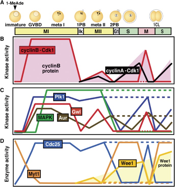
In starfish, 1-MeAde induces immature, previously arrested oocytes to undergo the meiotic G2/M-phase transition. This step, which is hallmarked by germinal vesicle breakdown (GVBD; equivalent to nuclear envelope breakdown or NEBD in somatic cells), occurs with no requirement of new protein synthesis. Thereafter, the meiotic cell cycle proceeds in vitro in the absence of fertilization through the completion of meiosis I and II, resulting in the formation of mature haploid eggs arrested at the G1-phase (also called the female pronucleus stage) (Fig. 3A). Once fertilization occurs, this second arrest is released, and the embryonic mitotic cycle initiates.
Figure 3.
Dynamics of cell cycle regulators and their choreographers in starfish meiotic and early cleavage cycles. (A) Fully grown immature starfish oocytes are arrested at prophase of meiosis I (MI), which is equivalent to G2-phase in somatic cells. This arrest is characterized by the presence of a large nucleus called the germinal vesicle (GV). Once these immature oocytes are isolated into seawater and treated with 1-MeAde (1-methyladenine), the starfish maturation-inducing hormone, meiosis resumes as hallmarked by GV breakdown (GVBD), followed by two consecutive M-phases, MI and meiosis II (MII), without an intervening S-phase. After the completion of MII, mature haploid eggs arrest at G1-phase unless fertilization occurs. Starfish oocytes are fertilizable throughout the meiotic cell cycle (even at prophase I or G1-phase), whereas physiological fertilization possibly occurs in late MI. After the completion of MII, fertilized eggs start to undergo cleavage cycles consisting of alternating S- and M-phases. The cell cycle dynamics of various regulators are schematically shown in B–D. meta I and meta II, metaphase of MI and MII, respectively; Ik, interkinesis period; 1PB and 2PB, the first and second polar body; 1CL, the first cleavage. (B) Cyclin B protein is already present in immature oocytes. By contrast, cyclin A protein (and Wee1 protein shown in D) is undetectable in immature oocytes and starts to accumulate near the end of MI (shaded areas). After meiotic resumption, the protein levels of cyclins A and B cycle along with the cell cycle, peaking at each metaphase. The Cdk1 level remains constant throughout the entire process. Accordingly, Cdk1 activity is represented by cyclin B-Cdk1 in MI, and largely by both cyclin B-Cdk1 and cyclin A-Cdk1 in and after MII. (C, D) In contrast to cyclins, the protein levels of Cdc25, Myt1, Greatwall kinase (Gwl), mitogen-activated protein kinase (MAPK), polo-like kinase 1 (Plk1), and Aurora (Aur) remain constant throughout the entire process. All of Gwl, Plk1, Aur, and MAPK are activated downstream of cyclin B-Cdk1 at meiotic resumption. The meiotic cycles are characterized by the unique dynamics in activities of MAPK and Plk1, and hence Cdc25 and Myt1 during the MI to MII transition and after the completion of MII (during G1-phase arrest) in unfertilized eggs (dotted lines). In contrast, Gwl activity correlates with cyclin B-Cdk1 activity. PP2A-B55 and PP1 activities are assumed to roughly mirror those of cyclin B-Cdk1 and Gwl. See the text for details.
To provide the necessary background, our laboratory first described the dynamics of key cell cycle regulators in starfish oocytes and eggs (Fig. 3B–D). For this, we isolated the cDNAs of starfish homologs of these cell cycle proteins and raised antibodies against each of their recombinant proteins, because commercially available antibodies failed to crossreact with most starfish proteins. Starting from cyclin B,84,85) these proteins included cell cycle engine elements (cyclin A, cyclin E, Cdk1, and Cdk2),86,87) their direct regulators (Cdc25, Myt1, and Wee1),88–90) their choreographers (Plk1, Aurora, Greatwall kinase [Gwl], B55 regulatory subunit of protein phosphatase 2A [PP2A-B55], cAMP-regulated phosphoprotein 19 [Arpp19], and Mos),90–94) DNA replication machinery elements (Cdc6, Mcm2, Mcm7, and Cdc45),87) and signal transduction elements (Akt/protein kinase B [PKB], PDK1, and TOR).89,95,96)
Fortunately, the suite of starfish cell cycle regulators is simpler than that of many other organisms because starfish characteristically do not have multiple subtypes of these molecules (e.g., cyclin B1 and B2, or Cdc25A, B, and C in mammals), thereby simplifying the analysis. These regulators can be categorized into two groups. The first group consists of proteins expressed in G2 phase-arrested immature starfish oocytes, and includes cyclin B, Cdk1, Cdc25, Myt1, Plk1, Aurora, Gwl, mitogen-activated protein kinase (MAPK),97) Cdc6, and Cdc45. With the exception of cyclin B, whose amount varies cyclically according to the cell cycle stage, the levels of all the other proteins in this group do not change throughout meiotic maturation. The other group consists of proteins that are undetectable in G2 phase-arrested immature oocytes and, therefore, must be newly synthesized after meiotic resumption. Mos protein accumulates immediately after GVBD, but others such as cyclin A, cyclin E, Cdk2, and Wee1 start to accumulate later at the end of meiosis I.
The cyclin B-Cdk1 complex is already present in G2 phase-arrested immature oocytes, and its activity cycles so that it peaks at each metaphase (Fig. 3B). The activities of the cyclin B-Cdk1 positive regulators (Cdc25, Plk1, Aur, and Gwl) cycle essentially in parallel with that of cyclin B-Cdk1, whereas the activities of the negative regulators (Myt1 and Wee1) are mirror images of the cyclin B-Cdk1 pattern (Fig. 3C, D). However, the features most characteristic of the meiotic cell cycle are that activities of Plk1 and Cdc25 remain high while Myt1 activity remains low at the ends of both meiosis I and II (namely, all three of these proteins remain phosphorylated), even though cyclin B-Cdk1 loses its activity at these times. It should be noted that these behaviors of Plk1, Cdc25, and Myt1 parallel the activity of MAPK, which is activated downstream of Mos.
4.2. Meiotic resumption.
4.2.1. Hormonal signaling that leads to cyclin B-Cdk1 activation.
Figure 2A illustrates the importance of the “initial activator” in triggering the cyclin B-Cdk1 activation process. In the somatic cells of higher eukaryotes, Aurora A, Plk1, cyclin A-Cdk1/2 and Cdc25B may all be involved in tipping the balance between Wee1/Myt1 and Cdc25 activities (for a review Ref. 79), and redundant or stochastic processes involving these proteins may in some uncharacterized fashion serve as the trigger (for reviews Refs. 71, 73). The major reason for the difficulty in identifying the initial activator is that cell cycle progression in proliferating somatic cells is essentially autonomous. By contrast, extracellular MIH controls the G2/M-phase transition in oocytes, and hence the initial activator may be identified at meiotic resumption.
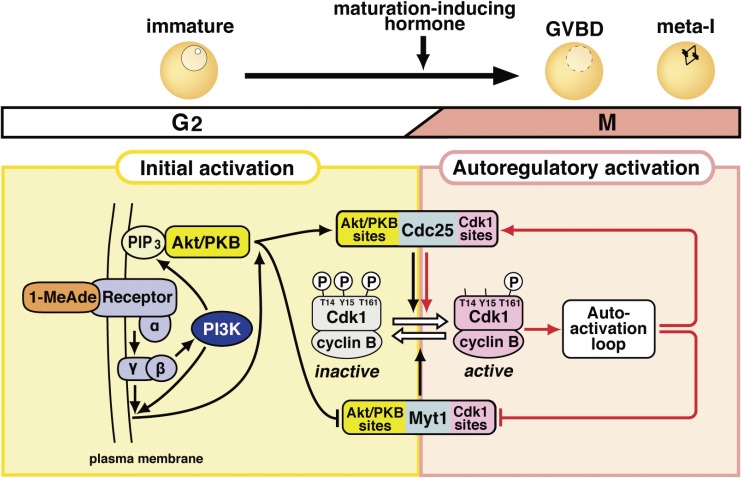
The starfish oocyte represents a unique system in which the initial activator is in fact characterized (Fig. 4). At meiotic resumption, this trigger cannot require activation of Plk1,90) Aurora,92) or MAPK,97) because all of these enzyme activities are absolutely downstream of, and not required for, cyclin B-Cdk1 activation. Eventually, we identified the initial activator in starfish oocytes as the kinase Akt/PKB (Fig. 4).89) Akt/PKB is activated downstream of 1-MeAde in a cyclin B-Cdk1-independent fashion, as expected from our previous studies.88) Akt/PKB directly phosphorylates both Cdc25 and Myt1 to reverse the balance of their activities, resulting in the net removal of inhibitory phosphorylations on Cdk1 (for reviews Refs. 10, 13). Thereafter, as will be addressed in Section 5.2, cyclin B-Cdk1 is further activated via the autoactivation loop as in somatic cells (Figs. 2A and 4).
Figure 4.
Signaling pathway leading to the activation of cyclin B-Cdk1 at the meiotic G2/M-phase transition in starfish oocytes. This pathway consists of the initial activation of cyclin B-Cdk1 and its subsequent autoregulatory activation, as shown in Fig. 2A. The initial activation pathway may be characteristic of the starfish oocyte system, whereas the autoregulatory activation pathway is largely conserved. The putative 1-MeAde receptor on the oocyte surface couples with heterotrimeric G-protein, from which the Gβγ complex is released to cause the initial activation of cyclin B-Cdk1 via two parallel pathways. In one pathway, Gβγ activates phosphoinositide 3-kinase (PI3K) to produce phosphatidylinositol 3,4,5-triphosphate (PIP3), depending on which Akt/protein kinase B (PKB) is activated. Akt/PKB directly phosphorylates Myt1 and Cdc25 for downregulation and upregulation, respectively. In the other pathway, Gβγ along with PI3K contributes, via unknown molecule(s), to the phosphorylation of Cdc25 and Myt1 on residues phosphorylated by Akt/PKB (Akt/PKB sites). These initial phosphorylations on the Akt/PKB sites, which are accomplished by possible cooperation of these two pathways, tip and reverse the balance between Cdc25 and Myt1 activities, leading to activation of a small population of cyclin B-Cdk1. The autoactivation loop then starts the activation of a much larger population of cyclin B-Cdk1 (see Fig. 9 for details). In the autoregulatory activation, Cdc25 and Myt1 are directly phosphorylated largely by cyclin B-Cdk1 (Cdk1 sites).
How does 1-MeAde induce the activation of Akt/PKB? Through its putative G protein-coupled receptor (GPCR) localized on the oocyte surface,25,98) 1-MeAde stimulates the dissociation of the heterotrimeric G protein consisting of the Gα(i)βγ complex into the Gβγ complex and Gα(i).99,100) The Gβγ complex then activates phosphoinositide 3-kinase (PI3K) to generate phosphatidylinositol 3,4,5-triphosphate (PIP3) at the plasma membrane.101) In a PIP3-dependent manner, Akt/PKB is phosphorylated by phosphoinositide-dependent kinase 1 (PDK1) on its activation loop95) and by target of rapamycin complex 2 (TORC2; or PDK2) on its C-terminal hydrophobic motif.96) These two kinds of phosphorylation lead to Akt/PKB activation (Fig. 4). Thus, meiotic resumption in starfish oocytes now represents a model in which the signal transduction pathway that links extracellular maturation-inducing hormone to the intracellular cell cycle regulators is characterized at least to some extent.
4.2.2. Emerging pathways involved in cyclin B-Cdk1 activation.
We have recently uncovered other pathways that may also lead from 1-MeAde towards the initial activation of cyclin B-Cdk1 at meiotic resumption. One of these mechanisms likely works in parallel with the Akt/PKB signaling pathway (Fig. 4), while the other may set a threshold for the activation of cyclin B-Cdk1.102)
Daisaku Hiraoka in my lab found that a Gβγ mutant, which was unable to activate PI3K (and hence Akt/PKB), enhanced the constitutively active PI3K-dependent initial phosphorylation of Cdc25 and Myt1 on residues phosphorylated by Akt/PKB.102) Considering that several AGC kinases (a subgroup of Ser/Thr protein kinases that are related to cyclic adenosine monophosphate (cAMP)-dependent protein kinase A [PKA], protein kinase G [PKG], and protein kinase C [PKC]) which are activated downstream of PI3K (for reviews Refs. 103, 104) have overlapping target consensus sequences, it is possible that Akt/PKB and another unidentified kinase, whose activation depends on both PI3K and the Gβγ mutant, cooperatively induce the initial phosphorylation of Cdc25 and Myt1 (Fig. 4).102)
Evidence for the existence of yet another pathway leading from 1-MeAde was our recent surprising finding that sub-threshold doses of 1-MeAde can transiently activate cyclin B-Cdk1, presumably via activation of the putative cell surface receptor and associated intracellular signaling pathway. The partially activated cyclin B-Cdk1 triggers negative feedback to induce the dephosphorylation of Cdc25 and Myt1 on the residues phosphorylated by Akt/PKB, inactivating this pool of cyclin B-Cdk1 and cancelling the sub-threshold noise signal.102) The molecular identity of the phosphatase involved in this cyclin B-Cdk1-dependent negative feedback remains unknown.
We assume that 1-MeAde dose-dependent competition exists between the cyclin B-Cdk1-dependent negative feedback loop and the two parallel signaling pathways that function downstream of Gβγ and lead to the initial phosphorylation of Cdc25 and Myt1. The point at which the latter two pathways becomes predominant over the negative feedback loop likely constitutes the threshold for the 1-MeAde dose. At supra-threshold doses of 1-MeAde, sufficient active cyclin B-Cdk1 accumulates to start the autoregulatory activation loop, leading to full activation of cyclin B-Cdk1.
4.3. The meiosis I to II transition.
4.3.1. The Mos–MAPK pathway.
We have shown that subsequent to meiotic resumption, both the transition from meiosis I to II and the subsequent arrest at G1-phase in starfish oocytes are regulated by the same molecular module, the Mos–MAPK signaling pathway (Fig. 5).
Figure 5.
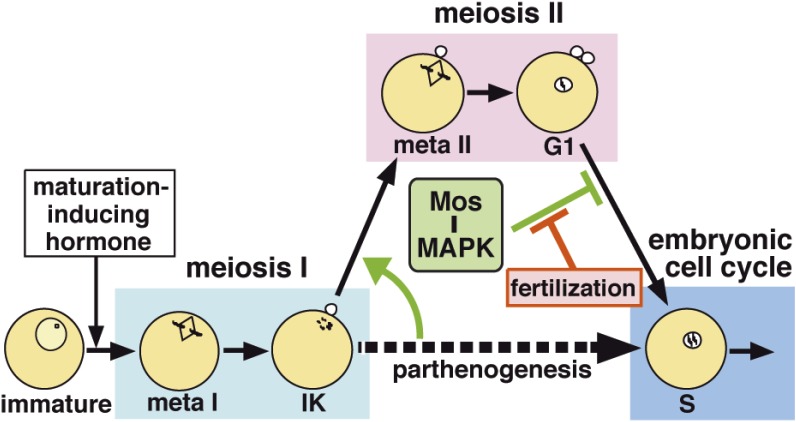
Mos–MAPK signaling ensures the successful transition from meiosis I to II to prevent parthenogenetic activation in starfish oocytes. At the end of meiosis I, the oocyte already has the ability to enter the embryonic mitotic cycle in the absence of fertilization (i.e., parthenogenesis). Mos–MAPK signaling, however, represses this ability in two ways. First, at the end of meiosis I, Mos–MAPK signaling causes swift activation of cyclin B-Cdk1 to force entry into meiosis II without an intervening S-phase. Subsequently, Mos–MAPK signaling prevents return to the embryonic mitotic cycle after the completion of meiosis II, resulting in the suppression of parthenogenesis. Once fertilization occurs, this prevention is cancelled, leading to the start of the embryonic mitotic cycle. Thus, Mos–MAPK signaling halves the ploidy and maintains the haploid state of oocytes until fertilization.
After the 1-MeAde stimulus, cyclin B-Cdk1 induces de novo synthesis and accumulation of Mos (also known as MAPK kinase kinase/MAPKKK), which in turn activates MEK (or MAPK kinase/MAPKK), MAPK, and its immediate downstream target Rsk (p90 ribosomal S6 kinase, p90Rsk).91,97,105) Thereafter, the Mos–MAPK–Rsk pathway remains activated even after completion of the meiotic cell cycle, unless fertilization occurs (Fig. 3C). Upon fertilization, Mos is degraded and hence the Mos–MAPK–Rsk pathway is shut down, initiating the alternating S- and M-phases of the embryonic mitotic cycles. Consistent with this scheme, if meiosis resumes while the Mos–MAPK–Rsk pathway is suppressed, DNA replication (S-phase) occurs immediately after meiosis I, and the embryonic mitotic cycle initiates in the absence of fertilization, as if parthenogenesis occurred.91,97,105)
These findings demonstrated that although starfish oocytes at the end of meiosis I can enter the embryonic mitotic cycle, the Mos–MAPK–Rsk pathway represses this ability, forcing entry into meiosis II without an S-phase. Mos–MAPK–Rsk further prevents return to the embryonic mitotic cycle after the completion of meiosis II until Mos is degraded at fertilization (Fig. 5) (for a review Ref. 10). Overall, the Mos–MAPK pathway both promotes the meiosis I to II transition and inhibits the subsequent meiosis to mitosis transition.
4.3.2. Meiosis I to II transition-specific regulation for cyclin B-Cdk1 activation.
The skipping of S-phase during the meiosis I to II transition requires that cyclin B-Cdk1 is immediately activated after exit from meiosis I.106–108) The Mos–MAPK–Rsk pathway accomplishes this task by maintaining Myt1 inactive and Cdc25 active on exit from meiosis I. Rsk directly phosphorylates Myt1 and Cdc25, leading to decreased inhibitory Tyr phosphorylation of Cdk1. Secondarily, MAPK, along with cyclin B-Cdk1 that has residual activity upon exit from meiosis I, and the newly assembled cyclin A-Cdk1 that has emerging activity upon entry into meiosis II, together maintain Plk1 activity, which in turn keeps Myt1 suppressed and Cdc25 activated.90) Thus, several processes work together to ensure the activation of newly assembled cyclin B-Cdk1 at entry into meiosis II (see Fig. 3(b) in Ref. 10).
4.4. G1-phase arrest.
4.4.1. Dual-lock to the start of the embryonic mitotic cycle.
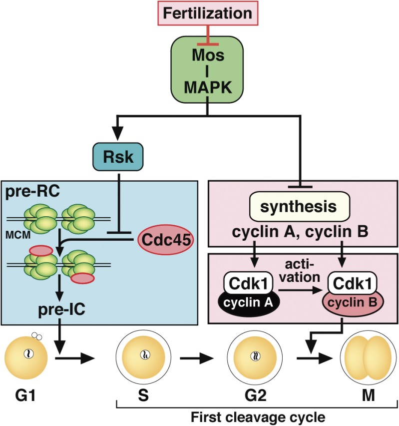
How does the Mos–MAPK pathway prevent the start of the embryonic mitotic cell cycle in unfertilized mature starfish eggs at G1-phase, given that these eggs lack a functional cell cycle checkpoint that could block cell cycle progression?109) We proposed a novel dual-lock model for G1-phase arrest (Fig. 6), in which the pathways functioning downstream of MAPK diverge to block the start of the embryonic mitotic cycle.110) A Rsk-dependent pathway prevents entry into S-phase, while a Rsk-independent pathway prevents entry into the first mitotic M-phase. Consistent with this model, in G1 phase-arrested starfish eggs, inactivation of Rsk initiated S-phase, but M-phase did not follow when MAPK remained active. By contrast, MAPK inactivation initiated M-phase, even when S-phase was inhibited by constitutively active Rsk.105,110) Fertilization induces Mos degradation, thus releasing the dual-lock and initiating the embryonic mitotic cycle. Insofar as we know, this is the first demonstration showing that the Mos–MAPK cascade separates into Rsk-dependent and Rsk-independent pathways to arrest the cell cycle.
Figure 6.
Dual-lock for G1-phase arrest in unfertilized mature starfish eggs. Unless fertilized, mature starfish eggs arrest at G1-phase after the completion of meiosis II. This arrest is accomplished through two separate pathways that function downstream of Mos–MAPK signaling. One is a Rsk-mediated pathway that prevents entry into S-phase by inhibiting Cdc45 loading onto the DNA replication machinery. The initiation of DNA replication is thus blocked by Rsk at the pre-replicative complex (pre-RC) stage, prior to the subsequent pre-initiation complex (pre-IC) stage just before the actual start of DNA replication. The other is an Rsk-unmediated pathway that prevents entry into the first mitotic M-phase by inhibiting new protein synthesis of cyclin A and cyclin B. Due to the absence of a DNA replication checkpoint, this dual-lock mechanism is required for G1 arrest. Upon fertilization, Mos degradation releases the dual-lock, resulting in the start of the embryonic mitotic cycle. MCM, MCM complex.
4.4.2. Rsk as a regulator of the replication machinery.
When G1 phase-arrested mature starfish eggs are fertilized, the initial S-phase preceding the first mitotic division does not require de novo protein synthesis, implying that all elements needed for DNA replication are already available in the unfertilized mature eggs. In eukaryotic cells, initiation of DNA replication is controlled by the strictly ordered assembly of various protein complexes onto the replication origins (for a review Ref. 111). However, the literature provided no information about the stage at which the initiation complex for DNA replication is arrested in unfertilized G1 phase-arrested eggs. We thus wanted to clarify when during replication complex assembly Rsk arrests starfish eggs. We eventually defined the arrest stage as the transition point from the pre-RC (the pre-replicative complex) to the pre-IC (the pre-initiation complex) (Fig. 6, left);87) in other words, chromatin is already loaded with the Mcm complex in G1 phase-arrested eggs. Rsk inactivation is necessary and sufficient for the further loading of Cdc45 onto chromatin, a step that supports the formation of the pre-IC and the subsequent initiation of DNA replication.
Our findings constituted the first description of the state of the DNA replication machinery in unfertilized mature eggs arrested at G1-phase. Our results further established a novel role for Rsk as a negative regulator of the pre-RC to pre-IC transition, although the direct target of Rsk has yet to be identified. Notably, the initiation of DNA replication in somatic cells is also regulated at the pre-RC stage by the G1 checkpoint,112) although that form of regulation is independent of Rsk.
4.4.3. MAPK as an inhibitor of cyclin synthesis.
In contrast with S-phase, the start of the embryonic M-phase requires the synthesis of proteins such as cyclins A and B; the reason is that translation of these proteins is repressed downstream of MAPK in G1 phase-arrested starfish eggs (Fig. 6, right).110) It remains unclear how MAPK suppresses translation initiation, but this process likely occurs after poly(A) tail elongation, because no further elongation of cyclin A and B mRNAs is detectable after release from G1 phase-arrest.110) Once fertilization releases the MAPK-dependent suppression, each cyclin accumulates and associates with Cdk1. Cyclin A-Cdk1 becomes active without inhibitory phosphorylation of Cdk1 by Wee1 and Myt1, but cyclin B-Cdk1 remains inactive due to this inhibitory phosphorylation. The active cyclin A-Cdk1 then inactivates Wee1 and Myt1 via Plk1, leading to the activation of cyclin B-Cdk1 and entry into the first embryonic M-phase (Fig. 6, right).86,90,113)
Curiously, the synthesis of cyclins A and B occurs upon entry into meiosis II and continues until metaphase of meiosis II,86,91,114) while cyclin synthesis is then repressed during G1-phase arrest. It currently appears paradoxical that both meiosis II synthesis and G1-phase repression depend on MAPK, which is fully active throughout these periods. Further studies are needed to clarify how the effect of MAPK is reversed at completion of meiosis II.
4.5. Conserved and diverse mechanisms of meiotic cell cycle control.
The stage of the second meiotic arrest differs in many species, implying the existence of a variety of molecular mechanisms regulating the meiotic cell cycle in oocytes. Nonetheless, some common principles are apparent. To highlight these recurrent themes, I compare here our findings on the meiotic cell cycle control in starfish with the situations in other metazoans.
4.5.1. Diverse identities of the initial activator at meiotic resumption.
In vertebrate oocytes, meiotic G2-phase arrest is maintained by high levels of cAMP and PKA, while downregulation of PKA is likely to be necessary and sufficient for meiotic resumption.115,116) Here, I compare and contrast the situation in mouse and frog oocytes, but readers should keep in mind that in starfish oocytes, cAMP and PKA are likely not involved in the meiotic G2-phase arrest (for a review Ref. 117).
In mouse oocytes arrested at meiotic G2-phase, cAMP is produced downstream of the constitutively active Gs-coupled orphan receptor (GPR3) present at the oocyte surface,118) whereas phosphodiesterase (PDE3A), which hydrolyses cAMP, is inhibited by cyclic guanosine monophosphate (cGMP) which is produced in the surrounding follicular cells (cumulus granulosa cells) and passes into the oocyte through gap junctions119) (for a review Ref. 35). Both mechanisms produce high levels of cAMP, thereby activating PKA, which then phosphorylates both Cdc25B for downregulation and Wee1B for upregulation to maintain the G2-phase arrest, because cyclin B1-Cdk1 is already present at sufficient levels for meiotic resumption (for reviews Refs. 116, 120).
To stimulate meiotic resumption in mammalian oocytes, the LH surge in large part decreases the production of cGMP by guanylyl cyclase (NPR2) in granulosa cells, which decreases the supply of cGMP to the oocyte121) (for a review Ref. 35). Reduced level of cGMP in turn increases cAMP hydrolysis by PDE3. Consequently, PKA is downregulated and both Cdc25B and Wee1B are dephosphorylated on their PKA target sites, possibly by protein phosphatase 1 (PP1), resulting in the initial activation of pre-existing cyclin B1-Cdk1 (for a review Ref. 116). Once cAMP levels decrease, no de novo protein synthesis is required for cyclin B1-Cdk1 activation until pro-metaphase.122)
In frog oocytes, there is some evidence that as in mammals a GPR3-related GPCR participates in maintaining meiotic G2-phase arrest by producing cAMP (see Ref. 123); however, the critical target(s) of PKA remain elusive (see Refs. 124, 125). For meiotic resumption, PKA downregulation is most likely necessary and sufficient,115) but one recent report even has asserted that release from the G2-phase arrest is independent of PKA activity.123) In any case, after progesterone stimulation, new protein synthesis either of Mos or cyclin B1 is needed to initially activate the pre-existing cyclin B2-Cdk1.126–128) The newly assembled cyclin B1-Cdk1, which escapes inhibition by Myt1, likely triggers the initial activation of cyclin B2-Cdk1 through inactivating phosphorylation of Myt1.30,129) The Mos–MAPK pathway then cooperatively promotes the initial activation of cyclin B2-Cdk1 through activating phosphorylation of Cdc25.30,129) Thereafter, much a larger population of cyclin B2-Cdk1 is activated through the conserved autoregulatory activation loop (see below). However, the molecular link from progesterone to de novo protein synthesis of Mos and cyclin B1 remains unclear.30)
It is thus clear that the mechanisms of meiotic resumption vary considerably in starfish, frog, and mouse oocytes. Different signals (1-MeAde, progesterone, cGMP) come from outside of the oocyte (see Fig. 1A). The signaling pathways inside the oocytes are also diverse; in starfish these pathways center positively on phosphorylation of the Akt/PKB target sites, while in mice and probably frogs PKA is the major negative mediator. Different events downstream of these kinases, such as the synthesis of cyclins and Mos, or the dephosphorylation/phosphorylation of Cdk regulatory enzymes, may assume more importance in some species than others. Regardless of these many differences, it must be remembered that the hormonal signaling processes in these various species all share the same final effector, cyclin B-Cdk1.
4.5.2. Conserved roles for the Mos–MAPK pathway.
Mos, originally identified as the product of the c-mos proto-oncogene, is expressed specifically during meiotic maturation in vertebrate oocytes.130,131) Although Mos was initially thought to be expressed in vertebrates only, our finding of the first invertebrate Mos in starfish91) prompted subsequent phylogenetic surveys. These found that mos genes are conserved but restricted to metazoans, namely from Cnidaria to Chordata, with the exception of Porifera (sponges) and Nematoda (Caenorhabditis elegans) (for a review Ref. 14).132) Another exception is fruit fly (Drosophila) mos, which is highly divergent compared with other metazoan mos and is not essential for meiosis.133)
Based on extensive studies in Xenopus oocytes, Noriyuki Sagata proposed three separate roles for the Mos–MAPK pathway in meiotic cell cycle control:9) First, Mos is the initial activator of cyclin B-Cdk1 at meiotic resumption;126,134) second, it governs the meiosis I to II transition;106) and third, it acts as the so-called cytostatic factor/CSF,32) which ensures meta-II arrest until release by fertilization135) (for a review Ref. 136). In each of these instances, Mos activity is mediated by MAPK. However, the first of these proposed roles for the Mos–MAPK pathway (as the initial activator for cyclin B-Cdk1) is not conserved in the oocytes of mice and most of invertebrates including starfish (for reviews Refs. 14, 137), and it does not appear to be valid even in frog oocytes.127,128)
By contrast, the second role for the Mos–MAPK pathway (as the coordinator of the meiosis I to II transition) is most likely conserved throughout metazoans (Fig. 7) (for reviews Refs. 9, 10, 14, 137), at least in terms of the action of the downstream kinase MAPK. In addition to detailed studies in starfish, ascidians, and frogs, MAPK remains active during the meiosis I to II transitions in all organisms examined so far (including Spisula, Urechis, and sea cucumber).
Figure 7.
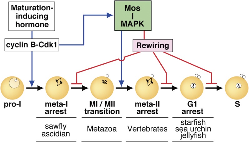
Mos–MAPK signaling governs meiotic cell cycle progression to accomplish the half-reduction of genomic ploidy in metazoan oocytes. At meiotic resumption, diverse hormonal signaling processes in various animal species center on activation of cyclin B-Cdk1 in all cases. Mos–MAPK is then activated downstream of cyclin B-Cdk1. Mos–MAPK positively regulates the meiosis I to II transition by avoiding S-phase and by facilitating entry into meiosis II in most metazoans (blue line). On the other hand, Mos–MAPK causes, via its downstream rewiring, a second meiotic arrest at diverse, species-specific stages (meta-I, meta-II, or G1; representative examples are indicated at each arrest) until fertilization (red lines). Thus, the conserved Mos–MAPK module is central to the production of haploid eggs for successive generations.
With regard to the third role for Mos, the molecular mechanism responsible for the meta-II arrest in frog oocytes is now well understood (for a review Ref. 138). Its central effector molecule is an APC/C inhibitor, Emi1-related protein 1 (Erp1, or so-called Emi2).139) Downstream of the Mos–MAPK pathway, Rsk directly phosphorylates Erp1, whose stability and inhibitory activity on the APC/C are in turn increased. The result is that cyclin B-Cdk1 activity is maintained at an elevated level to allow for meta-II arrest.140–143) Upon fertilization, the released Ca2+ activates CaMK-II to phosphorylate Erp1, thereby facilitating its subsequent phosphorylation by Plk1. Phosphorylated Erp1 then undergoes SCF-dependent degradation, and the APC/C is released from its inhibition, leading to the exit from meta-II arrest.144,145) Of note, these observations indicate that Mos degradation is not essential for the exit from meta-II arrest.
The second meiotic arrest, which maintains maturing/mature oocytes in a resting stage prior to fertilization, occurs at different stages in different organisms. Our own studies demonstrated that, as mentioned above, the same Mos–MAPK module participates in the G1 arrest in starfish oocytes. Later studies in oocytes from various metazoans including ascidians, sawflies, and jellyfish revealed that the Mos–MAPK pathway is involved in all second meiotic arrests, whether they occur during meta-I, meta-II, or G1-phase (detailed in Refs. 14, 137). To reconcile these findings, we proposed the idea that “rewiring” occurs downstream of the Mos–MAPK core module to define the specificity of the stage of the second arrest (Fig. 7).14)
The fact that the Mos–MAPK pathway is generally inactivated after fertilization presents a fascinating conundrum for organisms whose oocytes undergo meta-I arrest: how can the Mos–MAPK pathway accomplish the meiosis I to II transition even in these species (see Fig. 7)? To address this question, starfish oocytes have again provided an interesting clue. Starfish oocytes can be fertilized in vitro throughout meiotic maturation. Regardless of the timing of fertilization, however, the Mos–MAPK pathway remained active until oocytes reached meiosis II and then it was shut down, as if the “signal” for Mos degradation was “memorized” but not executed until completion of the meiosis I to II transition.97) Further studies are needed to clarify the molecular mechanisms corresponding to this hypothetical “signal” and the preservation of its “memory”.
4.5.3. An integrated view of meiotic cell cycle control.
The meiotic cell cycle, which produces haploid genomes, requires the cooperation of diverse and conserved modules (Fig. 7). After a hormonal stimulus, diverse pathways lead to the initial activation of cyclin B-Cdk1 at meiotic resumption in oocytes, depending on the animal species. In all cases, the Mos–MAPK pathway is then activated downstream of cyclin B-Cdk1. The Mos–MAPK pathway facilitates and coordinates entry into meiosis II by activating cyclin B-Cdk1 so swiftly that S-phase is avoided. Diverse circuitry downstream of the Mos–MAPK pathway is responsible for the second meiotic arrest at different organism-dependent stages until fertilization. Thus, the Mos–MAPK pathway is central to the half-reduction of genomic ploidy in oocytes: It alters the mitotic cell cycle to the meiotic cell cycle and it also prevents the meiosis-to-mitosis transition.
5. A reconsideration of MPF
5.1. An enigma: Cyclin B-Cdk1 is not synonymous with MPF.
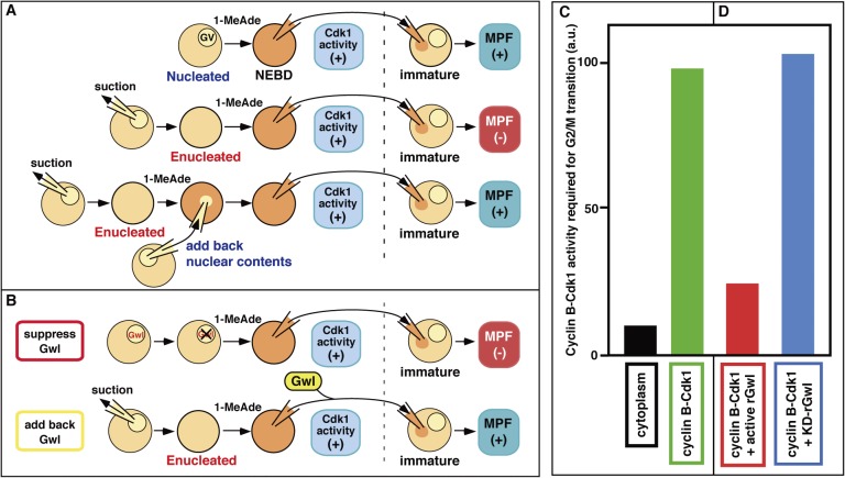
Although it has long been generally accepted that MPF and cyclin B-Cdk1 are synonymous terms, some of our early observations in starfish oocytes suggested that the situation might be more complicated.146) In particular, we noticed in 1976 that MPF is undetectable from enucleated oocytes treated with 1-MeAde. But consistently, MPF was restored when nuclear material was added (Fig. 8A).147) These observations in starfish (later confirmed by another group148)) contrasted significantly to the situation in frogs,32) where MPF is detectable from enucleated as well as nucleated oocytes after progesterone treatment. At least in starfish oocytes, however, it appeared that MPF requires a nuclear factor(s).
Figure 8.
MPF is not synonymous with cyclin B-Cdk1 and instead consists of both cyclin B-Cdk1 and Gwl. (A) Nuclear material is required for MPF. In the starfish system, MPF is undetectable from enucleated donor oocytes, whereas cyclin B-Cdk1 is invariably activated after 1-MeAde treatment. MPF is restored when the nuclear material is added back to the donor enucleated oocytes. MPF (+) and (−), detectable and undetectable, respectively. (B) Greatwall kinase (Gwl) is essential for MPF. When Gwl activity is suppressed in the nuclei of donor oocytes by injection of its neutralizing antibodies, MPF is undetectable, even though cyclin B-Cdk1 is invariably activated. Conversely, injection of Gwl into enucleated oocytes restores MPF. (C) One order of magnitude excess levels of cyclin B-Cdk1 activity are required to induce GVBD in the microinjection assay, when purified cyclin B-Cdk1 is compared with cyclin B-Cdk1 found in cytoplasmic MPF. (D) For GVBD induction by microinjection, the addition of Gwl reduces the required activity of purified cyclin B-Cdk1 approximately to that contained in cytoplasmic MPF. rGwl, active recombinant Gwl; KD, kinase-dead form as a control. Reproduced from Fig. 2 in Ref. 13 with some modifications.
The enigma arose later, after findings in many systems suggested that MPF is cyclin B-Cdk1. Surprisingly, we demonstrated that cyclin B-Cdk1 is localized specifically in the cytoplasm of immature starfish oocytes arrested at G2 phase.85) We also verified on this occasion that cyclin B-Cdk1, which can be measured as histone H1 kinase activity, is activated fully and invariably, even in enucleated starfish oocytes exposed to 1-MeAde (see also Refs. 93, 149). Thus, in starfish oocytes full activation of cyclin B-Cdk1 does not require the nucleus but full activation of MPF does. These observations indicated that MPF and cyclin B-Cdk1/M phase-specific histone H1 kinase are not synonymous at least in starfish oocytes, and that MPF consists of cyclin B-Cdk1 and (an) unidentified nuclear factor(s). Therefore, a true understanding of MPF clearly requires identification of its nuclear component(s).
5.2. Amplification of MPF and autoregulatory activation of cyclin B-Cdk1.
In retrospect, a clue towards solving the enigma that MPF does not equate to cyclin B-Cdk1 can be found in the first demonstrations of MPF in the oocytes of frogs32) and starfish:34) Intriguingly, in the absence of de novo protein synthesis, the MPF activity contained in the cytoplasm of mature oocytes did not decrease even after its serial transfer into immature oocytes (Fig. 1B, lower box). This observation implies that inactive MPF is present in immature oocytes and that it can be activated by active MPF, although the initial activation of MPF solely depends on maturation-inducing hormonal signaling. This autocatalytic activation property of MPF was designated as “amplification” by Masui.32) Based on enucleation experiments, we suggested that nuclear factor(s) is required for the amplification of MPF in starfish oocytes.147) However, for nearly three decades, our efforts to find its molecular identity were unsuccessful.
The amplification/autocatalytic activation of MPF appeared to be highly analogous to the autoregulatory activation of cyclin B-Cdk1 (Fig. 2A). Although it was found soon after the identification of cyclin B-Cdk1, it took more than one and a half decades to clarify molecular components involved in the autoactivation loop, because the underlying process is complicated even though it is highly conserved (Fig. 9). A major obstacle is that the cyclin B-Cdk1 autoactivation loop includes an antagonizing action against PP2A-B55,150–152) which counteracts the phosphorylation of Cdc25 and Wee1/Myt1 by cyclin B-Cdk1 (for reviews Refs. 73, 153, 154). A key element involved in turning off PP2A-B55 is Arpp19 or its close relative α-endosulfine (Ensa). Both proteins are directly phosphorylated and activated by Gwl,155,156) whereas this Gwl-mediated effect can be opposed by PP2A-B55.157)
Figure 9.
Molecular pathway for autoregulatory activation of cyclin B-Cdk1, and its core kinases, cyclin B-Cdk1 and Gwl, that constitute MPF. The autoactivation loop consists of two kinases, cyclin B-Cdk1 and Gwl, that promote activation of cyclin B-Cdk1, and two phosphatases, PP2A-B55 and PP1, that counteract activation of these two kinases. Once cyclin B-Cdk1 is initially activated, it causes, on one hand, suppression of the major antagonizing phosphatase PP2A-B55 via Arpp19 in two steps: First, direct phosphorylation of Arpp19 by initially activated cyclin B-Cdk1 converts Arpp19 into an inhibitor of PP2A-B55, and subsequent phosphorylation of Arpp19 on another residue by Gwl, which is activated downstream of cyclin B-Cdk1, enhances this conversion. On the other hand, initially activated cyclin B-Cdk1 directly phosphorylates and inhibits phosphatase PP1 which antagonizes the autoactivation of Gwl. Collectively, the cyclin B-Cdk1–Arpp19 pathway and the cyclin B-Cdk1–Gwl–Arpp19/Ensa pathway synergistically inhibit PP2A-B55 to support phosphorylation of Cdc25 and Myt1/Wee1 by cyclin B-Cdk1, resulting in the autoregulatory activation of cyclin B-Cdk1. The authentic, classical MPF is the system (composed of cyclin B-Cdk1 and Gwl) that initiates and accomplishes the autoregulatory activation of cyclin B-Cdk1 under intracellular circumstances in which the initial activation of cyclin B-Cdk1 is not allowed. Gwl contributes to MPF by accelerating the suppression of PP2A-B55 that counteracts the autoregulatory activation of cyclin B-Cdk1. Reproduced from Fig. 6 in Ref. 13 with some updates.
Gwl was first identified in the fruit fly Drosophila as a nuclear protein required for proper chromosome condensation and M-phase progression.158,159) It was subsequently shown to participate in the autoregulatory activation of cyclin B-Cdk1.160) Activation of Gwl depends on its direct phosphorylation by cyclin B-Cdk1 and its subsequent autophosphorylation.161) These two Gwl activation steps are opposed by PP2A-B55 and by PP1,162) whereas PP1 is directly phosphorylated and inactivated by cyclin B-Cdk1.163)
Cyclin B-Cdk1 can also block PP2A-B55 through an alternative pathway that bypasses Gwl. Here, cyclin B-Cdk1 directly phosphorylates Arpp19 on a conserved site that is different from the one targeted by Gwl but that also leads (somewhat more weakly) to the inhibition of PP2A-B55.94) The cyclin B-Cdk1–Arpp19 bypass might start the autoactivation loop immediately after the initial activation of cyclin B-Cdk1. Subsequently, the cyclin B-Cdk1–Gwl–Arpp19/Ensa pathway would act synergistically to autoactivate cyclin B-Cdk1. In this way, cyclin B-Cdk1-driven phosphorylation of Cdc25 and Myt1/Wee1 can be maintained to attain swift, robust, and complete activation of cyclin B-Cdk1 (Fig. 9). Thus, mitotic protein phosphorylation in vivo requires not only activation of the appropriate kinase but also inhibition of the antagonizing phosphatase.164,165)
5.3. A solution: MPF is comprised of cyclin B-Cdk1 and Gwl.
The events described above are well conserved in starfish oocytes, with the exception that Arpp19 is present but Ensa is not. Of particular interest to us, Gwl is localized in the nucleus of immature oocytes arrested at G2-phase, and it is activated immediately after cyclin B-Cdk1 activation at meiotic resumption (Fig. 3), whereas Arpp19 is largely cytoplasmic.93,94) We thus hypothesized that Gwl may be the nuclear component of MPF. Consistent with this idea, a previous study suggested that okadaic acid, an inhibitor of PP2A and PP1, mimics the nuclear factor.166)
To verify this hypothesis, we raised an antibody against starfish Gwl that inhibits its kinase activity and, accordingly, its activation as well, and we developed a method that allows entry of the antibody into the nucleus after its introduction into the cytoplasm.93) Using this approach, we demonstrated that MPF is undetectable from starfish oocytes in which Gwl activity is inhibited, even when the oocytes possess a nucleus and cyclin B-Cdk1 is fully activated by 1-MeAde treatment. By contrast, the introduction of recombinant Gwl into 1-MeAde-treated enucleated starfish oocytes restored MPF (Fig. 8B). We concluded that Gwl is the decades-long-unknown nuclear factor required for MPF; in other words, MPF is composed of both cyclin B-Cdk1 and Gwl (Fig. 9).93)
Our findings clarified a puzzling observation we made more than 20 years ago that contradicted the simple equating of purified cyclin B-Cdk1 with MPF. How then does Gwl function as a component of MPF? We had found that to induce the meiotic G2/M-phase transition by injection into immature starfish oocytes, purified cyclin B-Cdk1 needed to be added in approximately ten-fold excess to that present in the minimal amount of donor cytoplasmic MPF that could induce the transition (Figs. 1B and 8C).88,93) However, the addition of Gwl reduces the required activity of purified cyclin B-Cdk1 in the injection assay to approximately that present in cytoplasmic MPF (Fig. 8D).93) Of note, this reduction by Gwl has a physiological function beyond its contribution to cyclin B-Cdk1 activation. Although as just mentioned a ten-fold excess of purified cyclin B-Cdk1 can induce NEBD/GVBD, the subsequent formation of the meiotic spindle is abortive. However, the reduction by Gwl restores meiotic spindle formation.93)
The reader should recall that all the historical experimental data that equated MPF with M phase-specific histone H1 kinase and cyclin B-Cdk1 could be obtained only with frog oocytes or their extracts as a recipient of the MPF assay (see Ref. 59). Why was only the frog system used as a recipient? We found that the answer is simple: Gwl similarly reduces the required activity of purified cyclin B-Cdk1 in the injection assay using frog oocytes as well;93) and Gwl is mostly localized in the cytoplasm of immature frog oocytes arrested at G2-phase,93) even though Gwl is generally regarded as a nuclear protein. These two features could have made it easier to detect MPF in recipient frog oocytes. That is, even if the purified MPF preparation did not contain Gwl, the cytoplasmic localization of Gwl in recipient oocytes most likely reduced the required activity of purified cyclin B-Cdk1 upon injection of the purified MPF into the cytoplasm. The cytoplasmic localization of Gwl also explains why MPF was detectable from enucleated frog oocytes after progesterone treatment.32)
Taken together, these results show that Gwl is a critical constituent of MPF that is crucial for the physiological function of cyclin B-Cdk1.
5.4. Reconciling MPF, M phase-specific histone H1 kinase, and cyclin B-Cdk1.
A clear discrepancy exists between the currently well-accepted view that MPF = cyclin B-Cdk1 and our conclusion that MPF = cyclin B-Cdk1 + Gwl93) (for a review Ref. 13). Complicating the relationships between these terms further, Gwl is nonessential for cyclin B-Cdk1 activation at M-phase entry in oocytes of starfish93) and mice,167) as well as in mammalian somatic cells,168,169) although Gwl is necessary for further M-phase progression after NEBD in every case. These findings may be explained by the direct phosphorylation of Arpp19 by cyclin B-Cdk1, which bypasses Gwl in suppressing PP2A-B55.94)
The reason for this discrepancy is a semantic but nonetheless important difference in the definition of MPF. Today, MPF (referred to as “current MPF” below) is assumed to represent an activity that is already present in cells as an activated form of cyclin B-Cdk1. Originally, however, MPF (referred to as “classical MPF” below) was defined as a transferable activity that can activate cyclin B-Cdk1 that is in an inactive form in recipient cells (see Fig. 1B). In spite of these differences in definition, it is surprising that the molecular identities of the classical MPF and the current MPF are different because the autoregulatory activation of cyclin B-Cdk1 in cells should make them indistinguishable.
When a cell physiologically enters M-phase in the absence of active cyclin B-Cdk1 and Gwl, the initial activator acts as a trigger to reverse the balance between Cdc25 and Myt1/Wee1, resulting in the initial activation of cyclin B-Cdk1 (Figs. 2A and 9). This initially activated cyclin B-Cdk1 may begin to directly phosphorylate and suppress phosphatases, such as PP1 and PP2A-B55, that antagonize it. Accumulation of a small amount of activated cyclin B-Cdk1 starts the autoregulatory activation loop, which accomplishes full activation of cyclin B-Cdk1 by further suppressing the opposing phosphatases (e.g., through Gwl) and further enhancing the reversal of the Cdc25-Myt1/Wee1 balance. It is thus assumed that the autoregulatory activation loop is preceded by a “priming step” which is accomplished by the initial activator and the initially activated cyclin B-Cdk1.
By contrast with these normal events, in the case of classical MPF, the introduced active cyclin B-Cdk1 needs to start the autoregulatory activation loop under intracellular circumstances in which the priming step is skipped. The absence of the priming step most likely explains why the classical MPF requires Gwl, and also why, in the absence of Gwl, an extraordinary excess of purified cyclin B-Cdk1 activity is required for entry into M-phase in the classical MPF assay (i.e., in the microinjection assay). Classical MPF is thus the system (composed of cyclin B-Cdk1 and Gwl) that initiates the autoregulatory activation loop for cyclin B-Cdk1 under intracellular circumstances that do not allow the initial activation of cyclin B-Cdk1. In contrast, the current MPF simply represents the fully activated cyclin B-Cdk1 already present in a cell.
Another reason for the confusion between classical MPF and current MPF lies in the historical origins of the in vitro histone H1 kinase assay. As was described in section 3.2, the study of the M phase-specific histone H1 kinase began independently of that of classical MPF. Because of the similarities in their patterns of cell cycle-dependent changes in activity, and because both purified frog MPF and cyclin B-Cdk1 were shown to have histone H1 kinase activity, all of these were assumed to be the same entity. This assumption was so powerful that some inconsistent observations, for example those in enucleated starfish oocytes,149) were pushed aside and ignored. With our current knowledge, we can conclude that M phase-specific histone H1 kinase is indeed synonymous with cyclin B-Cdk1 and the current MPF, but it is not the same thing as classical MPF, which requires the activities of M phase-specific histone H1 kinase and Gwl.
6. Concluding remarks: Lessons from starfish oocytes
The study of starfish oocyte maturation is a born- and developed-in-Japan research field owing to the identification by Haruo Kanatani and colleagues of 1-MeAde as the first bona fide MIH in metazoan oocytes. My group entered the study of starfish MPF to elucidate the molecular mechanisms underlying 1-MeAde-induced meiotic maturation. Over the years, starfish have proven to be highly useful in elucidating many fundamental principles about the nature of MPF, the universal inducer of M-phase in eukaryotic cells. These principles include the historical convergence of “current” MPF with M phase-specific histone H1 kinase and cyclin B-Cdk1, and the much later realization that the authentic, classical MPF (i.e., a transferable M phase-inducing activity) consists both of cyclin B-Cdk1 and Gwl.
Comparisons between control of the meiotic cell cycle in 1-MeAde-treated starfish oocytes and in other systems, particularly frog oocytes, established the key concepts that the universal coordinator of the meiotic cell cycle is the Mos–MAPK pathway; and that the most conserved role of this pathway is to accomplish the meiotic I to II transition which results in the reduction of ploidy to half, a fundamental aspect of meiosis. Although rewiring downstream of the Mos–MAPK pathway executes the second meiotic arrest at diverse stages (meta-I, meta-II, and G1) depending on the species, it is noteworthy that in all cases the Mos–MAPK pathway converts the mitotic cell cycle into the meiotic cell cycle and ultimately blocks the meiosis-to-mitosis transition.
Current trends in life sciences focus greatly on research in mammals, particularly those related to human diseases, and to studies in so-called model systems such as the nematode C. elegans, the fruit fly Drosophila, and the frog Xenopus. However, biological systems are amazingly adaptive, and focus on any one organism leaves the overall picture in question. For example, C. elegans lacks obvious gwl170) and mos132) genes, Drosophila does not require mos for meiosis,133) and Xenopus oocytes localize Gwl in the cytoplasm.93) As described here, the starfish oocyte system has been invaluable for uncovering the conserved fundamentals of cell reproduction and the reduction of genomic ploidy during meiosis. Other starfish oocyte studies, such as those by the group of Peter Lenart and Jan Ellenberg, have had important, general impacts on the role of nuclear actin171) and the behavior of meiotic centrioles.172) Very recently, Kazuyoshi Chiba and colleagues found an interesting regulatory mechanism of poly(A)-tail elongation for protein synthesis during starfish oocyte maturation.173) My own group is now focusing our research on two areas: the identification of the 1-MeAde receptor and characterization of the starfish Asterina pectinifera genome (in collaboration with Takehiko Itoh). Based on the experiences recounted here, we anticipate that the completion of these studies will open new frontiers that will extend well beyond the starfish.
Acknowledgements
The author sincerely thanks all of his colleagues who have worked on the various research projects described here over the course of 25 years at the Tokyo Institute of Technology, particularly Shin-ichi Hisanaga, Kazunori Tachibana, Keita Ohsumi, Eiichi Okumura, Masatoshi Hara, and Daisaku Hiraoka. I also greatly appreciate Kazuyoshi Chiba of Ochanomizu University for giving me the opportunity to continue my research after retirement from the TIT. The author is cordially grateful to Mike L. Goldberg of Cornell University for incredibly generous and thoughtful critical reading of the manuscript. The studies discussed here were supported by grants-in-aid from the MEXT, JSPS, JST, Japan, and HFSP.
Profile
Takeo Kishimoto was born in Hyogo Prefecture in 1947. He graduated from the Department of Chemistry, Faculty of Science, the University of Tokyo in 1971. He then moved to the Zoological Institute, Graduate School of Science at the same university, started his graduate study under the supervision of Associate Professor Haruo Kanatani of the Ocean Research Institute, and received a Ph.D. degree in 1976. After a post-doctoral fellowship from the Japan Society for the Promotion of Science, he joined Prof. Kanatani’s laboratory at the National Institute for Basic Biology as a Research Associate in 1978. In 1987, he was promoted in July to Associate Professor at the same institute, and then in December to Professor at Tokyo Institute of Technology (Biological Laboratory, Faculty of Science, and then Laboratory of Cell and Developmental Biology in the new Faculty of Bioscience and Biotechnology in 1990). In 2013, he retired from Tokyo Institute of Technology, and became Professor Emeritus. He has been a Visiting Professor of Ochanomizu University since 2012. He has been continuously engaged in the study of MPF (maturation/M-phase promoting factor) in starfish oocytes, but his research field expanded from reproductive endocrinology of oocyte maturation to cell biology of M-phase control conserved in mitosis and meiosis of all eukaryotic cells. He was a Member of the Science Council of Japan from 2011 to 2017. He was honored to receive the Zoological Society Prize from the Zoological Society of Japan in 1993, the Prince Hitachi Prize for Comparative Oncology, Japan in 2005, the Gregor Johann Mendel Honorary Medal for Merit in the Biological Sciences from the Academy of Sciences of the Czech Republic in 2010, and the National Prize of Purple Ribbon Medal, Japan in 2011.
Abbreviations:
- APC/C
anaphase-promoting complex/cyclosome
- Arpp19
cyclic adenosine monophosphate-regulated phosphoprotein 19
- cAMP
cyclic adenosine monophosphate
- cdc
cell division cycle
- Cdk
cyclin-dependent kinase
- cGMP
cyclic guanosine monophosphate
- Ensa
α-endosulfine
- Erp1
Emi1-related protein 1
- GPCR
G protein-coupled receptor
- GSS
gonad-stimulating substance
- GVBD
germinal vesicle breakdown
- Gwl
Greatwall kinase
- LH
luteinizing hormone
- MAPK
mitogen-activated protein kinase
- 1-MeAde
1-methyladenine
- meta-I
metaphase of meiosis I
- meta-II
metaphase of meiosis II
- MIH
maturation-inducing hormone
- MIS
maturation-inducing substance
- MPF
maturation- or M phase-promoting factor
- NEBD
nuclear envelope breakdown
- PDE
phosphodiesterase
- PDK1
phosphoinositide-dependent kinase 1
- PI3K
phosphoinositide 3-kinase
- PIP3
phosphatidylinositol 3,4,5-triphosphate
- PKA
cAMP-dependent protein kinase
- PKB
protein kinase B
- PP1
protein phosphatase 1
- PP2
protein phosphatase 2
- pre-IC
pre-initiation complex
- pre-RC
pre-replicative complex
- Rsk
p90 ribosomal S6 kinase
- TORC2
target of rapamycin complex 2
This article commemorates the half-centennial anniversary of the finding of 1-methyladenine in starfish by the late Haruo Kanatani and colleagues as the first bona fide maturation-inducing hormone in metazoan oocytes.
References
- 1).Schleiden M.J. (1838) Beitraege zur Phytogenesis. Muller’s Arch. Anat. Physiol. Wiss. Med. 136–176. [Google Scholar]
- 2).Schwann, T. (1839) Mikroskopische Untersuchungen ueber die Uebereinstimmung in der Struktur und dem Wachsthum der Thiere und Pflanzen. Verlag der Sander’schen Buchhandlung, Berlin. [PubMed] [Google Scholar]
- 3).Virchow, R. (1859) Die Cellular Pathologie in ihrer Begrundung auf physiologische und pathologosche Gewebelehre. Verlag von August Hirschwald, Berlin. [Google Scholar]
- 4).Nurse P. (2000) A long twentieth century of the cell cycle control and beyond. Cell 100, 71–78. [DOI] [PubMed] [Google Scholar]
- 5).Howard A., Pelc S.R. (1953) Synthesis of desoxyribonucleic acid in normal and irradiated cells and its relationship to chromosome breakage. Heredity (London) Suppl. 6, 261–273. [Google Scholar]
- 6).Flemming, W. (1882) Zellsubstanz, Kem und Zelltheilung. F. C. W. Vogel, Leipzig. [Google Scholar]
- 7).Farmer J.B., Moore J.E.S. (1905) On the maiotic phase (reduction-divisions) in animals and plants. Q. J. Microsc. Sci. 48, 489–557. [Google Scholar]
- 8).Paweletz N. (2001) Walter Flemming: pioneer of mitosis research. Nat. Rev. Mol. Cell Biol. 2, 72–75. [DOI] [PubMed] [Google Scholar]
- 9).Sagata N. (1996) Meiotic metaphase arrest in animal oocytes: its mechanisms and biological significance. Trends Cell Biol. 6, 22–28. [DOI] [PubMed] [Google Scholar]
- 10).Kishimoto T. (2003) Cell-cycle control during meiotic maturation. Curr. Opin. Cell Biol. 15, 654–663. [DOI] [PubMed] [Google Scholar]
- 11).Sanchez F., Smitz J. (2012) Molecular control of oogenesis. Biochim. Biophys. Acta 1822, 1896–1912. [DOI] [PubMed] [Google Scholar]
- 12).Conti M., Hsieh M., Zamah A.M., Oh J.S. (2012) Novel signaling mechanisms in the ovary during oocyte maturation and ovulation. Mol. Cell. Endocrinol. 356, 65–73. [DOI] [PMC free article] [PubMed] [Google Scholar]
- 13).Kishimoto T. (2015) Entry into mitosis: a solution to the decades-long enigma of MPF (review). Chromosoma 124, 417–428. [DOI] [PMC free article] [PubMed] [Google Scholar]
- 14).Nishiyama, T., Tachibana, K. and Kishimoto, T. (2010) Cytostatic arrest: Post-ovulation arrest until fertilization in metazoan oocytes. In Oogenesis: The Universal Process (eds. Verlhac, M.H. and Villeneuve, A.). Wiley-Blackwell, UK, Chap. 14, pp. 357–384. [Google Scholar]
- 15).Chaet A.B., McConnaughy R.A. (1959) Physiologic activity of nerve extracts. Biol. Bull. 117, 407–408. [Google Scholar]
- 16).Kanatani H. (1973) Maturation-inducing substance in starfishes. Int. Rev. Cytol. 35, 253–298. [DOI] [PubMed] [Google Scholar]
- 17).Kanatani, H. (1985) Oocyte growth and maturation in starfish. In Biology of Fertilization, vol. 1 (eds. Metz, C.B. and Monroy, A.). Academic Press, San Diego, pp. 119–140. [Google Scholar]
- 18).Kanatani H., Ikegami S., Shirai H., Oide H., Tamura S. (1971) Purification of gonad-stimulating substance obtained from radial nerves of the starfish, Asterias amurensis. Dev. Growth Differ. 13, 151–164. [DOI] [PubMed] [Google Scholar]
- 19).Mita M., Yoshikuni M., Ohno K., Shibata Y., Paul-Prasanth B., Pitchayawasin S., et al. (2009) A relaxin-like peptide purified from radial nerves induces oocyte maturation and ovulation in the starfish, Asterina pectinifera. Proc. Natl. Acad. Sci. U.S.A. 106, 9507–9512. [DOI] [PMC free article] [PubMed] [Google Scholar]
- 20).Kanatani H. (1964) Spawning of starfish: Action of gamete-shedding substance obtained from radial nerves. Science 146, 1177–1179. [DOI] [PubMed] [Google Scholar]
- 21).Kanatani H., Shirai H. (1967) In vitro production of meiosis inducing substance by nerve extract in ovary of starfish. Nature 216, 284–286. [Google Scholar]
- 22).Schuetz A.W., Biggers J.D. (1967) Regulation of germinal vesicle breakdown in starfish oocytes. Exp. Cell Res. 46, 624–628. [DOI] [PubMed] [Google Scholar]
- 23).Hirai S., Kanatani H. (1971) Site of production of meiosis-inducing substance in ovary of starfish. Exp. Cell Res. 67, 224–227. [DOI] [PubMed] [Google Scholar]
- 24).Kanatani H., Shirai H., Nakanishi K., Kurokawa T. (1969) Isolation and identification of meiosis-inducing substance in starfish, Asterias amurensis. Nature 221, 273–274. [DOI] [PubMed] [Google Scholar]
- 25).Kanatani H., Hiramoto Y. (1970) Site of action of 1-methyladenine in inducing oocyte maturation in starfish. Exp. Cell Res. 61, 280–284. [DOI] [PubMed] [Google Scholar]
- 26).Kanatani H. (1970) On the substances controlling certain reproductive phenomena in starfishes. Pontif. Acad. Sci. Comment. 2, 1–36. [Google Scholar]
- 27).Masui Y. (1967) Relative roles of the pituitary, follicle cells, and progesterone in the induction of oocyte maturation in Rana pipiens. J. Exp. Zool. 166, 365–376. [DOI] [PubMed] [Google Scholar]
- 28).Schuetz A.W. (1967) Action of hormones on germinal vesicle breakdown in frog oocyte (Rana pipens) in vitro. J. Exp. Zool. 166, 347–354. [DOI] [PubMed] [Google Scholar]
- 29).Smith L.D., Ecker E.R., Suhtelney S. (1968) In vitro induction of physiological maturation in Rana pipiens oocytes from ovarian follicles. Dev. Biol. 17, 627–643. [DOI] [PubMed] [Google Scholar]
- 30).Haccard O., Jessus C. (2006) Oocyte maturation, Mos and cyclins. Cell Cycle 5, 1152–1159. [DOI] [PubMed] [Google Scholar]
- 31).Haccard O., Dupre A., Liere P., Pianos A., Eychenne B., Jessus C., et al. (2012) Naturally occurring steroids in Xenopus oocytes during meiotic maturation. Unexpected presence and role of steroid sulfates. Mol. Cell. Endocrinol. 362, 110–119. [DOI] [PubMed] [Google Scholar]
- 32).Masui Y., Markert C.L. (1971) Cytoplasmic control of nuclear behavior during meiotic maturation of frog oocytes. J. Exp. Zool. 177, 129–145. [DOI] [PubMed] [Google Scholar]
- 33).Smith L.D., Ecker R.E. (1971) The interaction of steroids with Rana pipiens oocytes in the induction of maturation. Dev. Biol. 25, 232–247. [DOI] [PubMed] [Google Scholar]
- 34).Kishimoto T., Kanatani H. (1976) Cytoplasmic factor responsible for germinal vesicle breakdown and meiotic maturation in starfish oocyte. Nature 260, 321–322. [DOI] [PubMed] [Google Scholar]
- 35).Jaffe L.A., Egbert J.R. (2017) Regulation of mammalian oocyte meiosis by intercellular communication within the ovarian follicle. Annu. Rev. Physiol. 79, 237–260. [DOI] [PMC free article] [PubMed] [Google Scholar]
- 36).Kishimoto T., Kuriyama R., Kondo H., Kanatani H. (1982) Generality of the action of various maturation-promoting factors. Exp. Cell Res. 137, 121–126. [DOI] [PubMed] [Google Scholar]
- 37).Wasserman W.J., Smith L.D. (1978) The cyclic behavior of a cytoplasmic factor controlling nuclear membrane breakdown. J. Cell Biol. 78, R15–R22. [DOI] [PMC free article] [PubMed] [Google Scholar]
- 38).Gerhart J., Wu M., Kirschner M. (1984) Cell cycle dynamics of an M-phase-specific cytoplasmic factor in Xenopus laevis oocytes and eggs. J. Cell Biol. 98, 1247–1255. [DOI] [PMC free article] [PubMed] [Google Scholar]
- 39).Sunkara P.S., Wright D.A., Rao P.N. (1979) Mitotic factors from mammalian cells induce germinal vesicle breakdown and chromosome condensation in amphibian oocytes. Proc. Natl. Acad. Sci. U.S.A. 76, 2799–2802. [DOI] [PMC free article] [PubMed] [Google Scholar]
- 40).Weintraub H., Buscaglia M., Ferrez M., Weiller S., Boulet A., Fabre F., et al. (1982) Mise en evidence d’une activite “MPF” chez Saccharomyces cerevisiae. C. R. Acad. Sci. Paris Ser. 3. 295, 787–790. [PubMed] [Google Scholar]
- 41).Tachibana K., Yanagishima N., Kishimoto T. (1987) Preliminary characterization of maturation-promoting factor from yeast Saccharomyces cerevisiae. J. Cell Sci. 88, 273–281. [DOI] [PubMed] [Google Scholar]
- 42).Kishimoto T., Yamazaki K., Kato Y., Koide S.S., Kanatani H. (1984) Induction of starfish oocyte maturation by maturation-promoting factor of mouse and surf clam oocytes. J. Exp. Zool. 231, 293–295. [Google Scholar]
- 43).Kishimoto T. (1988) Regulation of metaphase by a maturation-promoting factor. Dev. Growth Differ. 30, 105–115. [DOI] [PubMed] [Google Scholar]
- 44).Johnson R.T., Rao P.N. (1970) Mammalian cell fusion: Induction of premature chromosome condensation in interphse nuclei. Nature 226, 717–722. [DOI] [PubMed] [Google Scholar]
- 45).Miake-Lye R., Newport J., Kirschner M. (1983) Maturation-promoting factor induces nuclear envelope breakdown in cycloheximide-arrested embryos of Xenopus laevis. J. Cell Biol. 97, 81–91. [DOI] [PMC free article] [PubMed] [Google Scholar]
- 46).Gerhart J., Wu M., Cyert M., Kirschner M. (1985) M-phase promoting factors from eggs of Xenopus laevis. Cytobios 43, 335–347. [PubMed] [Google Scholar]
- 47).Wasserman W.J., Masui Y. (1976) A cytoplasmic factor promoting oocyte maturation: Its extraction and preliminary characterization. Science 191, 1266–1268. [DOI] [PubMed] [Google Scholar]
- 48).Wu M., Gerhart J.C. (1980) Partial purification and characterization of the manturation-promoting factor from eggs of Xenopus laevis. Dev. Biol. 79, 465–477. [DOI] [PubMed] [Google Scholar]
- 49).Adlakha R.C., Wright D.A., Sahasrabuddhe C.G., Davis F.M., Prashad N., Bigo H., et al. (1985) Partial purification and characterization of mitotic factors from HeLa cells. Exp. Cell Res. 160, 471–482. [DOI] [PubMed] [Google Scholar]
- 50).Kishimoto T., Kondo H. (1986) Extraction and preliminary characterization of manturation-promoting factor from starfish oocytes. Exp. Cell Res. 163, 445–452. [DOI] [PubMed] [Google Scholar]
- 51).Lohka M.J., Hayes M.K., Maller J.L. (1988) Purification of maturation-promoting factor, an intracellular regulator of early mitotic events. Proc. Natl. Acad. Sci. U.S.A. 85, 3009–3013. [DOI] [PMC free article] [PubMed] [Google Scholar]
- 52).Arion D., Meijer L., Brizuela L., Beach D. (1988) cdc2 is a component of the M phase-specific histone H1 kinase: evidence for identity with MPF. Cell 55, 371–378. [DOI] [PubMed] [Google Scholar]
- 53).Dunphy W.G., Brizuela L., Beach D., Newport J. (1988) The Xenopus cdc2 protein is a component of MPF, a cytoplamic regulator of mitosis. Cell 54, 423–431. [DOI] [PubMed] [Google Scholar]
- 54).Gautier J., Norbury C., Lohka M., Nurse P., Maller J. (1988) Purified maturation-promoting factor contains the product of a Xenopus homolog of the fission yeast cell cycle control gene cdc2+. Cell 54, 433–439. [DOI] [PubMed] [Google Scholar]
- 55).Draetta G., Luca F., Westendorf J., Brizuela L., Ruderman J., Beach D. (1989) cdc2 Protein kinase is complexed with both cyclin A and B: Evidence for proteolytic inactivation of MPF. Cell 56, 829–838. [DOI] [PubMed] [Google Scholar]
- 56).Labbe J.C., Capony J.P., Caput D., Cavadore J.C., Derancourt J., Kaghad M., et al. (1989) MPF from starfish oocytes at the first meiotic metaphase is a heterodimer containing one molecule of cdc2 and one molecule of cyclin B. EMBO J. 8, 3053–3058. [DOI] [PMC free article] [PubMed] [Google Scholar]
- 57).Meijer L., Arion D., Golsteyn R., Pines J., Brizuela L., Hunt T., et al. (1989) Cyclin is a component of the sea urchin egg M phase-specific histone H1 kinase. EMBO J. 8, 2275–2282. [DOI] [PMC free article] [PubMed] [Google Scholar]
- 58).Gautier J., Minshull J., Lohka M., Glotzer M., Hunt T., Maller J.L. (1990) Cyclin is a component of maturation-promoting factor from Xenopus. Cell 60, 487–494. [DOI] [PubMed] [Google Scholar]
- 59).Dunphy W.G., Newport J.W. (1988) Unraveling of mitotic control mechanisms. Cell 55, 925–928. [DOI] [PubMed] [Google Scholar]
- 60).Hunt T. (1989) Maturation promoting factor, cyclin and the control of M-phase. Curr. Opin. Cell Biol. 1, 268–274. [DOI] [PubMed] [Google Scholar]
- 61).Nurse P. (1990) Universal control mechanism regulating onset of M-phase. Nature 344, 503–508. [DOI] [PubMed] [Google Scholar]
- 62).Hartwell L.H., Culotti J., Reid B. (1970) Genetic control of the cell-division cycle in yeast. I. Detection of mutants. Proc. Natl. Acad. Sci. U.S.A. 66, 352–359. [DOI] [PMC free article] [PubMed] [Google Scholar]
- 63).Beach D., Durkacz B., Nurse P. (1982) Functionally homologous cell cycle control genes in budding and fission yeast. Nature 300, 706–709. [DOI] [PubMed] [Google Scholar]
- 64).Lee M.G., Nurse P. (1987) Complementation used to clone a human homologue of the fission yeast cell cycle control gene cdc2. Nature 327, 31–35. [DOI] [PubMed] [Google Scholar]
- 65).Draetta G., Brizuela L., Potashkin J., Beach D. (1987) Identification of p34 and p13, human homolog of the cell cycle regulators of fission yeast encoded by cdc2+ and suc1+. Cell 50, 319–325. [DOI] [PubMed] [Google Scholar]
- 66).Evans R., Rosenthal E.T., Youngblom J., Distal D., Hunt T. (1983) Cyclin: a protein specified by maternal mRNA in sea urchin eggs that is destroyed at each cleavage division. Cell 33, 389–396. [DOI] [PubMed] [Google Scholar]
- 67).Swenson K.I., Farrell K.M., Ruderman J.V. (1986) The clam embryo protein cyclin A induces entry into M phase and the resumption of meiosis in Xenopus oocytes. Cell 47, 861–870. [DOI] [PubMed] [Google Scholar]
- 68).Lake R.S., Salzman N.P. (1972) Occurrence and properties of a chromatin-associated F1-histone phosphokinase in mitotic chinese hamster cells. Biochemistry 11, 4817–4826. [DOI] [PubMed] [Google Scholar]
- 69).Picard A., Peaucellier G., LeBouffant F., LePeuch C., Doree M. (1985) Role of protein synthesis and proteases in production and inactivation of maturation-promoting activity during meiotic maturation of starfish oocytes. Dev. Biol. 109, 311–320. [DOI] [PubMed] [Google Scholar]
- 70).Lew D.J., Kornbluth S. (1996) Regulatory roles of cyclin dependent kinase phosphorylation in cell cycle control. Curr. Opin. Cell Biol. 8, 795–804. [DOI] [PubMed] [Google Scholar]
- 71).O’Farrell P.H. (2001) Triggering the all-or-nothing switch into mitosis. Trends Cell Biol. 11, 512–519. [DOI] [PMC free article] [PubMed] [Google Scholar]
- 72).Ferrell J.E., Jr., Pomerening J.R., Kim S.Y., Trunnell N.B., Xiong W., Huang C.Y.F., et al. (2009) Simple, realistic models of complex biological processes: Positive feedback and bistability in a cell fate switch and a cell cycle oscillator. FEBS Lett. 583, 3999–4005. [DOI] [PubMed] [Google Scholar]
- 73).Lindqvist A., Rodriguez-Bravo V., Medema R.H. (2009) The decision to enter mitosis: feedback and redundancy in the mitotic entry network. J. Cell Biol. 185, 193–202. [DOI] [PMC free article] [PubMed] [Google Scholar]
- 74).Glotzer M., Murray A., Kirschner M.W. (1991) Cyclin is degraded by the ubiquitin pathway. Nature 349, 132–138. [DOI] [PubMed] [Google Scholar]
- 75).Peters J.M. (2006) The anaphase promoting complex/cyclosome: a machine designed to destroy. Nat. Rev. Mol. Cell Biol. 7, 644–656. [DOI] [PubMed] [Google Scholar]
- 76).Nishiyama A., Tachibana K., Igarashi Y., Yasuda H., Tanahashi N., Tanaka K., et al. (2000) A non-proteolytic function of the proteasome is required for the dissociation of Cdc2 and cyclin B at the end of M-phase. Genes Dev. 14, 2344–2357. [DOI] [PMC free article] [PubMed] [Google Scholar]
- 77).Nasmyth K. (2001) A prize for proliferation. Cell 107, 689–701. [DOI] [PubMed] [Google Scholar]
- 78).Pines J., Hunter T. (1991) Cyclin-dependent kinases: a new cell cycle motif? Trends Cell Biol. 1, 117–121. [DOI] [PubMed] [Google Scholar]
- 79).Nigg E.A. (2001) Mitotic kinases as regulators of cell division and its checkpoints. Nat. Rev. Mol. Cell Biol. 2, 21–32. [DOI] [PubMed] [Google Scholar]
- 80).Malumbres M., Barbacid M. (2005) Mammalian cyclin-dependent kinases. Trends Biochem. Sci. 30, 630–641. [DOI] [PubMed] [Google Scholar]
- 81).Hartwell L.H., Weinert T.A. (1989) Checkpoints: Controls that ensure the order of cell cycle events. Science 246, 629–634. [DOI] [PubMed] [Google Scholar]
- 82).Bartek J., Lukas C., Lukas J. (2004) Checking DNA damage in S-phase. Nat. Rev. Mol. Cell Biol. 5, 792–804. [DOI] [PubMed] [Google Scholar]
- 83).London N., Biggins S. (2014) Signalling dynamics in the spindle checkpoint response. Nat. Rev. Mol. Cell Biol. 14, 735–747. [DOI] [PMC free article] [PubMed] [Google Scholar]
- 84).Tachibana K., Ishiura M., Uchida T., Kishimoto T. (1990) The starfish egg mRNA responsible for reinitiation of meiotic maturation encodes cyclin. Dev. Biol. 140, 241–252. [DOI] [PubMed] [Google Scholar]
- 85).Ookata K., Hisanaga S., Okano T., Tachibana K., Kishimoto T. (1992) Relocation and distinct subcellular localization of p34cdc2-cyclin B complex at meiosis reinitiation in starfish oocytes. EMBO J. 11, 1763–1772. [DOI] [PMC free article] [PubMed] [Google Scholar]
- 86).Okano-Uchida T., Sekiai T., Lee K., Okumura E., Tachibana K., Kishimoto T. (1998) In vivo Regulation of cyclin A/Cdc2 and cyclin B/Cdc2 through meiotic and early cleavage cycles in starfish. Dev. Biol. 197, 39–53. [DOI] [PubMed] [Google Scholar]
- 87).Tachibana K., Mori M., Matsuhira T., Karino T., Inagaki T., Nagayama A., et al. (2010) Initiation of DNA replication after fertilization is regulated by p90Rsk at pre-RC/pre-IC transition in starfish eggs. Proc. Natl. Acad. Sci. U.S.A. 107, 5006–5011. [DOI] [PMC free article] [PubMed] [Google Scholar]
- 88).Okumura E., Sekiai T., Hisanaga S., Tachibana K., Kishimoto T. (1996) Initial triggering of M-phase in starfish oocytes: a possible novel component of maturation-promoting factor besides cdc2 kinase. J. Cell Biol. 132, 125–135. [DOI] [PMC free article] [PubMed] [Google Scholar]
- 89).Okumura E., Fukuhara T., Yoshida H., Hanada S., Kozutsumi R., Mori M., et al. (2002) Akt inhibits Myt1 in the signalling pathway that leads to meiotic G2/M-phase transition. Nat. Cell Biol. 4, 111–116. [DOI] [PubMed] [Google Scholar]
- 90).Okano-Uchida T., Okumura E., Iwashita M., Yoshida H., Tachibana K., Kishimoto T. (2003) Distinct regulators for Plk1 activation in starfish meiotic and early embryonic cycles. EMBO J. 22, 5633–5642. [DOI] [PMC free article] [PubMed] [Google Scholar]
- 91).Tachibana K., Tanaka D., Isobe T., Kishimoto T. (2000) c-Mos forces the mitotic cell cycle to undergo meiosis II to produce haploid gametes. Proc. Natl. Acad. Sci. U.S.A. 97, 14301–14306. [DOI] [PMC free article] [PubMed] [Google Scholar]
- 92).Abe Y., Okumura E., Hosoya T., Hirota T., Kishimoto T. (2010) A single starfish Aurora kinase performs the combined functions of Aurora-A and Aurora-B in human cells. J. Cell Sci. 123, 3978–3988. [DOI] [PubMed] [Google Scholar]
- 93).Hara M., Abe Y., Tanaka T., Yamamoto T., Okumura E., Kishimoto T. (2012) Greatwall kinase and cyclin B-Cdk1 are both critical constituents of M-phase promoting factor. Nat. Commun. 3, 1059, doi:10.1038/ncomms2062. [DOI] [PMC free article] [PubMed] [Google Scholar]
- 94).Okumura E., Morita A., Wakai M., Mochida S., Hara M., Kishimoto T. (2014) Cyclin B-Cdk1 inhibits protein phosphatase PP2A-B55 via a Greatwall kinase-independent mechanism. J. Cell Biol. 204, 881–889. [DOI] [PMC free article] [PubMed] [Google Scholar]
- 95).Hiraoka D., Hori-Oshima S., Fukuhara T., Tachibana K., Okumura E., Kishimoto T. (2004) PDK1 is required for the hormonal signalling pathway leading to meiotic resumption in starfish oocytes. Dev. Biol. 276, 330–336. [DOI] [PubMed] [Google Scholar]
- 96).Hiraoka D., Okumura E., Kishimoto T. (2011) Turn motif phosphorylation negatively regulates activation loop phosphorylation in Akt. Oncogene 30, 4487–4497. [DOI] [PubMed] [Google Scholar]
- 97).Tachibana K., Machida T., Nomura Y., Kishimoto T. (1997) MAP kinase links the fertilization signal transduction pathway to the G1/S-phase transition in starfish eggs. EMBO J. 16, 4333–4339. [DOI] [PMC free article] [PubMed] [Google Scholar]
- 98).Shilling F., Chiba K., Hoshi M., Kishimoto T., Jaffe L.A. (1989) Pertussis toxin inhibits 1-methyladenine-induced maturation in starfish oocytes. Dev. Biol. 133, 605–608. [DOI] [PubMed] [Google Scholar]
- 99).Chiba K., Kontani K., Tadenuma H., Katada T., Hoshi M. (1993) Induction of starfish oocyte maturation by the βγ subunit of starfish G protein and possible existence of the subsequent effector in cytoplasm. Mol. Biol. Cell 4, 1027–1034. [DOI] [PMC free article] [PubMed] [Google Scholar]
- 100).Jaffe L.A., Gallo C.J., Lee R.H., Ho Y.K., Jones T.L. (1993) Oocyte maturation in starfish is mediated by the βγ-subunit complex of a G-protein. J. Cell Biol. 121, 775–783. [DOI] [PMC free article] [PubMed] [Google Scholar]
- 101).Sadler K.C., Ruderman J.V. (1998) Components of the signaling pathway linking the 1-methyladenine receptor to MPF activation and maturation in starfish oocytes. Dev. Biol. 197, 25–38. [DOI] [PubMed] [Google Scholar]
- 102).Hiraoka D., Aono R., Hanada S., Okumura E., Kishimoto T. (2016) Two new competing pathways establish the threshold for cyclin B-Cdk1 activation at the meiotic G2/M transition. J. Cell Sci. 129, 3153–3166. [DOI] [PMC free article] [PubMed] [Google Scholar]
- 103).Pearce L.R., Komander D., Alessi D.R. (2010) The nuts and bolts of AGC protein kinases. Nat. Rev. Mol. Cell Biol. 11, 9–22. [DOI] [PubMed] [Google Scholar]
- 104).Lien E.C., Dibble C.C., Toker A. (2017) PI3K signaling in cancer: beyond AKT. Curr. Opin. Cell Biol. 45, 62–71. [DOI] [PMC free article] [PubMed] [Google Scholar]
- 105).Mori M., Hara M., Tachibana K., Kishimoto T. (2006) p90Rsk is required for G1 phase arrest in unfertilized starfish eggs. Development 133, 1823–1830. [DOI] [PubMed] [Google Scholar]
- 106).Furuno N., Nishizawa M., Okazaki K., Tanaka H., Iwashita J., Nakajo N., et al. (1994) Suppression of DNA replication via Mos function during meiotic divisions in Xenopus oocytes. EMBO J. 13, 2399–2410. [DOI] [PMC free article] [PubMed] [Google Scholar]
- 107).Ohsumi K., Sawada W., Kishimoto T. (1994) Meiosis-specific cell cycle regulation in maturing Xenopus oocytes. J. Cell Sci. 107, 3005–3013. [DOI] [PubMed] [Google Scholar]
- 108).Iwabuchi M., Ohsumi K., Yamamoto T.M., Sawada W., Kishimoto T. (2000) Residual Cdc2 activity remaining at meiosis I exit is essential for meiotic M-M transition in Xenopus oocyte extracts. EMBO J. 19, 4513–4523. [DOI] [PMC free article] [PubMed] [Google Scholar]
- 109).Yamada H., Hirai S., Ikegami S., Kawarada Y., Okuhara E., Nagano H. (1985) The fate of DNA originally existing in the zygote nucleus during achromosomal cleavage of fertilized echinoderm eggs in the presence of aphidicolin: Microscopic studies with anti-DNA antibody. J. Cell. Physiol. 124, 9–12. [DOI] [PubMed] [Google Scholar]
- 110).Hara M., Mori M., Wada T., Tachibana K., Kishimoto T. (2009) Start of the embryonic cell cycle is dually locked in unfertilized starfish eggs. Development 136, 1687–1696. [DOI] [PubMed] [Google Scholar]
- 111).Tognetti S., Riera A., Speck C. (2015) Switch on the engine: how the eukaryotic replicative helicase MCM2-7 becomes activated. Chromosoma 124, 13–26. [DOI] [PubMed] [Google Scholar]
- 112).Diffley J.F. (2004) Regulation of early events in chromosome replication. Curr. Biol. 14, R778–R786. [DOI] [PubMed] [Google Scholar]
- 113).Tachibana K., Hara M., Hattori Y., Kishimoto T. (2008) Cyclin B-Cdk1 controls pronuclear union in interphase. Curr. Biol. 18, 1308–1313. [DOI] [PubMed] [Google Scholar]
- 114).Lapasset L., Pradet-Balade B., Verge V., Lozano J.C., Oulhen N., Cormier P., et al. (2008) Cyclin B synthesis and rapamycin-sensitive regulation of protein synthesis during starfish oocyte meiotic divisions. Mol. Reprod. Dev. 75, 1617–1626. [DOI] [PubMed] [Google Scholar]
- 115).Maller J.M., Krebs E.G. (1977) Progesterone-stimulated meiotic cell division in Xenopus oocytes: induction by regulatory subunit and inhibition by catalytic subunit of adenosine 3′:5′-monophosphate-dependent protein kinase. J. Biol. Chem. 252, 1712–1718. [PubMed] [Google Scholar]
- 116).Adhikari D., Liu K. (2014) The regulation of maturation promoting factor during prophase I arrest and meiotic entry in mammalian oocytes. Mol. Cell. Endocrinol. 382, 480–487. [DOI] [PubMed] [Google Scholar]
- 117).Meijer L., Guerrier P. (1984) Maturation and fertilization in starfish oocytes. Int. Rev. Cytol. 86, 129–196. [DOI] [PubMed] [Google Scholar]
- 118).Mehlmann L., Saeki Y., Tanaka S., Brennan T.J., Evsikov A.V., Pendola F.L., et al. (2004) The Gs-linked receptor GPR3 maintains meiotic arrest in mammalian oocytes. Science 306, 1947–1950. [DOI] [PubMed] [Google Scholar]
- 119).Norris R.P., Ratzan W.J., Freudzon M., Mehlmann L.M., Krall J., Movsesian M.A., et al. (2009) Cyclic GMP from the surrounding somatic cells regulates cyclic AMP and meiosis in the mouse oocyte. Development 136, 1869–1878. [DOI] [PMC free article] [PubMed] [Google Scholar]
- 120).Homer H. (2013) The APC/C in female mammalian meiosis I. Reproduction 146, 61–71. [DOI] [PubMed] [Google Scholar]
- 121).Robinson J.W., Zhang M., Shuhaibar L.C., Norris R.P., Geerts A., Wunder F., et al. (2012) Luteinizing hormone reduces the activity of the NPR2 guanylyl cyclase in mouse ovarian follicles, contributing to the cyclic GMP decrease that promotes resumption of meiosis in oocytes. Dev. Biol. 366, 308–316. [DOI] [PMC free article] [PubMed] [Google Scholar]
- 122).Hashimoto N., Kishimoto T. (1988) Regulation of meiotic metaphase by a cytoplasmic maturation-promoting factor during mouse oocyte maturation. Dev. Biol. 126, 242–252. [DOI] [PubMed] [Google Scholar]
- 123).Nader N., Courjaret R., Dib M., Kulkarni R.P., Machaca K. (2016) Release from Xenopus oocyte prophase I meiotic arrest is independent of a decrease in cAMP levels or PKA activity. Development 143, 1926–1936. [DOI] [PubMed] [Google Scholar]
- 124).Duckworth B.C., Weaver J.S., Ruderman J.V. (2002) G2 arrest in Xenopus oocytes depends on phosphorylation of cdc25 by protein kinase A. Proc. Natl. Acad. Sci. U.S.A. 99, 16794–16799. [DOI] [PMC free article] [PubMed] [Google Scholar]
- 125).Dupre A., Daldello E.M., Nairn A.C., Jessus C., Haccard O. (2014) Phosphorylation of ARPP19 by protein kinase A prevents meiosis resumption in Xenopus oocytes. Nat. Commun. 5, 3318, doi:10.1038/ncomms4318. [DOI] [PMC free article] [PubMed] [Google Scholar]
- 126).Sagata N., Oskarsson M., Copeland T., Brumbaugh J., Vande Woude G.F. (1988) Function of c-mos proto-oncogene product in meiotic maturation in Xenopus oocytes. Nature 335, 519–525. [DOI] [PubMed] [Google Scholar]
- 127).Dupre A., Jessus C., Ozon R., Haccard O. (2002) Mos is not required for the initiation of meiotic maturation in Xenopus oocytes. EMBO J. 21, 4026–4036. [DOI] [PMC free article] [PubMed] [Google Scholar]
- 128).Haccard O., Jessus C. (2006) Redundant pathways for Cdc2 activation in Xenopus oocyte: either cyclin B or Mos synthesis. EMBO Rep. 7, 321–325. [DOI] [PMC free article] [PubMed] [Google Scholar]
- 129).Gaffre M., Martoriati A., Belhachemi N., Chambon J.P., Houliston E., Jessus C., et al. (2011) A critical balance between cyclin B synthesis and Myt1 activity controls meiosis entry in Xenopus oocytes. Development 138, 3735–3744. [DOI] [PubMed] [Google Scholar]
- 130).Oskarsson M., Mcclements W.L., Blair D.G., Maizel J.V., Vande Woude G.F. (1980) Properties of a normal mouse cell DNA sequence (sarc) homologous to the src sequence of Moloney sarcoma virus. Science 207, 1222–1224. [DOI] [PubMed] [Google Scholar]
- 131).Sagata N. (1997) What does Mos do in oocytes and somatic cells. BioEssays 19, 13–21. [DOI] [PubMed] [Google Scholar]
- 132).Amiel A., Leclere L., Robert L., Chevalier S., Houliston E. (2009) Conserved functions for Mos in eumetazoan oocyte maturation revealed by studies in a cnidarian. Curr. Biol. 19, 305–311. [DOI] [PubMed] [Google Scholar]
- 133).Ivanovska I., Lee E., Kwan K.M., Fenger D.D., Orr-Weaver T.L. (2004) The Drosophila MOS ortholog is not essential for meiosis. Curr. Biol. 14, 75–80. [DOI] [PubMed] [Google Scholar]
- 134).Sagata N., Daar I., Oskarsson M., Showalter S.D., Vande Woude G.F. (1989a) The product of the mos proto-oncogene as a candidate ‘initiator’ for oocyte maturation. Science 245, 643–646. [DOI] [PubMed] [Google Scholar]
- 135).Sagata N., Watanabe N., Vande Woude G.F., Ikawa Y. (1989b) The c-mos proto-oncogene product is a cytostatic factor responsible for meiotic arrest in vertebrate eggs. Nature 342, 512–518. [DOI] [PubMed] [Google Scholar]
- 136).Masui Y. (2000) The elusive cytostatic factor in the animal egg. Nat. Rev. Mol. Cell Biol. 1, 228–232. [DOI] [PubMed] [Google Scholar]
- 137).Costache V., McDougall A., Dumollard R. (2014) Cell cycle arrest and activation of development in marine invertebrate deuterostomes. Biochem. Biophys. Res. Commun. 450, 1175–1181. [DOI] [PubMed] [Google Scholar]
- 138).Hormanseder E., Tischer T., Mayer T.U. (2013) Modulation of cell cycle control during oocyte-to-embryo transitions. EMBO J. 32, 2191–2203. [DOI] [PMC free article] [PubMed] [Google Scholar]
- 139).Schmidt A., Duncan P.I., Rauh N.R., Sauer G., Fry A.M., Nigg E.A., et al. (2005) Xenopus polo-like kinase Plx1 regulates XErp1, a novel inhibitor of APC/C activity. Genes Dev. 19, 502–513. [DOI] [PMC free article] [PubMed] [Google Scholar]
- 140).Nishiyama T., Ohsumi K., Kishimoto T. (2007) XErp1 phosphorylation by p90Rsk is required for cytostatic factor arrest in Xenopus eggs. Nature 446, 1096–1099. [DOI] [PubMed] [Google Scholar]
- 141).Inoue D., Ohe M., Kanemori Y., Nobui T., Sagata N. (2007) A direct link of the Mos-MAPK pathway to Erp1/Emi2 in meiotic arrest of Xenopus laevis eggs. Nature 446, 1100–1104. [DOI] [PubMed] [Google Scholar]
- 142).Isoda M., Sako K., Suzuki K., Nishino K., Nakajo N., Ohe M., et al. (2011) Dynamic regulation of Emi2 by Emi2-bound Cdk1/Plk1/CK1 and PP2A-B56 in meiotic arrest of Xenopus eggs. Dev. Cell 21, 506–519. [DOI] [PubMed] [Google Scholar]
- 143).Sako K., Suzuki K., Isoda M., Yoshikai S., Senoo C., Nakjo N., et al. (2014) Emi2 mediates meiotic MII arrest by competitively inhibiting the binding of Ube2S to the APC/C. Nat. Commun. 5, 3667, doi:10.1038/ncomms4667. [DOI] [PubMed] [Google Scholar]
- 144).Rauh N.R., Schmidt A., Bormann J., Nigg E.A., Mayer T.U. (2005) Calcium triggers exit from meiosis II by targeting the APC/C inhibitor XErp1 for degradation. Nature 437, 1048–1052. [DOI] [PubMed] [Google Scholar]
- 145).Nishiyama T., Yoshizaki N., Kishimoto T., Ohsumi K. (2007) Transient activation of calcineurin is essential to initiate embryonic development in Xenopus laevis. Nature 449, 341–345. [DOI] [PubMed] [Google Scholar]
- 146).Kishimoto T. (1996) Starfish maturation-promoting factor. Trends Biochem. Sci. 21, 35–37. [PubMed] [Google Scholar]
- 147).Kishimoto T., Hirai S., Kanatani H. (1981) Role of germinal vesicle material in producing maturation-promoting factor in starfish oocyte. Dev. Biol. 81, 177–181. [DOI] [PubMed] [Google Scholar]
- 148).Picard A., Doree M. (1984) The role of the germinal vesicle in producing maturation-promoting factor (MPF) as revealed by the removal and transplantation of nuclear material in starfish oocytes. Dev. Biol. 104, 357–365. [DOI] [PubMed] [Google Scholar]
- 149).Picard A., Labbe J.C., Doree M. (1988) The cell cycle can occur in starfish oocytes and embryos without the production of transferable MPF (maturation-promoting factor). Dev. Biol. 128, 129–135. [DOI] [PubMed] [Google Scholar]
- 150).Mochida S., Ikeo S., Gannon J., Hunt T. (2009) Regulated activity of PP2A-B55δ is crucial for controlling entry into and exit from mitosis in Xenopus egg extracts. EMBO J. 28, 2777–2785. [DOI] [PMC free article] [PubMed] [Google Scholar]
- 151).Castilho P.V., Williams B.C., Mochida S., Zhao Y., Goldberg M.L. (2009) The M phase kinase Greatwall (Gwl) promotes inactivation of PP2A/B55delta, a phosphatase directed against CDK phosphosites. Mol. Biol. Cell 20, 4777–4789. [DOI] [PMC free article] [PubMed] [Google Scholar]
- 152).Vigneron S., Brioudes E., Burgess A., Labbe J.C., Lorca T., Castro A. (2009) Greatwall maintains mitosis through regulation of PP2A. EMBO J. 28, 2786–2793. [DOI] [PMC free article] [PubMed] [Google Scholar]
- 153).Mochida S., Hunt T. (2012) Protein phosphatases and their regulation in the control of mitosis. EMBO Rep. 13, 197–203. [DOI] [PMC free article] [PubMed] [Google Scholar]
- 154).Qian J., Winkler C., Bollen M. (2013) 4D-networking by mitotic phosphatases. Curr. Opin. Cell Biol. 25, 697–703. [DOI] [PubMed] [Google Scholar]
- 155).Gharbi-Ayachi A., Labbe J.C., Burgess A., Vigneron S., Strub J.M., Brioudes E., et al. (2010) The substrate of Greatwall kinase, Arpp19, controls mitosis by inhibiting protein phosphatase 2A. Science 330, 1673–1677. [DOI] [PubMed] [Google Scholar]
- 156).Mochida S., Maslen S.L., Skehel M., Hunt T. (2010) Greatwall phosphorylates an inhibitor of protein phosphatase 2A that is essential for mitosis. Science 330, 1670–1673. [DOI] [PubMed] [Google Scholar]
- 157).Williams B.C., Filter J.J., Blake-Hodek K.A., Wadzinski B.E., Fuda N.J., Shalloway D., et al. (2014) Greatwall-phosphorylated Endosulfine is both an inhibitor and a substrate of PP2A-B55 heterotrimers. eLife 3, e01695, doi:10.7554/eLife.01695. [DOI] [PMC free article] [PubMed] [Google Scholar]
- 158).Yu J., Fleming S.L., Williams B., Williams E.V., Li Z., Somma P., et al. (2004) Greatwall kinase: a nuclear protein required for proper chromosome condensation and mitotic progression in Drosophila. J. Cell Biol. 164, 487–492. [DOI] [PMC free article] [PubMed] [Google Scholar]
- 159).Archambault V., Zhao X., White-Cooper H., Carpenter A.T.C., Glover D.M. (2007) Mutations in Drosophila Greatwall/Scant reveal its role in mitosis and meiosis and interdependence with Polo kinase. PLoS Genet. 3, e200, doi:10.1371/journal.pgen.0030200. [DOI] [PMC free article] [PubMed] [Google Scholar]
- 160).Yu J., Zhao Y., Li Z., Galas S., Goldberg M.L. (2006) Greatwall kinase participates in the Cdc2 autoregulatory loop in Xenopus egg extracts. Mol. Cell 22, 83–91. [DOI] [PubMed] [Google Scholar]
- 161).Blake-Hodek K.A., Williams B.C., Zhao Y., Castilho P.V., Chen W., Mao Y., et al. (2012) Determinants for activation of the atypical AGC kinase Greatwall during M phase entry. Mol. Cell. Biol. 32, 1337–1353. [DOI] [PMC free article] [PubMed] [Google Scholar]
- 162).Heim A., Konietzny A., Mayer T.U. (2015) Protein phosphatase 1 is essential for Greatwall inactivation at mitotic exit. EMBO rept. 16, 1501–1510. [DOI] [PMC free article] [PubMed] [Google Scholar]
- 163).Wu J.Q., Guo J.Y., Tang W., Yang C.S., Freel C.D., Chen C., et al. (2009) PP1-mediated dephosphorylation of phosphoproteins at mitotic exit is controlled by inhibitor-1 and PP1 phosphorylation. Nat. Cell Biol. 11, 644–651. [DOI] [PMC free article] [PubMed] [Google Scholar]
- 164).Hegarat N., Rata S., Hochegger H. (2016) Bistability of mitotic entry and exit switches during open mitosis in mammalian cells. Bioessays 38, 627–643. [DOI] [PubMed] [Google Scholar]
- 165).Mochida S., Rata S., Hino H., Nagai T., Novak B. (2016) Two bistable switches govern M phase entry. Curr. Biol. 26, 3361–3367. [DOI] [PMC free article] [PubMed] [Google Scholar]
- 166).Picard A., Labbe J.C., Barakat H., Cavadore J.C., Doree M. (1991) Okadaic acid mimics a nuclear component required for cyclin B-cdc2 kinase microinjection to drive starfish oocytes into M phase. J. Cell Biol. 115, 337–344. [DOI] [PMC free article] [PubMed] [Google Scholar]
- 167).Adhikari D., Diril M.K., Busayavalaska K., Risal S., Nakagawa S., Lindkvist R., et al. (2014) Mastl is required for timely activation of APC/C in meiosis I and Cdk1 reactivation in meiosis II. J. Cell Biol. 206, 843–845. [DOI] [PMC free article] [PubMed] [Google Scholar]
- 168).Alvarez-Fernandez M., Sanchez-Martinez R., Sanz-Castillo B., Gan P.P., Sanz-Flores M., Trakala M., et al. (2013) Greatwall is essential to prevent mitotic collapse after nuclear envelope breakdown in mammals. Proc. Natl. Acad. Sci. U.S.A. 110, 17374–17379. [DOI] [PMC free article] [PubMed] [Google Scholar]
- 169).Cundell M.J., Bastos P.N., Zhang T., Holder J., Gruneberg U., Novak B., et al. (2013) The BEG (PP2A-B55/ENSA/Greatwall) pathway ensures cytokinesis follows chromosome separation. Mol. Cell 52, 393–405. [DOI] [PMC free article] [PubMed] [Google Scholar]
- 170).Kim M.Y., Bucciarelli E., Morton D.G., Williams B.C., Blake-Hodek K., Pellacani C., et al. (2012) Bypassing the Greatwall-Endosulfine pathway: Plasticity of a pivotal cell-cycle regulatory module in Drosophila melanogaster and Caenorhabditis elegans. Genetics 191, 1181–1197. [DOI] [PMC free article] [PubMed] [Google Scholar]
- 171).Lenart P., Bacher C.P., Daigle N., Hand A.R., Eils R., Terasaki M., et al. (2005) A contractile nuclear actin network drives chromosome congression in oocytes. Nature 436, 812–818. [DOI] [PubMed] [Google Scholar]
- 172).Borrego-Pinto J., Somogyi K., Karreman M.A., Konig J., Muller-Reichert T., Bettencourt-Dias M., et al. (2016) Distinct mechanisms eliminate mother and daughter centrioles in meiosis of starfish oocytes. J. Cell Biol. 212, 815–827. [DOI] [PMC free article] [PubMed] [Google Scholar]
- 173).Ochi H., Chiba K. (2016) Hormonal stimulation of starfish oocytes induces partial degradation of the 3′ termini of cyclin B mRNAs with oligo(U) tails, followed by poly(A) elongation. RNA 22, 822–829. [DOI] [PMC free article] [PubMed] [Google Scholar]


