Abstract
Background:
Presence of teeth in a neonate is a rare occurrence due to the disturbance in the biological chronology of teeth. Although uncommon, these teeth if present are found to have several clinical implications.
Aims:
This study aimed to describe the clinical characteristics and the treatment outcome of natal and neonatal teeth from a hospital setting.
Materials and Methods:
This retrospective study was carried out by reviewing the hospital records of babies with natal or neonatal teeth in a tertiary hospital in Tamil Nadu between January 1, 2012, and December 31, 2014. Babies with complete clinical data along with their follow-up records were selected and results were analyzed.
Results:
Complete clinical data of 33 babies with a total of 52 teeth were included, of which 28 teeth were natal and 24 teeth were neonatal. All the teeth were located in the mandibular primary incisor region and majority were in pairs. A positive family history was present in eight cases. Extractions were carried out only in cases where the teeth were found to be extremely loose or interfering with feeding. The only local complication noted in this study was Riga–Fede disease.
Conclusions:
The findings of this study suggest that natal and neonatal teeth may have a possible hereditary basis. All the teeth were noted to be prematurely erupted primary teeth rather than supernumerary teeth. Both dentists and pediatricians need to be aware of the clinical implications of these teeth and that they should be retained unless they are symptomatic.
Keywords: Natal teeth, neonatal teeth, primary teeth, supernumerary teeth
Introduction
Natal and neonatal teeth are rare dental anomalies seen in the oral cavity of a newborn baby. These teeth are a result of a biological disturbance in the chronology of teeth, the etiology of which is still not understood.[1] The clinical implications are significant, as these teeth not only impact on the physical needs of the baby, but may also evoke distress in the parents.
“Natal teeth” are teeth which are present at the time of birth, while “neonatal teeth” are those which erupt during the neonatal period (up to 30 days of age).[2] Spouge and Feasby also defined “early infancy teeth” as those teeth that erupt between 1 and 3½ months of age.[3]
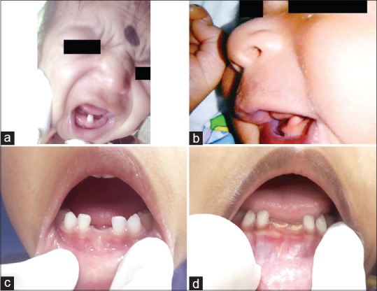
Natal teeth have been reported to be more frequent than neonatal teeth and the incidence of both is reported to range from 1:2000 to 1:3500.[4] While there are no clearly established gender differences in prevalence, some authors have found slight predilection for females.[2,4,5,6] These teeth are commonly located in the mandibular incisors[7] and are usually paired. Numerous studies have found 90% of the natal and neonatal teeth to be primary teeth, with only 10% being supernumerary.[6] Based on the clinical characteristics, Spouge and Feasby have classified natal and neonatal teeth as “mature” teeth with fully developed shape and morphology or “immature” teeth where the structure and development are incomplete.[3] Depending on the degree of maturity, these may be small, conical, and hypoplastic or may even resemble a normal tooth[8] [Figure 1a]. Common complications associated with these teeth include sublingual ulcerations, refusal to feed, inhalation of loose teeth, and laceration of the mother's breasts during feeding.[2] It is suggested that a well-implanted natal or neonatal tooth should be left in the arch; extraction is indicated only when it is extremely mobile, causes injury to the baby, or when there is a risk of aspiration.[9]
Figure 1.

Clinical photographs of (a) A fully erupted natal tooth in the mandibular anterior region in a newborn. (b) A soft-tissue swelling with an unerupted tooth in the mandibular anterior region in a newborn. (c) A 3-year-old baby with missing natal teeth in 71 and 81 regions and with no space loss. (d) A 4-year-old baby with hypoplastic natal teeth in 71 and 81 region
Limited population-based studies have been published on the clinical aspects of natal and neonatal teeth in India. A retrospective study carried out by Basavanthappa et al. found all 17 natal and neonatal teeth to be supernumerary teeth.[10]
Therefore, the aim of this study was to describe the clinical characteristics and the treatment outcome of natal and neonatal teeth from a hospital setting in southern part of Tamil Nadu.
Materials and Methods
A retrospective analysis of case records of babies born in a tertiary hospital in Tamil Nadu and the babies referred to the Dental and Oral Surgery department with natal and neonatal teeth, between January 1, 2012, and December 31, 2014, was carried out. Clinical data such as age, gender, antenatal and neonatal details, and other associated physical findings were noted. Details regarding the natal and neonatal teeth including their location, associated complaints, local complications, and the treatment given were recorded. Follow-up data of these children were also obtained. Any charts with incomplete data were excluded from the study. The study was carried out after obtaining approval from the Institutional Review Board (No: 10885).
Statistical analysis
The clinical data were entered into Microsoft Excel 2013 and descriptive statistical analysis was performed. Data were summarized using mean ± standard deviation for continuous variables and frequency along with percentage for categorical variables. The mean number of teeth between gender and gestational age was compared using independent t-test. P < 0.05 was considered to be statistically significant. All the analyses were done using Statistical Package for the Social Sciences, Version 21, IBM Analytics (Bangalore, India).
Results
The study sample comprised of 33 babies (15 males; 18 females), with a total of 52 natal and neonatal teeth. Eighteen babies presented with 28 natal teeth (54%) and 15 babies with 24 neonatal teeth (46%), of which 38 teeth (73%) were noted in pairs. All of these 52 natal and neonatal teeth were located in the mandibular central incisor region.
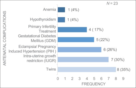
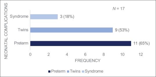
Of the 32 mothers, 23 mothers (72%) had antenatal complications, of which 10 mothers (43%) had more than one complication [Figure 2]. Among the 33 babies, 17 babies (51%) had some neonatal complications and 16 babies (48%) required nursery admissions. Eleven babies (33%) were preterm with a gestational age < 37 weeks and three had associated clinical syndromes including Down syndrome, suspected Carpenter's syndrome with craniosynostosis, and Aicardi–Goutier syndrome with hypothyroidism [Figure 3]. A positive family history of natal and neonatal teeth was present in eight cases which included one set of monozygotic twins. No statistical difference was noted in the mean number of teeth between gender (P = 0.305) and gestational age (P = 1.000).
Figure 2.

Distribution of antenatal complications
Figure 3.

Distribution of neonatal complications
Of the total, 36 teeth (69%) were visible of which 25 teeth (48%) were recorded to be loose. The remaining 16 teeth (31%) were covered with mucosa which also included two cases of soft-tissue swelling on the mandibular anterior ridge [Figure 1b] which eventually ruptured with a tooth.
Of the 52 teeth, extraction of 34 teeth (65%) had been carried out, while the remaining teeth (35%) were kept under observation. All the 25 loose teeth were extracted to reduce the risk of aspiration, which was a major concern, especially among those babies who required neonatal nursery care. No case of aspiration, however, was noted. The remaining nine teeth were removed due to feeding difficulty. With regard to complications secondary to the tooth, refusal to feed was recorded in 2 babies who had presented with a sublingual ulcer 1 month after birth. Among the 32 mothers, only one experienced difficulty due to breast lacerations.
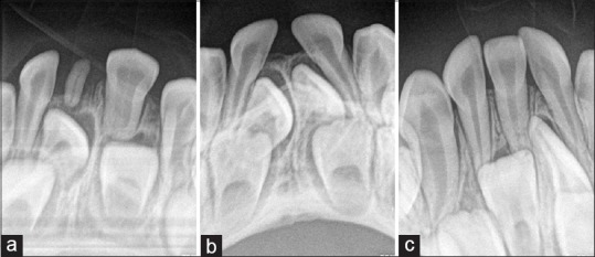
The follow-up records noted that out of the 52 natal and neonatal teeth, 40 teeth (77%) were clinically missing [Figure 1c] and 6 (11%) were hypoplastic [Figure 1d]. Two pairs of neonatal teeth (8%) with normal tooth structure were present and early eruption of permanent tooth with contralateral primary tooth in place was noted in two cases (4%). A residual natal tooth [Figure 4a] was noted in one case and a complete space loss [Figure 4b and c] was recorded in two cases.
Figure 4.

Intraoral periapical radiographic images of (a) A residual natal tooth in 81 region along with fused teeth in 71 and 72 regions. (b) A 4-year-old baby with missing natal teeth in 71 and 81 regions and with complete space loss. (c) A 3-year-old baby with missing natal tooth in 71 region and with complete space loss
Discussion
Natal and neonatal teeth are a rare occurrence and may be associated with anxiety and culturally prevalent misconceptions. Of the total 52 teeth, 28 were natal teeth (18 cases) and 24 were neonatal teeth (15 cases) and all were located in mandibular central incisor region. Many studies have been reported that natal and neonatal teeth are commonly found in the mandibular central incisor region, including that by Bodenhoff, who found 85% of them to be mandibular incisors, 11% to be maxillary incisors, 3% to be mandibular canines and molars, and 1% to be maxillary canine or molars.[7] This is possibly due to the fact that the mandibular incisors are the first teeth to erupt.[2,5,7]
In this study, majority of the natal and neonatal teeth were in pair and were similar to that reported in previous studies.[5,6] The greater frequency of natal teeth as compared to neonatal teeth may be because these are usually discovered by a pediatrician during the routine examination of the newborn, while neonatal teeth are accidentally seen by the mother.[2] In this study, no gender difference was noted although some studies have found predilection for females.[2,5]
Although the etiology is still unknown, several factors have been identified that result in a disturbance of the biological chronology of the teeth. These include the superficial position of the tooth germ, infection or malnutrition, eruption accelerated by febrile incidents or hormonal stimulation,[1] hereditary transmission of a dominant autosomal gene,[11,12] osteoblastic activity inside the germ area,[13] and hypovitaminosis.[14]
Hals has suggested that the abnormal superficial position of the tooth germs is in turn due to a hereditary factor.[11] A positive family history was present in eight cases in the present study, which included four siblings (including one set of twins), 2 fathers, 1 maternal grandmother, and 1 maternal aunt. Interestingly, 2 natal teeth were noted in a set of preterm monozygous twin girls. There have been reports of rare occurrences of natal teeth in twins.[15,16]
Some of the predisposing factors for the development of natal and neonatal teeth noted in literature include poor maternal health, endocrine disturbances, fever during pregnancy, and congenital syphilis.[2] In this study, majority of the mothers (72%) were noted with some antenatal complications such as twin gestation (35%), intrauterine growth retardation (30%), gestational diabetes mellitus (22%), pregnancy-induced hypertension, and eclampsia (26%).
We also noted 51% of the babies with neonatal complications, of which 48% of the babies required nursery admission. No clinical significance was noted between the number of teeth and gestational age. While we found three babies to have Down syndrome, Carpenter's syndrome, and Aicardi–Goutier syndrome, other studies have reported the occurrence of natal teeth in Ellis–van Creveld syndrome,[17] pachyonychia congenita, Hallermann–Streiff syndrome,[18] Pierre–Robin sequence, cleft lip and palate, Pfeiffer syndrome, ectodermal dysplasia, craniofacial dysostosis, Sotos syndrome, epidermolysis-bullosa simplex including Van der Woude syndrome, Down syndrome,[19] and Walker–Warburg syndrome.[20] The pair of neonatal teeth in the child with Down syndrome was noted to have normal tooth structure and morphology.
Hypermobility and refusal to feed are common symptoms associated with natal and neonatal teeth. Riga–Fede disease, noted only in two cases in this study, involves sublingual ulceration which may interfere with the feeding of the baby and thus result in nutritional deficiency and failure to gain weight.[21] Among the 36 teeth (69%) that were visible, 25 teeth (48%) were recorded as extremely mobile and were extracted. The soft-tissue swelling noted in 2 babies were self-limiting and excision was not done as the parents opted for a conservative management. The swelling was reported to have eventually ruptured, exposing a tooth. Published studies by Kates et al. and Wang et al. have reported cyst-like mass although excision was carried out in both the cases.[6,22]
Majority of the symptomatic natal and neonatal teeth were extracted in this study. It is important to assess the general condition of the baby before extraction. At birth, intramuscular Vitamin K injection was routinely administered to all babies born in this hospital and no record of excessive bleeding was noted in any case after extraction. During the process of tooth removal, care must be taken to avoid injury to the gingiva and should be alert to the risk of aspiration.[14]
The follow-up age of the study sample ranged from 18 months to 5 years. Six teeth were found to be discolored and hypoplastic. The enamel of natal and neonatal teeth was hypomineralized and therefore prone to wear and discoloration.[23] Residual natal/neonatal teeth have been previously reported[24,25] and curettage of the underlying tissue following extraction has been recommended.[26] Curettage was not routinely done in any of our cases and residual natal tooth was noted in one case with natal tooth. Space loss was found in two cases; Gardnier reported space loss in nine cases, although the space was regained when permanent incisors erupted.[27] Studies have found that 95% of the natal and neonatal teeth are primary teeth and only 5% are supernumerary teeth.[28] Radiographs have been recommended to differentiate between primary and supernumerary teeth.[29] In this study, all the natal and neonatal teeth were found to be primary mandibular central incisors. Previous studies have reported majority of the natal and neonatal teeth to be primary teeth[2,6,7,8,22] although this is not in agreement with the findings of Basavanthappa et al. who found all 17 natal/neonatal teeth to be supernumerary teeth.[10]
The following limitations were noted in this study: being a retrospective study, the true incidence of natal and neonatal teeth could not be estimated. Babies who had incomplete records or who did not report for follow-up could not be included. While etiology cannot be established, some factors associated with this dental anomaly were identified.
Conclusion
Natal and neonatal teeth in this study were found to be precociously erupted primary teeth. The most frequent location of these teeth was the mandibular incisor region. There appears to be a possible hereditary basis with a positive family history present in many cases. It also appears to be associated with antenatal and neonatal complications. Dentists and pediatricians should consider these teeth as prematurely erupted primary teeth rather than supernumerary teeth and not regard them as a normal phenomenon. Extraction is recommended if the teeth are extremely mobile and are a risk to the baby. Larger prospective study will help in a better understanding of the natal and neonatal teeth.
Declaration of patient consent
The authors certify that they have obtained all appropriate patient consent forms. In the form the patient(s) has/have given his/her/their consent for his/her/their images and other clinical information to be reported in the journal. The patients understand that their names and initials will not be published and due efforts will be made to conceal their identity, but anonymity cannot be guaranteed.
Financial support and sponsorship
Nil.
Conflicts of interest
There are no conflicts of interest.
References
- 1.Bigeard L, Hemmerle J, Sommermater JI. Clinical and ultrastructural study of the natal tooth: Enamel and dentin assessments. ASDC J Dent Child. 1996;63:23–31. [PubMed] [Google Scholar]
- 2.Massler M, Savara BS. Natal and neonatal teeth; a review of 24 cases reported in the literature. J Pediatr. 1950;36:349–59. doi: 10.1016/s0022-3476(50)80105-1. [DOI] [PubMed] [Google Scholar]
- 3.Spouge JD, Feasby WH. Erupted teeth in the newborn. Oral Surg Oral Med Oral Pathol. 1966;22:198–208. [Google Scholar]
- 4.Chow MH. Natal and neonatal teeth. J Am Dent Assoc. 1980;100:215–6. doi: 10.14219/jada.archive.1980.0061. [DOI] [PubMed] [Google Scholar]
- 5.Allwright WC. Natal and neonatal teeth. Br Dent J. 1958;105:163–72. [Google Scholar]
- 6.Kates GA, Needleman HL, Holmes LB. Natal and neonatal teeth: A clinical study. J Am Dent Assoc. 1984;109:441–3. doi: 10.14219/jada.archive.1984.0415. [DOI] [PubMed] [Google Scholar]
- 7.Bodenhoff J, Gorlin RJ. Natal and neonatal teeth: Folklore and fact. Pediatrics. 1963;32:1087–93. [PubMed] [Google Scholar]
- 8.Rusmah M. Natal and neonatal teeth: A clinical and histological study. J Clin Pediatr Dent. 1991;15:251–3. [PubMed] [Google Scholar]
- 9.Toledo AO. Pediatric Dentistry: Fundamentals for Practice Clinic. Sao Paulo: Premier; 1996. Growth and development: Notions of odontopediatric interest; pp. 17–40. [Google Scholar]
- 10.Basavanthappa NN, Kagathur U, Basavanthappa RN, Suryaprakash ST. Natal and neonatal teeth: A retrospective study of 15 cases. Eur J Dent. 2011;5:168–72. [PMC free article] [PubMed] [Google Scholar]
- 11.Hals H. Natal and neonatal teeth. Oral Surg Oral Med Oral Pathol. 1957;10:509–21. doi: 10.1016/0030-4220(57)90011-7. [DOI] [PubMed] [Google Scholar]
- 12.Bodenhoff J. Natal and neonatal teeth. Dent Abstr. 1960;5:485–8. [Google Scholar]
- 13.Jasmin JR, Clergeau-Guerithault S. A scanning electron microscopic study of the enamel of neonatal teeth. J Biol Buccale. 1991;19:309–14. [PubMed] [Google Scholar]
- 14.Anderson RA. Natal and neonatal teeth: Histologic investigation of two black females. ASDC J Dent Child. 1982;49:300–3. [PubMed] [Google Scholar]
- 15.Prabhakar AR, Ravi GR, Raju OS, Kurthukoti J, Shubha AB. Neonatal tooth in fraternal twins: A case report. Int J Clin Pediatr Dent. 2009;2:40–4. doi: 10.5005/jp-journals-10005-1028. [DOI] [PMC free article] [PubMed] [Google Scholar]
- 16.Hersh JH, Verdi GD. Natal teeth in monozygotic twins with van der Woude syndrome. Cleft Palate Craniofac J. 1992;29:279–81. doi: 10.1597/1545-1569_1992_029_0279_ntimtw_2.3.co_2. [DOI] [PubMed] [Google Scholar]
- 17.Weiss H, Crosett AD., Jr Chondroectodermal dysplasia; report of a case and review of the literature. J Pediatr. 1955;46:268–75. doi: 10.1016/s0022-3476(55)80280-6. [DOI] [PubMed] [Google Scholar]
- 18.Robotta P, Schafer E. Hallermann-Streiff syndrome: Case report and literature review. Quintessence Int. 2011;42:331–8. [PubMed] [Google Scholar]
- 19.Ndiokwelu E, Adimora GN, Ibeziako N. Neonatal teeth association with Down's syndrome. A case report. Odontostomatol Trop. 2004;27:4–6. [PubMed] [Google Scholar]
- 20.Venkatesh C, Adhisivam B. Natal teeth in an infant with congenital hypothyroidism. Indian J Dent Res. 2011;22:498. doi: 10.4103/0970-9290.87088. [DOI] [PubMed] [Google Scholar]
- 21.Hegde RJ. Sublingual traumatic ulceration due to neonatal teeth (Riga-Fede disease) J Indian Soc Pedod Prev Dent. 2005;23:51–2. doi: 10.4103/0970-4388.16031. [DOI] [PubMed] [Google Scholar]
- 22.Wang CH, Lin YT, Lin YJ. A survey of natal and neonatal teeth in newborn infants. J Formos Med Assoc. 2017;116:193–6. doi: 10.1016/j.jfma.2016.03.009. [DOI] [PubMed] [Google Scholar]
- 23.Bjuggren G. Premature eruption in the primary dentition – A clinical and radiological study. Sven Tandlak Tidskr. 1973;66:343–55. [PubMed] [Google Scholar]
- 24.Ooshima T, Mihara J, Saito T, Sobue S. Eruption of tooth-like structure following the exfoliation of natal tooth: Report of case. ASDC J Dent Child. 1986;53:275–8. [PubMed] [Google Scholar]
- 25.Tsubone H, Onishi T, Hayashibara T, Sobue S, Ooshima T. Clinico-pathological aspects of a residual natal tooth: A case report. J Oral Pathol Med. 2002;31:239–41. doi: 10.1034/j.1600-0714.2002.310408.x. [DOI] [PubMed] [Google Scholar]
- 26.King NM, Lee AM. Prematurely erupted teeth in newborn infants. J Pediatr. 1989;114:807–9. doi: 10.1016/s0022-3476(89)80142-8. [DOI] [PubMed] [Google Scholar]
- 27.Gardiner H. Erupted teeth in the newborn. Proc R Soc Med. 1961;54:504–6. [PMC free article] [PubMed] [Google Scholar]
- 28.Howkins C. Congenital teeth. Br Dent Assoc. 1932;53:402–5. [Google Scholar]
- 29.Almeida CM, Gomide MR, Nishiyama CK. Dente natal/neonatal. Odontol Clin. 1997;7:43–5. [Google Scholar]


