INTRODUCTION
Communicating science to peers and students often involves constructing clear, concise flow diagrams and illustrations as well as writing narratives (1, 2). Diverse methods for learning, including the use of diagrams of complex biological pathways (2), help increase the number of active retrieval pathways, lengthen memory, and improve recall efficiency (3). Diagrams vary in their complexity, which should match the audience’s familiarity with the topic. High school, premedical, and medical students who reviewed a brief set of comic strips or a comic chapter book on the anatomy of the digestive system had greater recall of the organs’ functions than control students (4). A group of tenth-grade biology students who studied diagrams enhanced their restudy and recall of biology concepts (5). A different group of tenth-grade biology students improved their comprehension of scientific diagrams by participating in workbook-focused instruction on conventions of diagrams and discussions led by a teacher (6). Since even physicians have been shown to prefer flowcharts and flow diagrams for learning and recalling clinical guidelines (7), science communicators should consider flowcharts and diagrams as important tools for teaching or refreshing science concepts with students of most ages and levels. While we present tips and tools for teaching students in grades 6 through 12 and college, all scientists and communicators can use the same tips and tools (tables, figures, and URLs) to design effective diagrams that communicate complex processes in simple and engaging visual ways.
Both flowcharts and flow diagrams can help students and readers who learn through seeing comprehend the relationships between objects or steps. Some people can remember details from a picture with text for a longer time than details from prose: for example, pictures with text in patient leaflets improve recall, comprehension, and medication adherence by the general public (8). Flowcharts and flow diagrams commonly use brief text and graphic elements to give an overview of a multistep process, a theory, or comparisons (Table 1). Although some students have better spatial cognition than others, all students who actively construct diagrams discover how to “follow the arrows” (9, 10). Students who self-completed diagrams with both text and graphic elements but not diagrams with only graphic elements could transfer inferences to a different scientific field (11). Furthermore, teacher instruction with a workbook that explains the common meanings of symbols and graphic elements in the diagrams from their biology textbook improved students’ comprehension (6).
TABLE 1.
Common uses of flowcharts and flow diagrams.
| Content and Examples | |
|---|---|
|
| |
| Flowcharts | Flow Diagrams |
1. Linear progression of steps
|
1. Complex set of inputs, and interactions, in a process or theory
|
2. Group of steps that are repeated
|
2. Comparison of two similar but distinct processes, or comparison of two substances affecting a process
|
3. Relationships between members of group
|
3. An organ, or organelle, shown at different magnifications
|
The recently described Scientific Process Flowchart Assessment method can help evaluate students’ comprehension and visualization of the scientific process (12). Questions in prose help assess students’ grasp of concept definitions and facts but not how students organize the knowledge and relate it to similar fields (12). Diagrams can help students organize the knowledge (10) and apply it to other fields. Students who create flow diagrams may form a better-integrated or deeper understanding of the topic (10) because they need to notice all of the elements and their functions (13). In addition, flowcharts and flow diagrams are easy to understand for someone even with limited knowledge of the language; effective diagrams help students and scientists convey their research to peers and international colleagues at scientific meetings. Because students developing their own flowcharts may be able to more rapidly interpret flowcharts made by others (9), we encourage teachers to make construction or use of flowcharts a weekly activity, possibly as part of their preparation for laboratory exercises.
PROCEDURE
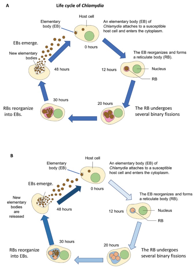
Teachers and professors can ask their students to prepare a flowchart of the procedure as pre-laboratory preparation rather than writing a description. Because people read English from left to right and top to bottom, steps in a flowchart also progress from top to bottom for more rapid comprehension and retention (14). Since many students do not know the conventions of diagrams, teachers can download and use an effective workbook developed by Cromley et al. (6) (workbook available at http://hdl.handle.net/2142/97891) for teaching them to tenth-grade biology students. Cromley et al. (6, 11) provide an additional workbook focused on genetics, and two focused on atoms and proteins at the same URL. (When you use them, please provide feedback to Dr. Cromley.) Table 2 lists the basic conventions of diagrams—both text and graphic elements—with examples shown in Figures 1 and 2. The life cycle (Fig. 1) proceeds in a clockwise direction. Fig. 1A is typical of diagrams found in scientific articles: the listing of the hours after infection and the blue arrows show the passage of time and the sequence of events. In comparison, Figure 1B uses extra variation in colors of the conventions to emphasize the progression of the infectious process (arrows go from light to dark blue) and differentiate between the two types of infectious particles (EB = brown circle versus RB = beige circle). The changes in Figure 1B can help readers comprehend the life cycle more quickly. To be effective, multiple flowcharts and diagrams in a single manuscript or slide presentation should use the same conventions (e.g., same shape, size, color, filling) only for identical variables or processes (2). Students, communicators, and scientists can portray similar but distinct variables or processes with related graphic elements (e.g., gradated colored arrows, different colored elements for different stages of infectious particles in Fig. 1B).
TABLE 2.
Explanation and examples of conventions of diagrams.
| Conventions of Diagrams—Prose | Examples in Figure 1 |
|---|---|
| Title | |
| • Is at the top | The title at the top: “Life cycle of Chlamydia” |
| • Tells key idea of diagram | |
| Caption | |
| • Is next to the figure number; often located below a figure | “FIGURE 1. Flow diagram showing the life cycle of Chlamydia.” |
| • Expands on key idea of diagram (what to notice) | Provides description of each panel of figure: A), B) |
| • May include abbreviations | EB = elementary body of Chlamydia; RB = reticulate body of Chlamydia. |
| Labels | |
| • Inside diagram | |
| -- Naming labels: Name parts of things | “Elementary body (EB)”, “Nucleus” |
| -- Explanatory labels: Describe what is happening in a part of the diagram | Six explanatory labels are present in Figure 1A. The first one is located at 1 o’clock. |
| -- Labels of passage of time: List amount of time that has passed between two events | The infection starts at 0 hours and progresses (clockwise) with events described at 12 hours, 20 hours, 30 hours, and 48 hours. |
| Legend | |
| • Identifies what any symbols used represent | |
|
| |
| Conventions of Diagrams—Graphic Elements | Examples in Figures 1 or 2 |
|
| |
| Arrows | |
| • Single shape, color, and size should mean same thing | Figure 1: The five blue process arrows show the sequence of events during Chlamydia infection. Note that the length of the arrows does not correlate with the length of elapsed time. |
| • Common to have same type of arrow mean related process instead of same process | |
| -- Process arrows: Indicate a sequence of events | |
| -- Divergent arrows: Show two processes that occur at same time OR that two possibilities exist but only one occurs | |
| Cycle or circle | |
| • Start is located at 12 o’clock | Figure 1: The life cycle is drawn as a circle. |
| • Proceeds clockwise | Figure 1. Drawings of an infected cell as it progresses through all the stages of infection. |
| • Drawings or illustrations of animals, humans, organs, cells, microbes | |
| Color | |
| • Color of symbols and graphic elements depicts relationship. OR |
Figure 1B: Change in color of arrows—from light blue at beginning of infection to dark blue at release of infectious EBs—shows direction and correlates with passage of time. |
| • Same color: Color of object is same in nature and in diagram OR |
Figure 1. The beige color of the depicted cytoplasm of the infected cells is close to the true color observed under the light microscope |
| • After staining and in diagrams. E.g., photographs of stained tissue where certain cells are stained, (e.g., a specific microbe, protein, or RNA using Gram stain, immunohistochemistry, or in situ hybridization, respectively) OR |
|
| • False color: Color of object is changed to contrast it with background or other related biological part | Figure 1: The contents of the cell, the EBs, and RBs use false color to make them easier to see. |
| Magnification | |
| • Zoom-in: Like a magnifying glass; shows a magnified part of an object | |
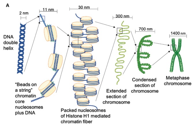
| • Zoom-out: Like stepping back from a leaf to see a forest; shows the object at lower magnification and as part of a bigger structure. | Figure 2. |
EB = elementary body; RB = reticulate body.
FIGURE 1.
Flow diagrams showing the life cycle of Chlamydia, a pathogenic organism. A) Original diagram. B) Revised diagram uses similar conventions of diagrams and some modifications have been made to improve comprehension. EB = elementary body of Chlamydia; RB = reticulate body of Chlamydia.
FIGURE 2.
A flow diagram that zooms out (reduces magnification) from a DNA helix at high resolution through several more-compact structures to full condensation and into a metaphase chromosome.
Flow diagrams can show the relationship of a small part to a large part. For example, Figure 2 shows a model of chromosome condensation. It displays a naked DNA helix, and zooms out in five steps to show the structures in which the DNA lives, up to a condensed metaphase chromosome. This model does not provide the mechanisms and proteins involved in going from the DNA helix to the condensed metaphase chromosome. Thus, the designer needs to decide not only what to include, but also what to leave out to avoid too much information and clutter (14). To avoid audience overload in diagrams for slide presentations, diagram creators can group similar variables or processes together and show the relationships between four groups or fewer on a slide (14). Adding animation to highlight group X can help focus the audience’s attention on a specific point during the oral presentation. Since students who draw and use diagrams, especially those containing text (11), can more easily comprehend and apply the displayed scientific concepts in future biological and microbiological knowledge and research (10, 15), scientists and communicators may wish to include at least naming and explanatory labels, graphic elements, and a detailed legend (2) in their flowcharts and diagrams.
Several vendors sell templates of biological shapes and some flow diagrams with a non-exclusive license for use of modified works. To save time and maintain quality, we modified two PowerPoint templates from the scientific illustration toolkits for presentations and publications from MOTIFOLIO to make flow diagrams for our figures. Scientists and communicators should consider using unambiguous conventions, especially unambiguous graphic elements, in their diagrams to optimize the audience’s focus at the start and support the appropriate flow of their attention to subsequent parts.
CONCLUSION
Flowcharts and diagrams help many people remember a sequence of events and recall interactions in a complex process. Teachers, professors, and science communicators can display information for their students and audiences via flowcharts in class discussions, on handouts, in articles, during continuing education programs, and in slide presentations. Explaining the conventions of diagrams in the figure legends and clearly indicating the sequence in which to read them can help all audiences better assimilate the relationships the flow diagram conveys. Students who use and make flowcharts as part of weekly laboratories may become more adept in applying complex concepts (15). Students of all ages, colleagues, and readers likely will appreciate these insights and enhanced communication skills.
ACKNOWLEDGMENTS
The authors declare that there are no conflicts of interest.
REFERENCES
- 1.Vandenbroucke JP, von Elm E, Altman DG, Gotzsche PC, Mulrow CD, Pocock SJ, Poole C, Schlesselman JJ, Egger M STROBE Initiative. Strengthening the reporting of observational studies in epidemiology (STROBE): explanation and elaboration. Int J Surg. 2014;12:1500–1524. doi: 10.1016/j.ijsu.2014.07.014. [DOI] [PubMed] [Google Scholar]
- 2.O’Hara L, Livigni A, Theo T, Boyer B, Angus T, Wright D, Chen SH, Raza S, Barnett MW, Digard P, Smith LB, Freeman TC. Modelling the structure and dynamics of biological pathways. PLOS Biol. 2016;14:e1002530. doi: 10.1371/journal.pbio.1002530. [DOI] [PMC free article] [PubMed] [Google Scholar]
- 3.Congleton A, Rajaram S. The origin of the interaction between learning method and delay in the testing effect: the roles of processing and conceptual retrieval organization. Mem Cognit. 2012;40:528–539. doi: 10.3758/s13421-011-0168-y. [DOI] [PubMed] [Google Scholar]
- 4.Kim J, Chung MS, Jang HG, Chung BS. The use of educational comics in learning anatomy among multiple student groups. Anat Sci Educ. 2017;10:79–86. doi: 10.1002/ase.1619. [DOI] [PubMed] [Google Scholar]
- 5.Bergey BW, Cromley JG, Kirchgessner ML, Newcombe NS. Using diagrams versus text for spaced restudy: effects on learning in 10th grade biology classes. Br J Educ Psychol. 2015;85:59–74. doi: 10.1111/bjep.12062. [DOI] [PubMed] [Google Scholar]
- 6.Cromley JG, Perez TC, Fitzhugh SL, Newcombe NS, Wills TW, Tanaka JC. Improving students’ diagram comprehension with classroom instruction. J Experiment Educ. 2013;81:511–537. doi: 10.1080/00220973.2012.745465. [DOI] [Google Scholar]
- 7.Kang MK, Kim BK, Kim TW, Kim SH, Kang HR, Park HW, Chang YS, Kim SS, Min KU, Kim YY, Cho SH. Physicians’ preferences for asthma guidelines implementation. Allergy Asthma Immunol Res. 2010;2:247–253. doi: 10.4168/aair.2010.2.4.247. [DOI] [PMC free article] [PubMed] [Google Scholar]
- 8.Katz MG, Kripalani S, Weiss BD. Use of pictorial aids in medication instructions: a review of the literature. Am J Health Syst Pharm. 2006;63:2391–2397. doi: 10.2146/ajhp060162. [DOI] [PubMed] [Google Scholar]
- 9.von Kotzebue L, Gerstl M, Nerdel C. Common mistakes in the construction of diagrams in biological contexts. Res Sci Educ. 2015;45:193–213. doi: 10.1007/s11165-014-9419-9. [DOI] [Google Scholar]
- 10.Hoskins SG, Stevens LM, Nehm RH. Selective use of the primary literature transforms the classroom into a virtual laboratory. Genetics. 2007;176:1381–1389. doi: 10.1534/genetics.107.071183. [DOI] [PMC free article] [PubMed] [Google Scholar]
- 11.Cromley JG, Bergey BW, Fitzhugh SL, Newcombe N, Wills TW, FST, Tanaka JC. Effects of three diagram instruction methods on transfer of diagram comprehension skills: the critical role of inference while learning. Learn Instruct. 2013;26:45–58. doi: 10.1016/j.learninstruc.2013.01.003. [DOI] [Google Scholar]
- 12.Wilson KJ, Rigakos B. Scientific process flowchart assessment (SPFA): a method for evaluating changes in understanding and visualization of the scientific process in a multidisciplinary student population. CBE Life Sci Educ. 2016;15(4) doi: 10.1187/cbe.16-06-0196. pii:ar63. [DOI] [PMC free article] [PubMed] [Google Scholar]
- 13.Scheiter K, Schleinschok K, Ainsworth S. Why sketching may aid learning from science texts: contrasting sketching with written explanations. Top Cogn Sci. 2017;9:866–882. doi: 10.1111/tops.12261. [DOI] [PubMed] [Google Scholar]
- 14.Kosslyn SM. Better power point quick fixes based on how your audience thinks. Oxford Press; New York: 2011. [Google Scholar]
- 15.Hoskins SG, Lopatto D, Stevens LM. The C.R.E.A.T.E. approach to primary literature shifts undergraduates’ self-assessed ability to read and analyze journal articles, attitudes about science, and epistemological beliefs. CBE Life Sci Educ. 2011;10:368–378. doi: 10.1187/cbe.11-03-0027. [DOI] [PMC free article] [PubMed] [Google Scholar]


