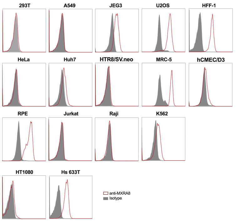
Extended Data Figure 5. Surface expression of MXRA8 in different human cell lines.
Human cell lines were tested for MXRA8 surface expression by flow cytometry: 293T (embryonic kidney), A549 (lung adenocarcinoma), JEG3 (placental choriocarcinoma), U2OS (osteosarcoma), HFF-1 (foreskin fibroblasts), HeLa (cervical carcinoma), Huh7 (hepatocarcinoma), HTR8/SV.neo (trophoblast progenitor), MRC-5 (lung carcinoma), hCMEC/D3 (cerebral microvascular endothelial cells), RPE (retinal pigment epithelial cell), Jurkat (T cell lymphoma), Raji (B cell lymphoma), K562 (eryrtholeukemia), HT1080 (fibrosarcoma), and Hs 633T (fibrosarcoma) cells. Representative data are shown of two independent experiments. Gray histograms, isotype control mAb; red histograms, anti-MXRA8 mAb.

