
Figure 2.
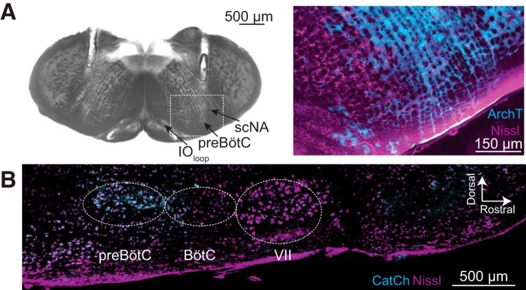
. A, Bright field image of a transverse section from an adult Dbx1;ArchT mouse at the level the preBötC, as indicated by the loop of the inferior olive (IOloop) and the semi-compact division of the nucleus ambiguus (scNA). Parallel tracks of implanted fiber optics are visible from the dorsal border of the tissue section into the intermediate reticular formation dorsal to the preBötC. The selection box was imaged using fluorescence microscopy to show ArchT (cyan) protein expression in the preBötC in detail, Nissl staining (magenta) included for contrast. B, Parasagittal section from an adult Dbx1;CatCh mouse. Nissl (magenta) shows anatomic landmarks including the facial (VII) cranial nucleus, Bötzinger complex (BötC), and the preBötC. CatCh (cyan) expression is limited to the preBötC.