Abstract
Background and aims
Initial clinical management decision in patients with acute gastrointestinal bleeding (GIB) is often based on identifying high- and low-risk patients. Little is known about the role of lactate measurement in the triage of patients with acute GIB. We intended to assess if lactate on presentation is predictive of need for intervention in patients with acute GIB.
Patients and methods
We performed a single-center, retrospective, cross-sectional study including patients ≥18 years old presenting to emergency with acute GIB between January 2014 and December 2014. Intensive care unit (ICU) admission, inpatient endoscopy (upper endoscopy and/or colonoscopy), and packed red blood cell (PRBC) transfusion were assessed as outcomes. Analyses included univariate and multivariate logistic regression analyses.
Results
Of 1,237 patients with acute GIB, 468 (37.8%) had venous lactate on presentation. Of these patients, 165 (35.2%) had an elevated lactate level (>2.0 mmol/L). Patients with an elevated lactate level were more likely to be admitted to ICU than patients with a normal lactate level (adjusted odds ratio [AOR] 2.96, 95% confidence interval [CI] 1.74–5.01; p<0.001). Patients with an elevated lactate level were more likely to receive PRBC transfusion (AOR 3.65, 95% CI 1.76–7.55; p<0.001) and endoscopy (AOR 1.64, 95% CI 1.02–2.65; p=0.04) than patients with a normal lactate level.
Conclusion
Elevated lactate level predicts the need for ICU admissions, transfusions, and endoscopies in patients with acute GIB. Lactate measurement may be a useful adjunctive test in the triage of patients with acute GIB.
Keywords: venous lactate, ICU admissions, endoscopy, acute gastrointestinal bleeding
Introduction
Acute gastrointestinal bleeding (GIB) is a common medical emergency with significant morbidity, mortality, and cost. In 2012, GIB accounted for >500,000 US hospital discharges, 27,732 deaths, and a cost of ~$5 billion dollars.1 Appropriate risk stratification of these patients can reduce resource utilization and costs without adversely affecting patient outcomes.2
Initial clinical management decision in patients presenting to emergency department (ED) with acute GIB is often based on identifying high- and low-risk patients. Patients with high risks of adverse outcomes, such as death or rebleeding, are more likely to benefit from early, aggressive management, whereas patients with lower risks may be considered for early hospital discharge or even outpatient management.3–7 Various risk-scoring systems, both for upper and lower GIB, have been developed to identify high-risk patients.5–12 These scoring systems are based on clinical, laboratory, and endoscopic findings. Emergent endoscopy and, therefore, endoscopic findings are often unavailable at the time of initial assessment. Therefore, risk-scoring systems based on clinical and laboratory parameters may be more useful to a clinician at the time of initial assessment. Patient demographic characteristics, medical history, comorbidities, use of certain medications, and clinical and laboratory findings on presentation have been found to predict the severity of acute GIB in prior studies.3–13
Venous lactate is predictive of the severity of illness and risk of mortality in patients with sepsis.14,15 A few studies have evaluated the role of initial venous lactate in predicting outcomes in patients with acute GIB.16–19 This study was designed to assess whether venous lactate on presentation was predictive of need for intervention in patients with acute GIB.
Patients and methods
A retrospective cross-sectional study was performed including patients ≥18 years old who presented to ED of Banner University Medical Center Tucson at the Main Campus (479 beds) and South Campus (161 beds) with acute GIB between January 2014 and December 2014. Patients were identified from the ED encounter database using International Classification of Diseases, Ninth Revision (ICD-9) codes representing acute GIB (Table S1). Similar diagnostic codes have been used in prior studies to identify patients with GIB.7,11 Patients without venous lactate on presentation were excluded.
Patient’s demographic information, medical history, and clinical and laboratory data were collected from the clinical data warehouse (CDW). The CDW is the University of Arizona Health Sciences’ centralized, standardized, integrated repository of data, which includes data extracted from the electronic health record (EHR) of Banner University Medical Center Tucson. Study data were collected and managed using Research Electronic Data Capture (REDCap), an electronic data capture tool hosted at the University of Arizona.20
Patient characteristics included age, gender, ethnicity, language, insurance status, and comorbidities as described by the Charlson Comorbidity Index.21 Other clinical and laboratory variables included history of prior GIB, history of alcohol use, history of smoking, use of nonsteroidal anti-inflammatory drug (NSAID), use of aspirin, use of anticoagulant, presentation with syncope, bright red blood per rectum, melena, abdominal pain, hematemesis, altered mental status, ascites, initial heart rate and systolic blood pressure, initial hemoglobin, platelet count, prothrombin time as international normalized ratio (INR), creatinine, and venous lactate. Additionally, we collected data on time of presentation, day of presentation, and month of presentation. The range of normal venous lactate level was 0.5–2.0 mmol/L. Accordingly the venous lactate was predefined as normal (0.5–2.0 mmol/L) or elevated (>2.0 mmol/L).
Intensive care unit (ICU) admission, inpatient endoscopy (upper endoscopy and/or colonoscopy), and packed red blood cell (PRBC) transfusion (transfusion of at least a unit of PRBC) were assessed as outcomes (categorical outcome variables). Two groups were also compared for any difference in the length of hospital stay.
The data accessed were de-identified and did not require patient informed consent to access. The study was approved by the University of Arizona Institutional Review Board (protocol number: 1612054091).
Statistics
Univariate analysis was used to compare the outcomes and the study variables for the groups (patients who had normal vs elevated venous lactate level). All comparisons were unpaired, and tests of significance were two tailed. Continuous variables were compared using the Mann–Whitney U test and categorical variables using the chi-square test or Fisher’s exact test, where applicable. Multivariate logistic regression analysis was performed for the dependent categorical outcome variables using the variable venous lactate as the predictor of interest and other factors predictive of the severity of acute GIB in prior studies as covariates. The adjusted odds ratios (AORs), 95% confidence intervals (CIs), and p-values were reported for the variable venous lactate. We assessed the discriminative property of the predictive models by estimating their area under the receiver operating characteristic (ROC) curve. We also compared the length of hospital stay between the groups using multivariate linear regression analysis. The mean difference, 95% CI, and p-value were reported for the variable venous lactate.
The statistical analysis was performed using the statistical software package Stata, version 12 (StataCorp LP, College Station, TX, USA).
Results
A total of 1,237 patients with acute GIB were identified, 804 (65.0%) from Main Campus and 433 (35.0%) from South Campus. In all, 769 patients who did not have venous lactate on presentation were excluded. In the final analysis, there were 468 patients with an elevated lactate level in 165 (35.3%) patients.
Of this study population, the median age was 59.5 years and 46.6% were females. Most patients were Caucasian (54.5%) or Hispanic (32.3%), and almost all patients were English-speaking (91.0%). The type of health insurance included Medicare (49.4), Medicaid (33.1%), private (13.5%), and no insurance (4.0%). Median Charlson Comorbidity Index score was 3 (Table 1).
Table 1.
Study population characteristics at initial presentation, including demographics and history variables (univariate analysis)
| Population characteristics | Total population (N=468) | Patents who had normal initial venous lactatea (n=303, 64.7%) | Patients who had elevated initial venous lactateb (n=165, 35.3%) | p-valuec |
|---|---|---|---|---|
| Age, median (IQR), years | 59.5 (47–71) | 62 (47–74) | 55 (46–67) | 0.01 |
| Gender, n (%) | <0.001 | |||
| Male | 250 (53.4) | 143 (47.2) | 107 (64.8) | |
| Female | 218 (46.6) | 160 (52.8) | 58 (35.2) | |
| Ethnicity/race, n (%) | 0.89 | |||
| Caucasian | 255 (54.5) | 163 (53.8) | 92 (55.7) | |
| Hispanic | 151 (32.3) | 100 (33.0) | 51 (30.9) | |
| African-American | 25 (5.3) | 15 (4.9) | 10 (6.1) | |
| Other | 37 (7.9) | 25 (8.3) | 12 (7.3) | |
| Language, n (%) | 0.06 | |||
| English | 426 (91.0) | 269 (88.8) | 157 (95.2) | |
| Spanish | 34 (7.3) | 28 (9.2) | 6 (3.6) | |
| Other | 8 (1.7) | 6 (1.9) | 2 (1.2) | |
| Insurance, n (%) | 0.003 | |||
| Medicare | 231 (49.4) | 168 (55.4) | 63 (38.2) | |
| Medicaid | 155 (33.1) | 86 (28.4) | 69 (41.8) | |
| Private | 63 (13.5) | 39 (12.9) | 24 (14.5) | |
| Uninsured | 19 (4.0) | 10 (3.3) | 9 (5.5) | |
| History of prior GIB, n (%) | 98 (20.9) | 59 (19.5) | 39 (23.6) | 0.29 |
| History of alcohol use, n (%) | 276 (58.9) | 202 (66.7) | 74 (44.8) | <0.001 |
| History of smoking, n (%) | 126 (26.9) | 88 (29.0) | 38 (23.0) | 0.16 |
| History of NSAID use, n (%) | 84 (17.9) | 54 (17.8) | 30 (18.2) | 0.92 |
| Use of anticoagulant, n (%) | 48 (10.3) | 29 (9.6) | 19 (11.5) | 0.51 |
| Use of aspirin, n (%) | 112 (23.9) | 78 (25.7) | 34 (20.6) | 0.21 |
| Charlson comorbidity index, median (IQR) | 3 (1–5) | 3 (1–5) | 4 (1–5) | 0.47 |
Notes:
Normal venous lactate range 0.5–2.0 mmol/L.
Elevated venous lactate >2.0 mmol/L.
p-values cited compare patients with normal and elevated venous lactate on presentation. Bold values signify statistically significant p-values.
Abbreviations: IQR, interquartile range; GIB, gastrointestinal bleeding; NSAID, nonsteroidal anti-inflammatory drug.
A total of 128 (27.3%) patients were admitted to ICU. Inpatient endoscopy was performed on 167 (35.7%) patients. A total of 171 (36.5%) patients received transfusion. Median length of hospital stay was 3 days (Table 2).
Table 2.
Study population characteristics at initial presentation, including clinical features, laboratory values, time of presentation, and outcome variables (univariate analysis)
| Population characteristics | Total population (N=468) | Patients who had normal initial venous lactatea (n=303, 64.7%) | Patients who had elevated initial venous lactateb (n=165, 35.3%) | p-valuec |
|---|---|---|---|---|
| Syncope, n (%) | 11 (2.3) | 6 (1.9) | 5 (3.0) | 0.53 |
| Bright red blood per rectum, n (%) | 73 (15.6) | 55 (18.1) | 18 (10.9) | 0.04 |
| Hematemesis, n (%) | 82 (17.5) | 40 (13.2) | 42 (25.4) | 0.001 |
| Abdominal pain, n (%) | 128 (27.3) | 101 (33.3) | 27 (16.3) | <0.001 |
| Altered mental status, n (%) | 13 (2.8) | 4 (1.3) | 9 (5.4) | 0.01 |
| Ascites, n (%) | 41 (8.7) | 16 (5.3) | 25 (15.1) | <0.001 |
| Heart rate, median (IQR), per minute | 76 (67–86) | 75 (67–85) | 78 (68–90) | 0.09 |
| Systolic blood pressure, median (IQR), mmHg | 122 (110–137) | 122 (110–138) | 121 (109–135) | 0.39 |
| Hemoglobin, median (IQR), g/dL | 11.3 (8.8–13.6) | 11.6 (9.1–13.8) | 10.6 (8.3–13.1) | 0.02 |
| Platelet count, median (IQR), ×109/L | 215 (157–293) | 225 (168–299) | 188 (126–287) | 0.01 |
| INR, median (IQR) | 1.1 (1–1.5) | 1.1 (1.0–1.3) | 1.2 (1.1–1.7) | <0.001 |
| Creatinine, median (IQR), mg/dL | 0.9 (0.8–1.3) | 0.9 (0.8–1.2) | 1.0 (0.8–1.5) | 0.003 |
| Presentation during daytime (7 am–7 pm), n (%) | 334 (71.4) | 211 (69.6) | 123 (74.5) | 0.26 |
| ICU admission, n (%) | 128 (27.3) | 59 (19.5) | 69 (41.8) | <0.001 |
| Endoscopy, n (%) | 167 (35.7) | 87 (28.7) | 80 (48.5) | <0.001 |
| Medical therapy, n (%) | 366 (78.2) | 220 (72.6) | 146 (88.5) | <0.001 |
| PRBC transfusion, n (%) | 171 (36.5) | 90 (29.7) | 81 (49.1) | <0.001 |
| Length of hospital stay, median (IQR), days | 3 (2–6) | 3 (1–5) | 4 (2–8) | <0.001 |
Notes:
Normal venous lactate range 0.5–2.0 mmol/L.
Elevated venous lactate >2.0 mmol/L.
p-values cited compare patients with normal and elevated venous lactate on presentation. Bold values signify statistically significant p-values.
Abbreviations: IQR, interquartile range; INR, international normalized ratio; ICU, intensive care unit; PRBC, packed red blood cell.
Univariate analysis revealed no significant differences in regard to ethnicity, language, Charlson Comorbidity Index, history of prior GIB, history of smoking, use of NSAID, use of aspirin, and use of anticoagulant between the two groups. Patients with an elevated lactate level were younger and were less likely to have a history of alcohol use than patients with a normal lactate level. The two groups differed in regard to gender and insurance status (Table 1).
The two groups did not differ in regard to the initial heart rate, systolic blood pressure, presentation with syncope, and time, day, or month of presentation. Patients with an elevated lactate level were more likely to present with hematemesis, altered mental status, and ascites than patients with a normal lactate level. Compared to patients with a normal lactate level, patients with an elevated lactate level had lower hemoglobin and platelet count and higher INR and creatinine. Patients with an elevated lactate level were more likely to be admitted to ICU and receive transfusion and endoscopy than patients with a normal lactate level. The median length of hospital stay was significantly higher in patients with an elevated lactate level than in those with a normal lactate level (4 vs 3 days; p<0.001; Table 2).
Multivariate analysis
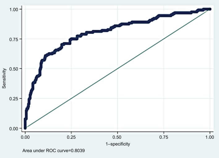
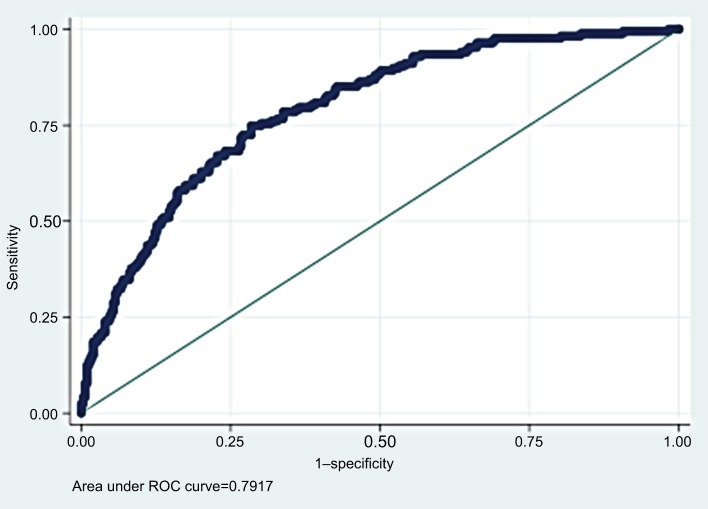
Multivariate logistic regression analysis identified venous lactate as a significant predictor of ICU admission, inpatient endoscopy, and PRBC transfusion. Patients with an elevated lactate level were more likely to be admitted to ICU than patients with a normal lactate level (AOR 2.96, 95% CI 1.74–5.01; p<0.001). Patients with an elevated lactate level were more likely to receive PRBC transfusion (AOR 3.65, 95% CI 1.76–7.55; p<0.001) and endoscopy (AOR 1.64, 95% CI 1.02–2.65; p=0.04) than patients with a normal lactate level (Table 3). The ROC areas for predicting ICU admission and endoscopy were 0.80 (95% CI 0.75–0.85; Figure 1) and 0.79 (95% CI 0.75–0.83; Figure 2), respectively.
Table 3.
AOR of elevated initial venous lactate for outcome variables in patients with acute GIB
| Outcome | AOR | 95% CI; p-value |
|---|---|---|
| ICU admission | 2.96 | 1.74–5.01; <0.001 |
| Inpatient endoscopy | 1.64 | 1.02–2.65; 0.04 |
| PRBC transfusion | 3.65 | 1.76–7.55; <0.001 |
Notes: Multivariate logistic regression model included elevated initial venous lactate (>2.0 mmol/L) as the predictor of interest. Other variables in the analysis included age, gender, ethnicity, smoking, alcohol use, use of NSAID, use of aspirin, Charlson Comorbidity Index, presentation with syncope, bright red blood per rectum, abdominal pain, hematemesis, altered mental status, ascites, initial heart rate and systolic blood pressure, initial hemoglobin, platelet count, prothrombin time as INR, and creatinine.
Abbreviations: AOR, adjusted odds ratio; GIB, gastrointestinal bleeding; CI, confidence interval; ICU, intensive care unit; PRBC, packed red blood cell; NSAID, nonsteroidal anti-inflammatory drug; INR, international normalized ratio.
Figure 1.
Area under the ROC curve of predictive model for ICU admission.
Abbreviations: ROC, receiver operating characteristic; ICU, intensive care unit.
Figure 2.
Area under the ROC curve of predictive model for inpatient endoscopy.
Abbreviation: ROC, receiver operating characteristic.
Multivariate linear regression analysis revealed significant difference in the length of hospital stay between the two groups (mean difference=1.37, 95% CI 0.12–2.63; p=0.03).
Discussion
We found that an elevated lactate level on presentation was an independent predictor of ICU admission, inpatient endoscopy, and PRBC transfusion in patients with acute GIB. These findings suggest that a single venous blood lactate measurement provides clinically useful information in patients with acute GIB and support the use of venous lactate measurement in the initial clinical management decision.
Prior studies have reported prognostic use of lactate measurement in predicting active bleeding or mortality in patients with acute GIB.16–19 Our study evaluated the usefulness of lactate measurement on resource utilizations (eg, ICU admission, length of hospital stay) and other patient-oriented outcomes (eg, need for transfusion and endoscopy) in patients with acute GIB. Identifying patients who need clinical treatment may be more important than identifying patients at risk for death as proper treatment can prevent complications and deaths.10 With increasingly limited health care resources, there has been a growing interest in cost-saving measures. Longer length of hospital stay, ICU admission, endoscopy, and PRBC transfusion have been recognized as key cost drivers in patients with acute GIB.9,22
We found that patients with an elevated lactate level on presentation were more likely to require ICU admission, transfusion, and inpatient endoscopy than patients with a normal lactate level. These findings are perhaps not surprising as an elevated lactate level in the setting of GIB is often associated with poor outcomes. Prior studies have reported increased mortality associated with an elevated lactate level in patients with acute GIB.16–18 Wada et al19 found that low lactate clearance was associated with an increased risk of active bleeding in patients with upper GIB. Active bleeding could partly explain higher rates of ICU admissions, transfusions, and endoscopies in patients with an elevated lactate level.
The findings of our study have important implications. Venous lactate measurement in conjunction with other prognostic factors may assist clinicians with prompt recognition of high-risk patients who require early, aggressive interventions, such as ICU admissions, emergent endoscopies, and transfusions. Early recognition of such patients may prevent poor outcomes. Conversely, low-risk patients with a normal lactate level may be considered for general medical floor admission or even early hospital discharge for outpatient management. Enhanced accuracy in triage can potentially lead to more efficient use of hospital resources, ultimately reducing cost and improving patient outcomes.2,23,24
Our study has several strengths. First, our study population was fairly large, ethnically diverse, and represented an urban US population. We believe that the findings of our study are likely applicable to other urban US tertiary hospitals. Second, we adjusted for several important demographic and clinical risk factors in our multivariate analysis to minimize confounding. Third, we analyzed the association of venous lactate with several clinically relevant outcomes in patients with acute GIB.
Our study has several limitations. A large subset of patients was excluded due to absence of ED venous lactate measurements, which could have caused selection bias. It is possible that clinicians are more likely to obtain venous lactate measurements in more severely ill patients than stable patients. Our study findings therefore may not be applicable to patients with acute GIB in general. Owing to the retrospective nature of our study, we did not evaluate the effectiveness of initial treatment measures on clinical parameters (eg, serial hematocrit, lactate clearance, repeat hemodynamic parameters), and these factors may have affected the outcomes (eg, ICU admission, inpatient endoscopy). In addition, we were unable to measure time from the onset of GIB to the venous lactate collection, which may limit interpretation of our data. We did not stratify our analysis based on the source of bleeding, as there was uncertainty in differentiating the source of bleeding based on the initial clinical findings. Furthermore, endoscopic findings were unavailable for the majority of the patients. However, a prior study found no significant difference in the distribution of upper and lower gastrointestinal sources between the low-risk and high-risk patients.4 Lastly, in contrast to previous studies among patients with acute upper GIB,25,26 endoscopy rate was lower in our study. Limited data exist on the utilization of colonoscopy in patients with acute lower GIB. Our study included patients with acute lower GIB, which may partly explain the lower endoscopy rate in our study. Another potential reason for the lower endoscopy rate could be the overdiagnosis of patients with acute GIB in our study. We utilized ED encounter database to identify patients with acute GIB. The ED diagnosis was mostly based on subjective complaint and objective data; rectal examination findings or endoscopic findings were missing in the majority of patients.
Conclusion
Elevated venous lactate on presentation predicts the need for ICU admission, transfusion, and endoscopy in patients with acute GIB. Our findings suggest that venous lactate measurement may be a useful adjunctive test in the triage of patients with acute GIB. Further prospective studies are needed to validate these findings.
Supplementary material
Table S1.
Diagnoses and corresponding ICD-9 codes to identify patients with acute GIB
| Diagnosis | ICD-9 code |
|---|---|
| Diverticulosis or diverticulitis of the colon with hemorrhage | 562.12, 562.13 |
| Angiodysplasia of the intestine with hemorrhage | 569.85 |
| Hemorrhage of the rectum and anus | 569.3 |
| Internal, external, or unspecified hemorrhoids with bleeding | 455.2, 455.5, 455.8 |
| Hematemesis | 578.0 |
| Hemorrhage of the gastrointestinal tract site unspecified | 578.9 |
| Blood in stool or melena | 578.1 |
| Esophageal varices with hemorrhage | 456.0, 456.20 |
| Ulcer of esophagus with bleeding | 530.21 |
| Esophageal hemorrhage unspecified | 530.82 |
| Duodenitis with hemorrhage | 535.61 |
| Gastritis with hemorrhage | 535.01 |
| Mallory–Weiss tear | 530.7 |
| Gastric ulcer, acute with hemorrhage±perforation | 531.0, 531.2 |
| Duodenal ulcer, acute with hemorrhage±perforation | 532.0, 532.2 |
| Peptic ulcer, acute with hemorrhage±perforation | 533.0, 533.2 |
| Gastrojejunal ulcer, acute with hemorrhage±perforation | 534.0, 534.2 |
| Angiodysplasia of the stomach or duodenum with hemorrhage | 537.83 |
| Diverticulosis or diverticulitis of the small intestine with hemorrhage | 562.02, 562.03 |
Abbreviations: ICD-9, International Classification of Diseases, Ninth Revision; GIB, gastrointestinal bleeding.
Acknowledgments
The authors thank Sasha Taleban, MD, for his review of the study proposal and insightful comments and Vern Pilling for his assistance with data retrieval. The abstract of this paper was presented at the World Congress of Gastroenterology at the American College of Gastroenterology (ACG) 2017, Orlando, FL, USA, as a poster presentation with interim findings. The poster’s abstract was published in “Poster Abstracts” in American College of Gastroenterology 112:S290; doi:10.1038/ajg.2017.302.
Footnotes
Disclosure
The authors report no conflicts of interest in this work.
References
- 1.Peery AF, Crockett SD, Barritt AS, et al. Burden of gastrointestinal, liver, and pancreatic diseases in the United States. Gastroenterology. 2015;149(7):1731–1741.e3. doi: 10.1053/j.gastro.2015.08.045. [DOI] [PMC free article] [PubMed] [Google Scholar]
- 2.Cipolletta L, Bianco MA, Rotondano G, Marmo R, Piscopo R. Outpatient management for low-risk nonvariceal upper GI bleeding: a randomized controlled trial. Gastrointest Endosc. 2002;55(1):1–5. doi: 10.1067/mge.2002.119219. [DOI] [PubMed] [Google Scholar]
- 3.Bordley DR, Mushlin AI, Dolan JG, et al. Early clinical signs identify low-risk patients with acute upper gastrointestinal hemorrhage. JAMA. 1985;253(22):3282–3285. [PubMed] [Google Scholar]
- 4.Kollef MH, Canfield DA, Zuckerman GR. Triage considerations for patients with acute gastrointestinal hemorrhage admitted to a medical intensive care unit. Crit Care Med. 1995;23(6):1048–1054. doi: 10.1097/00003246-199506000-00009. [DOI] [PubMed] [Google Scholar]
- 5.Rockall TA, Logan RF, Devlin HB, Northfield TC. Selection of patients for early discharge or outpatient care after acute upper gastrointestinal haemorrhage. National audit of acute upper gastrointestinal haemorrhage. Lancet. 1996;347(9009):1138–1140. doi: 10.1016/s0140-6736(96)90607-8. [DOI] [PubMed] [Google Scholar]
- 6.Das A, Wong RC. Prediction of outcome of acute GI hemorrhage: a review of risk scores and predictive models. Gastrointest Endosc. 2004;60(1):85–93. doi: 10.1016/s0016-5107(04)01291-x. [DOI] [PubMed] [Google Scholar]
- 7.Farooq FT, Lee MH, Das A, Dixit R, Wong RC. Clinical triage decision vs risk scores in predicting the need for endotherapy in upper gastrointestinal bleeding. Am J Emerg Med. 2012;30(1):129–134. doi: 10.1016/j.ajem.2010.11.007. [DOI] [PubMed] [Google Scholar]
- 8.Horibe M, Kaneko T, Yokogawa N, et al. A simple scoring system to assess the need for an endoscopic intervention in suspected upper gastrointestinal bleeding: a prospective cohort study. Dig Liver Dis. 2016;48(10):1180–1186. doi: 10.1016/j.dld.2016.07.009. [DOI] [PubMed] [Google Scholar]
- 9.Kollef MH, O’Brien JD, Zuckerman GR, Shannon W. BLEED: a classification tool to predict outcomes in patients with acute upper and lower gastrointestinal hemorrhage. Crit Care Med. 1997;25(7):1125–1132. doi: 10.1097/00003246-199707000-00011. [DOI] [PubMed] [Google Scholar]
- 10.Blatchford O, Murray WR, Blatchford M. A risk score to predict need for treatment for upper-gastrointestinal haemorrhage. Lancet. 2000;356(9238):1318–1321. doi: 10.1016/S0140-6736(00)02816-6. [DOI] [PubMed] [Google Scholar]
- 11.Strate LL, Orav EJ, Syngal S. Early predictors of severity in acute lower intestinal tract bleeding. Arch Intern Med. 2003;163(7):838–843. doi: 10.1001/archinte.163.7.838. [DOI] [PubMed] [Google Scholar]
- 12.Das A, Ben-Menachem T, Cooper GS, et al. Prediction of outcome in acute lower-gastrointestinal haemorrhage based on an artificial neural network: internal and external validation of a predictive model. Lancet. 2003;362(9392):1261–1266. doi: 10.1016/S0140-6736(03)14568-0. [DOI] [PubMed] [Google Scholar]
- 13.Barkun A, Bardou M, Marshall JK. Nonvariceal Upper GI Bleeding Consensus Conference Group. Consensus recommendations for managing patients with nonvariceal upper gastrointestinal bleeding. Ann Intern Med. 2003;139(10):843–857. doi: 10.7326/0003-4819-139-10-200311180-00012. [DOI] [PubMed] [Google Scholar]
- 14.Mikkelsen ME, Miltiades AN, Gaieski DF, et al. Serum lactate is associated with mortality in severe sepsis independent of organ failure and shock. Crit Care Med. 2009;37(5):1670–1677. doi: 10.1097/CCM.0b013e31819fcf68. [DOI] [PubMed] [Google Scholar]
- 15.Musikatavorn K, Thepnimitra S, Komindr A, Puttaphaisan P, Rojanasarntikul D. Venous lactate in predicting the need for intensive care unit and mortality among nonelderly sepsis patients with stable hemody-namic. Am J Emerg Med. 2015;33(7):925–930. doi: 10.1016/j.ajem.2015.04.010. [DOI] [PubMed] [Google Scholar]
- 16.Koch A, Buendgens L, Duckers H, et al. Bleeding origin, patient-related risk factors, and prognostic indicators in patients with acute gastrointestinal hemorrhages requiring intensive care treatment. A retrospective analysis from 1999 to 2010. Med Klin Intensivmed Notfmed. 2013;108(3):214–222. doi: 10.1007/s00063-013-0226-2. [DOI] [PubMed] [Google Scholar]
- 17.Shah A, Chisolm-Straker M, Alexander A, Rattu M, Dikdan S, Manini AF. Prognostic use of lactate to predict inpatient mortality in acute gastrointestinal hemorrhage. Am J Emerg Med. 2014;32(7):752–755. doi: 10.1016/j.ajem.2014.02.010. [DOI] [PubMed] [Google Scholar]
- 18.El-Kersh K, Chaddha U, Sinha RS, Saad M, Guardiola J, Cavallazzi R. Predictive role of admission lactate level in critically ill patients with acute upper gastrointestinal bleeding. J Emerg Med. 2015;49(3):318–325. doi: 10.1016/j.jemermed.2015.04.008. [DOI] [PubMed] [Google Scholar]
- 19.Wada T, Hagiwara A, Uemura T, Yahagi N, Kimura A. Early lactate clearance for predicting active bleeding in critically ill patients with acute upper gastrointestinal bleeding: a retrospective study. Intern Emerg Med. 2016;11(5):737–743. doi: 10.1007/s11739-016-1392-z. [DOI] [PubMed] [Google Scholar]
- 20.Harris PA, Taylor R, Thielke R, Payne J, Gonzalez N, Conde JG. Research electronic data capture (REDCap) – a metadata-driven methodology and workflow process for providing translational research informatics support. J Biomed Inform. 2009;42(2):377–381. doi: 10.1016/j.jbi.2008.08.010. [DOI] [PMC free article] [PubMed] [Google Scholar]
- 21.Charlson ME, Pompei P, Ales KL, MacKenzie CR. A new method of classifying prognostic comorbidity in longitudinal studies: development and validation. J Chronic Dis. 1987;40(5):373–383. doi: 10.1016/0021-9681(87)90171-8. [DOI] [PubMed] [Google Scholar]
- 22.Campbell HE, Stokes EA, Bargo D, et al. Costs and quality of life associated with acute upper gastrointestinal bleeding in the UK: cohort analysis of patients in a cluster randomised trial. BMJ Open. 2015;5(4):e007230. doi: 10.1136/bmjopen-2014-007230. [DOI] [PMC free article] [PubMed] [Google Scholar]
- 23.Dulai GS, Gralnek IM, Oei TT, et al. Utilization of health care resources for low-risk patients with acute, nonvariceal upper GI hemorrhage: an historical cohort study. Gastrointest Endosc. 2002;55(3):321–327. doi: 10.1067/mge.2002.121880. [DOI] [PubMed] [Google Scholar]
- 24.Stanley AJ, Ashley D, Dalton HR, et al. Outpatient management of patients with low-risk upper-gastrointestinal haemorrhage: multicentre validation and prospective evaluation. Lancet. 2009;373(9657):42–47. doi: 10.1016/S0140-6736(08)61769-9. [DOI] [PubMed] [Google Scholar]
- 25.Ananthakrishnan AN, McGinley EL, Saeian K. Outcomes of weekend admissions for upper gastrointestinal hemorrhage: a nationwide analysis. Clin Gastroenterol Hepatol. 2009;7(3):296e–302e. doi: 10.1016/j.cgh.2008.08.013. [DOI] [PubMed] [Google Scholar]
- 26.Abougergi MS, Travis AC, Saltzman JR. The in-hospital mortality rate for upper GI hemorrhage has decreased over 2 decades in the United States: a nationwide analysis. Gastrointest Endosc. 2015;81(4):882–888.e1. doi: 10.1016/j.gie.2014.09.027. [DOI] [PubMed] [Google Scholar]
Associated Data
This section collects any data citations, data availability statements, or supplementary materials included in this article.
Supplementary Materials
Table S1.
Diagnoses and corresponding ICD-9 codes to identify patients with acute GIB
| Diagnosis | ICD-9 code |
|---|---|
| Diverticulosis or diverticulitis of the colon with hemorrhage | 562.12, 562.13 |
| Angiodysplasia of the intestine with hemorrhage | 569.85 |
| Hemorrhage of the rectum and anus | 569.3 |
| Internal, external, or unspecified hemorrhoids with bleeding | 455.2, 455.5, 455.8 |
| Hematemesis | 578.0 |
| Hemorrhage of the gastrointestinal tract site unspecified | 578.9 |
| Blood in stool or melena | 578.1 |
| Esophageal varices with hemorrhage | 456.0, 456.20 |
| Ulcer of esophagus with bleeding | 530.21 |
| Esophageal hemorrhage unspecified | 530.82 |
| Duodenitis with hemorrhage | 535.61 |
| Gastritis with hemorrhage | 535.01 |
| Mallory–Weiss tear | 530.7 |
| Gastric ulcer, acute with hemorrhage±perforation | 531.0, 531.2 |
| Duodenal ulcer, acute with hemorrhage±perforation | 532.0, 532.2 |
| Peptic ulcer, acute with hemorrhage±perforation | 533.0, 533.2 |
| Gastrojejunal ulcer, acute with hemorrhage±perforation | 534.0, 534.2 |
| Angiodysplasia of the stomach or duodenum with hemorrhage | 537.83 |
| Diverticulosis or diverticulitis of the small intestine with hemorrhage | 562.02, 562.03 |
Abbreviations: ICD-9, International Classification of Diseases, Ninth Revision; GIB, gastrointestinal bleeding.


