Figure 1.
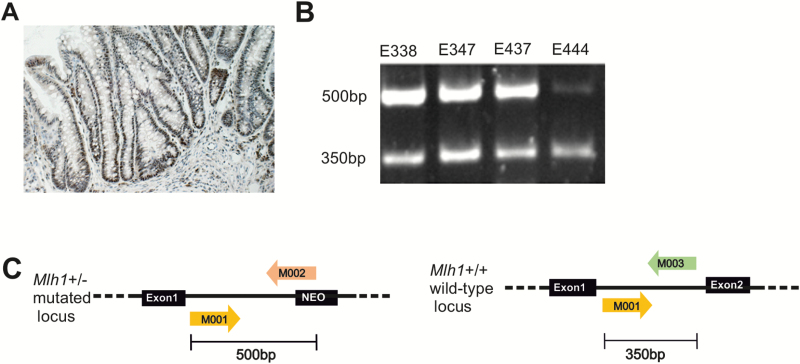
Mlh1 protein expression and loss of heterozygosity analyses. (A) An example of a colon carcinoma showing positive Mlh1 expression analysed by immunohistochemistry (mouse E402, tubular adenocarcinoma). (B) Four CRCs found in heterozygote Mlh1+/− mice showing that the normal Mlh1 allele (350 bp) was still present in tumours. (C) In Mlh1 heterozygote mice, one of the Mlh1 alleles is mutated by replacing the exon 2 with a neomycin cassette. Loss of Mlh1 heterozygosity was analysed using the genotyping primers M001, M002 and M003, which produce two different length fragments, 350 and 500 bp, that separate the normal (M001/M003) and the mutated allele (M001/M002), respectively.

