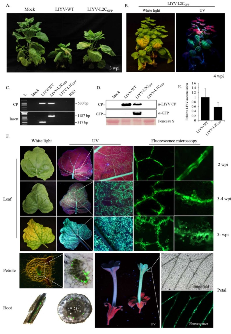
Figure 2.
Viral infection and GFP expression of LIYV-based “add-a-gene” expression vectors in Hc-Pro Nicotiana benthamiana plants. (A) Phenotypes of LIYV-WT and LIYV-L2CGFP infected N. benthamiana plants photographed at 3 weeks post inoculation. Mock indicates buffer-inoculated control. (B) LIYV-L2CGFP infected N. benthamiana plant photographed under white and UV light. (C) Detection of viral infection and insertion integrity by RT-PCR with total RNA extracted from upper non-inoculated leaves of LIYV-WT, LIYV-L1CGFP, and LIYV-L2CGFP agroinoculated plants. Two primer sets were used to amplify the sequence of LIYV CP (CP, 530 bp) and the sequence flanking the GFP cassette (Insert, 1187 bp), LIYV-WT without the insert was amplified as a control (317 bp). (D) Immunoblot analysis of LIYV CP and GFP accumulation in upper non-inoculated leaves using LIYV CP and GFP specific antibodies. The Ponceau S stained rubisco large subunit serves as a loading control. (E) Quantification of LIYV RNA1 accumulation in LIYV-WT and LIYV-L2CGFP infected plants by RT-qPCR. The PP2A transcript level of N. benthamiana was used as an internal control. Error bars denote standard errors from at least three biological replicates. (F) Systemic spread and distribution in leaves, petioles, roots and flowers of LIYV-L2CGFP infected plants monitored by GFP fluorescence. Leaf, progress of LIYV infection and accumulation along veins over time. GFP fluorescence was visualized under UV light and fluorescence microscopy with different magnification. GFP was confined to vascular tissues of petiole, root (left, root segment; right, root cross-section), and petal was seen under UV light and fluorescence microscopy. Dotted lines indicate enlarged areas.

