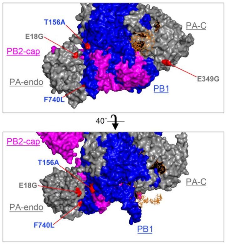
Figure 3.
CA/07 mouse adaptation mutations are surface-exposed in the ternary complex. CA/07-MA amino acid substitutions were mapped onto the 3D structure of bat influenza A/little yellow-shouldered bat/Guatemala/060/2010 (H17N10) bound to an RNA primer [49] (protein accession number: 4WSB). PA is grey, PB1 blue, PB2 magenta and the RNA strand orange. Relative positions of the C-terminal cap-binding domain of PB2 (PB2-cap), the N-terminal endonuclease domain of PA (PA-endo) and the C-terminal domain of PA (PA-C) are indicated. Locations of mutations in PB1 and PA identified by deep sequencing are highlighted in red. The image was generated using PyMOL Version 2.0.4. Views were rendered at Ray 2400 with 1000 dpi.

