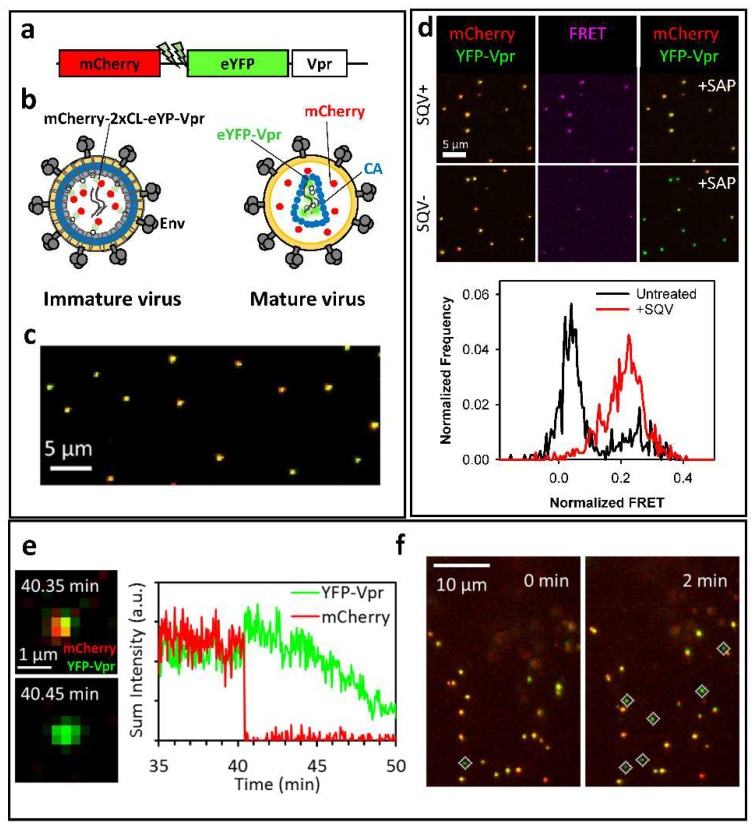
Figure 1.
Bi-functional fluorescence marker for HIV-1 protease activity and fusion. (a) Illustration of the bi-functional mCherry-2xCL-eYFP-Vpr marker; (b) Cartoon of immature and mature HIV-1 particles labeled with mCherry-2xCL-eYFP-Vpr. eYFP fluorescence is quenched in immature viral particles as a result of FRET between the eYFP and mCherry. eYFP fluorescence is enhanced in mature virions following the release of mCherry from the bi-functional marker; (c) A representative image of single viral particles bound to a cover glass showing a nearly perfect colocalization of mCherry and eYFP; (d) Immature viruses produced in the presence of saquinavir (SQV) exhibit high Forster Resonance Energy Transfer (FRET) between eYFP and mCherry (middle panel). Corresponding fluorescence images of single virions before (left) and after (right) saponin lysis (+SAP) are shown; (e) Single virus fusion is detected based upon a quick release of fluid-phase mCherry marker from eYFP-Vpr labeled mature HIV-1 core. The fluorescence intensity traces of the fusing particle are shown on the right; (f) Automated detection of post-fusion HIV-1 cores in CV1 cells as eYFP-Vpr-labeled labeled puncta negative for mCherry (marked by white diamonds). Data are adapted from Sood et al., 2017.

