

Abstract
The abnormal oocyte (abo) gene of Drosophila melanogaster is a peculiar maternal effect gene whose mutations cause a maternal-effect lethality that can be rescued by specific regions of heterochromatin during early embryogenesis. Here we show that abo encodes an evolutionary conserved chromosomal protein that localizes exclusively to the histone gene cluster and binds to the regulatory regions of such genes. We also show a significant increase of histone transcripts in eggs of abo mutant mothers and a partial rescue of the abo maternal-effect defect by deficiencies of the histone gene cluster. On the basis of these results, we suggest that the Abo protein functions specifically as a negative regulator of histone transcription and propose a molecular model to account for the ability of heterochromatin to partially rescue the abo maternal-effect defect. Our model proposes that increased doses of specific heterochromatic regions titrate out abnormally high levels of histones present in embryos from mutant abo mothers and that a balanced pool of histones is critical for normal embryogenesis in Drosophila.
The abnormal oocyte (abo) gene of Drosophila melanogaster is a euchromatic gene that, when mutant, causes a recessive maternal-effect defect that markedly reduces the viability of offspring (1). It has been shown that abo maternal-effect lethality occurs mainly during late embryogenesis, after cuticle deposition but before hatching, with some lethality occurring during larval stages. The lethal embryos show cuticular defects due to a failure to complete a regular gastrulation (2). The viability of these embryos can be rescued by a paternally contributed abo wild-type allele, suggesting that abo also has a zygotic function. The most striking aspect of the abo maternal effect is its genetic interaction with heterochromatin. An increase in the dosage of specific regions of heterochromatin, denoted ABO, to either the mutant mother or the zygote (1–4), increases embryonic survival rates.
To elucidate the nature of this gene and its peculiar interaction with heterochromatin, we have molecularly cloned and characterized the wild-type and mutant abo alleles and identified the abo protein product. We found that abo encodes a chromosomal protein that is exclusively localized to the histone-cluster region and binds to the regulatory regions containing the histone gene promoters. We also found that in eggs of abo mutant mothers, the amount of histone transcripts is greatly increased. Finally, we found that chromosomal deficiencies of the histone gene cluster partially rescue the abo maternal-effect defect. These results demonstrate that abo is a specific negative regulator of histone gene expression and suggest a molecular model to explain its interaction with heterochromatin.
Methods
Recombinant DNA Techniques.
A genomic library was constructed from abo1/abo1 adults in λ GEM-12 Genomic cloning Vector (Promega). All of the positive clones isolated by the screenings of genomic libraries were subcloned in pGEM7-Zf (Promega), and those isolated from cDNA libraries were subcloned in pGEM11zF (Promega). Clones were sequenced by using AmpliCycle Sequencing Kit (Perkin–Elmer).
To make the expression construct for enhanced green fluorescent protein (EGFP)-tagged Abo, a GFP gene fragment was fused to the 3′end of the abo gene by using the pP{GS[ry+, UASEGFP]} vector and the method described in ref. 5. The germline transformation was carried out according to ref. 6.
Isolation of RNA and Northern Blot Analyses.
For abo RNA blot analyses, Drosophila RNA samples were isolated by using the Ultraspec II RNA Isolation System, according to manufacturer's instruction (Biotecx Laboratories, Houston). PolyA+ RNA was selected by oligo(dT) chromatography. RNA samples were separated on a 1% agarose 3-(N-morpholino)propanesulfonic acid formaldehyde gel, transferred to a nylon membrane (Hybond N, Amersham Pharmacia), and hybridized with radiolabeled abo DNA probes (7). For histone RNA blot analysis, total RNA extracted from unfertilized eggs was loaded onto a 0.4-mm-thick, 6% polyacrylamide, 6 M urea gel, transferred to a nylon membrane (Hybond N, Amersham Pharmacia), and hybridized with radiolabeled B5 Histone DNA clone probes (7). Histone probes for each histone class were obtained as PCR amplified fragments of cDM5009 clone by using specific primers. Northern blots were quantitated by Bio-Rad Chemidoc scanning of the autoradiograph, with exposure in a linear range of film exposure. The software used for scanning was quantity one 4.2.1 (Bio-Rad).
Generation of Abo Antibodies and Indirect Immunofluorescence.
The pET System (Novagen) was used for cloning and expression of fusion protein; a coding region from nucleotide 756 to the 3′ end of the cDNA clone was inserted into BamHI-HindIII-digested pET29 vector. The correct reading frame of fusion protein was checked by sequencing. To produce polyclonal anti-Abo antibodies, mice were immunized with the Abo fusion protein. For immunofluorescence and sequential in situ hybridization, chromosomes from larvae salivary glands and brain were fixed and processed as described (8). Chromosome preparations were analyzed by using a computer-controlled Nikon (E1000) epifluorescence microscope equipped with a cooled charge-coupled device camera (Photometrics, Tucson, AZ). By using the Adobe photoshop program (Adobe Systems, Mountain View, CA), the fluorescent signals, recorded separately as gray-scale digital images, were pseudocolored and merged.
X-ChIP and PCR Analysis.
Crosslinked chromatin was prepared from Drosophila embryos (0–4 h old) or SL-2 culture cells (grown in serum-free medium; HyQ-CCM 3, HyClone), and immunoprecipitations were performed basically as described previously (9, 10). The final precipitated DNA was redissolved in 120 μl of TE buffer (10 mM Tris, pH 8; 1 mM EDTA) and stored at 4°C or used directly for PCR. The complete sequence of the D. melanogaster histone L-form repeated unit (11) was used to design 12 primer pairs of 20–25 bp length (melting temperature 64–68°C) that would amplify 400–500 bp fragments spanning the whole locus. PCR was performed in 40-μl reactions by using 2–3 μl of the template of the immunoprecipitated material or 200 ng of total genomic DNA from 0- to 4-h embryos or SL-2 culture cells, respectively, by using Taq polymerase and reaction buffer (Promega). For PCR amplifications, we used: (i) 94°C, 3 min, 1×; (ii) 94°C, 1 min, 58–65°C, 1 min, 72°C, 1 min, 34×; (iii) 94°C, 1 min, 58–65°C, 1 min, 72°C, 7 min; 1×. For individual primer pairs, annealing temperature and cycle number were adjusted until no signal was detected for the mock-immunoprecipitation (ip) DNA, but the amplification on the genomic template was not altered. Signals obtained with the abo-ip DNA under these conditions were considered significant. The amplified DNA (half of the PCR reaction) was separated on 1.5% agarose gels and visualized with ethidium bromide.
Results
Identification of the abo Gene.
We previously cloned a 9-kb BamHI fragment that was disrupted in a P-induced abo lethal allele (abo2) (2). We demonstrated that this fragment contained the abo gene by its ability to complement the maternal defect in transgenic flies. Southern blot analysis of wild-type and abo mutants and transcript analysis localized the abo coding region within a 2.3-kb EcoRI-XhoI fragment that mapped to 32C of the salivary gland polytene second chromosome map (2).
To identify the abo gene, we cloned the abo1 and abo2 mutations. We constructed phage genomic libraries of abo1/abo1 and abo2/abo+ flies (2) and screened them with the 2.3-kb fragment and P[ry+] probes, respectively. In addition, by using the 2.3-kb fragment, we screened two cDNA libraries obtained from wild-type 0- to 3-h embryos (12) and ovaries (13), respectively. By sequence analysis of the 2.3-kb fragment, the corresponding cDNAs, and the mutant clones, we defined the abo gene as a 1,974-bp sequence (Fig. 1A) containing a putative TATA box, a CAAT box, and an ORF, interrupted by a small intron, and producing a single 1.8-kb transcript (Fig. 1B). This transcript encodes a putative 509-aa protein that we called Abo (Fig. 1C). Fig. 1A shows that the abo1 mutation is due to the insertion of an incomplete Doc transposable element into the coding region of the abo gene producing a larger transcript than the wild type (Fig. 1B), whereas that abo2 mutation is caused by the P[ry+] insertion into the 5′ promoter region and does not produce a detectable transcript (Fig. 1B).
Figure 1.

The abo gene structure. (A) Genomic organization of the abo locus. The red triangle indicates the insertion points of P[ry+] in the 5′ noncoding region of the abo2 mutation. The green triangle indicates the insertion point of the Doc fragment in the first exon of the abo1 mutation. (B) Northern blot of total RNA of females (10 μg), with the indicated genotypes, hybridized with the clone that spans the region interrupted by the P[ry+] insertion in the abo2 mutation. An RNA transcript of 1.8 kb is present in the wild-type and abo2/Cy lanes, whereas an additional transcript of about 3 kb is present in the abo1/Cy lane. The presence of only the 3-kb transcript in the abo1/abo1 lane clearly shows that this transcript is made by the abo1 mutation. In the abo1/abo2 lane also, only the 3-kb transcript is present. This indicates that the abo2 mutation does not produce a detectable transcript. The intensity of signals is expressed as ratio of abo signal/rp49 control value. The same ratio value was found also in a second Northern blot experiment. (C) Alignment of the predicted amino acid sequences of the Drosophila Abo protein, Arabidopsis thaliana tDET1, Solanum lycopersicon tDET1 protein, Oryza sativa tDET1 protein, and unknown putative human and mouse proteins. The red letters indicate complete identity, the green letters strong similarity, and the blue letter weak similarity.
By a computer database search by using the blastp program, we failed to find any known protein motifs present in the conceptually translated Abo protein. However, as shown in Fig. 1C, we found 25.3% identity and 51.9% similarity to the DET1 protein, a nuclear located negative regulator of light-mediated gene expression in Arabidopsis (14), whose putative homologues are present also in Oryza sativa (GenBank accession no. BAB16336.1) and Lycopersicon esculentum (15). Intriguingly, we found also 24% identity and 44% similarity to the putative human hCP43420 protein from the Celera Human Report (www.celera.com) and to a putative mouse protein (GenBank accession no. BAB27766). Considering the evolutionary distance, the homology between these proteins appears significant. They share stretches of homology across their entire lengths and are very similar in charge, distribution of hydrophilic residues, and overall amino acid composition. In particular, the human and mouse proteins appear strikingly identical, with few differences in the nucleotide sequences of their encoding genes.
The Abo Protein Is Exclusively Localized to the Histone Cluster.
The homology with DET1 suggested that the Abo protein might also be a transcriptional regulator and therefore might bind specific target sequences. To test this, we used bacterially produced Abo protein as antigen to raise a polyclonal antibody in mice. As shown in Fig. 2, we obtained polyclonal anti-Abo antibodies that, when used on Western blots of nuclear extracts from both embryos and SL-2 cultured cells, showed a strong reacting band of the expected molecular mass (about 60 kDa). By using these antibodies, we immunostained both the polytene chromosomes from salivary glands and the mitotic chromosomes of neuroblasts from wild-type larvae. We observed a strong signal exclusively localized on the 39E region on polytene chromosomes (Fig. 3A). In mitotic metaphase chromosomes, a unique strong signal was present on the constriction on the base of the left arm of the second chromosome (Fig. 3D). In both cases, the signal was localized at the position of the histone gene cluster, as confirmed by sequential immunostaining with the anti-Abo antibodies and in situ hybridization of the cDm500 probe, which contains the histone cluster (16) (Fig. 3 B and D). Our results clearly demonstrate that the regions with exclusive binding affinity for Abo contain the histone clusters in both the polytenes and mitotic chromosomes. We obtained a confirmation of this conclusion by constructing an Abo tagged with the enhanced GFP variant form of the GFP and showing that the protein is strongly accumulated at the histone gene region (see Fig. 3C for an example).
Figure 2.

Western blot analysis of nuclear extracts from embryos and SL-2 cultured cells by using the polyclonal anti-Abo antibodies. In both the extracts, a prominent reacting band of about 60 kDa is clearly visible.
Figure 3.

Immunostaining of polytenes and mitotic chromosomes of D. melanogaster with antibodies directed against the Abo protein and sequential in situ hybridization with the histone probes. (A) The immunopattern on polytenes shows a single strong signal at the base of the second chromosome left arm. (B) The immunosignal (green signal) is clearly localized at the 39E region and colocalizes with the histone gene cluster (red signal), as shown in the merged figure (yellow signals). (C) Immunolocalization of Abo protein tagged with GFP by an anti-GFP antibody. Also in this case, the protein is strongly accumulated on the histone region. (D) The immunopattern on mitotic chromosomes also shows a single signal (green signal) at the secondary constriction at the base of the second chromosome left arm that clearly colocalizes with the histone gene cluster (red signal), as shown in the merged figure (yellow signal). Abo, Abo protein; His-C, histone cluster; X, 3, and 4 indicate the relative chromosome pairs; 2L and 2R indicate the left and right arms of the second chromosome, respectively; c, centromere.
To test the presence of Abo homologs in other Drosophila species, we performed immunostaining with anti-Abo antibodies and sequential in situ hybridization with histone genes probe on polytene chromosomes of Drosophila simulans (data not shown) and Drosophila virilis (Fig. 4). In both species, we found immunosignals exclusively localized on the histone gene clusters.
Figure 4.

Immunostaining of polytenes of D. virilis with antibodies directed against the Abo protein and sequential in situ hybridization with the histone probes. (A) The immunopattern on polytenes shows two signals (green signals) that colocalize with the two histone genes clusters (red signals), as shown by in situ hybridization in B.
To identify Abo-binding sites in the histone repeat unit, we applied the X-ChIP (formaldehyde-crosslinked-chromatin immunoprecipitation) method by using our polyclonal anti-Abo antibodies. We designed 12 overlapping primer pairs that amplify 400- to 500-bp fragments spanning the whole Drosophila histone repeat unit (see Fig. 5A) and used them to amplify the DNA immunoprecipitated from chromatin of early embryos (0–4 h old) and SL-2 cultured cells. We found binding of Abo protein in early embryos to the promoter regions of H2A-H2B (fragment abo-2) and H3-H4 (fragment abo-10) (Fig. 5B). In SL-2 cells, Abo binds to an additional site in the H1 promoter fragment abo-6 (Fig. 5C). These results show clearly that Abo protein binding is restricted to the three main regulatory regions of the repeat unit containing the histone gene promoters.
Figure 5.

Mapping of Abo-binding sites in the D. melanogaster histone repeat unit. (A) The histone L-form repeat unit. Red arrows represent the coding regions of the histone genes starting with the ATG, and blue circles indicate the position of TATA boxes. The location of the DNA fragments amplified by the 12 primer pairs (abo-1–12) are shown. The three fragments containing Abo-binding sites are in green. (B) PCR analysis of immunopurified DNA from embryonic chromatin (embryo–ChIP). For each primer pair, the amplification products using genomic DNA (g), mock immunoprecipitation (−), and anti-Abo immunoprecipitation (+) are shown. (C) PCR analysis of immunopurified DNA from SL-2 chromatin (SL-2-ChIP). Abbreviations as in B. The experiment was repeated three times with same results.
abo Is a Specific Negative Regulator of Histones.
The functional significance of the interaction of abo with the promoters of histone genes was addressed by a quantitation of histone transcripts in unfertilized eggs from heterozygote abo1/abo2 and abo1/abo+ mothers. The results showed that abo mutations affect histone transcription. We found much higher levels of H2A, H2B, in eggs from mutant mothers than in eggs from their heterozygous sisters. We also found that the amount of H3 and H4 transcripts was significantly higher, whereas variations in the amount of H1 transcripts were not detectable (Fig. 6). These results strongly suggest that abo is a negative regulator of histone genes. We further explored this possibility by testing the genetic effects of deficiencies of the entire histone gene cluster (17) on the abo1 maternal effect. The data on the survival of embryos from homozygous abo1 mothers carrying either one or two histone regions are shown in Table 1. These results clearly show that the histone deficiencies [Df(2)DS5 and Df(2)DS6] induce a strong suppression of the abo1 maternal-effect defect, thus giving strong support to the suggestion that Abo negatively regulates histone gene expression.
Figure 6.
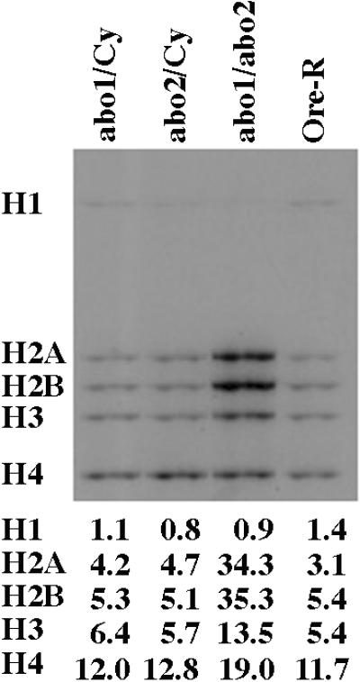
Northern blot analysis of histone gene transcripts in unfertilized eggs of wild-type and abo homozygous mutant females. As shown, the H2a+ H2b, H3, and H4 transcripts appear increased in the eggs from the abo1/abo2 mutant genotype with respect to those of wild-type or heterozygous females. The transcription of the H1 is not detectably affected by the abo mutation. The intensity of signals is expressed as a ratio of histone signal/rp49 control value. Quantitative results were consistent with those obtained in a second Northern blot.
Table 1.
Deficiencies of the histone gene cluster suppress the abo1 maternal effect defect
| Maternal genotype | No. eggs | Progeny | Survival (adults/eggs) | Relative survival (E/C) | |
|---|---|---|---|---|---|
| Df(2L)DS5,abo1/abo1 | E | 2905 | 969 | 0.33 | 0.40 |
| Df(2L)DS5,abo1/Cy | C | 2895 | 2400 | 0.83 | |
| Df(2L)DS6,abo1/abo1 | E | 1988 | 626 | 0.32 | 0.41 |
| Df(2L)DS6,abo1/Cy | C | 2000 | 1565 | 0.78 | |
| Df(2L)DS9,abo1/abo1 | E | 3450 | 570 | 0.17 | 0.19 |
| Df(2L)DS9,abo1/Cy | C | 3300 | 2907 | 0.88 |
The results of crosses of females bearing the indicated second chromosomes and the homologue Cy (C) or abo1 (E) by OR-R males. Df(2)DS5 and Df(2)DS6 are histone deficiencies; Df(2)DS9 does not affect the histone genes. C, control; E, experimental.
Discussion
Taken together, our studies reveal that abo is a negative regulator of H2A, H2B, H3, and H4 expression during oogenesis. Hence, the deleterious maternal-effect defect induced by the abo mutations is probably due to an excess of these histones. The regulation of histone expression has been extensively studied in different species (18, 19). The 5′ flanking regions contain cis elements that interact with transacting factors. These transacting factors differ among species and, more surprisingly, also differ among the different classes of histone genes. It has been proposed that the coordinate expression of the histone genes probably depends on the interaction of a protein complex with the different transacting factors (20). In this context, the uniqueness of the Abo protein location on the histone genes in different Drosophila species and its strong evolutionarily conservation suggest that this protein probably plays a basic role in regulating histone gene expression. However, differential histone gene expression in early embryogenesis of several species has been seen (21–23). In Drosophila, specific histone classes are also known to be differentially expressed. For example, it has been shown that the maternal histone H1 transcript is not translated in early embryogenesis (24) and is replaced by the HMG-D chromosomal protein (25). Intriguingly, the lack of any effect on H1 histone maternal transcription by the abo mutations and the lack of binding to its promoter by Abo in early embryos suggest that the regulation of histone H1 in both ovaries and embryos could not involve the abo gene. However, Abo does bind to the H1 promoter in SL-2 cells (representing late embryonic tissue), suggesting that Abo is probably involved in transcriptional regulation of histone H1 later in embryogenesis. Moreover, the differential enhancement of transcripts that we found in eggs from abo mutant mothers suggests that Abo could be more important for H2A and H2B repression than H3 and H4 repression during oogenesis.
The present data suggest a simple direct model for explaining an intriguing aspect of this gene, namely its interaction with the specific heterochromatic regions termed ABO elements. According to our model, homozygous abo mothers produce eggs with disproportionately high levels of H2A, H2B, H3, and H4 histones, which affects egg viability. Increasing doses of the ABO regions may titrate out these histones, reducing their negative effect. We predict that the abo and ABO-counteracting effects are produced by modulations in chromatin structure. Histones could be involved in such effects, as suggested by growing evidence showing that modified histones have differential chromosomal distributions, and hence they could play a role in the formation of heterochromatic domains (26). In fact, we have observed that H4 histone acetylated at lysine 4 and H3 histone methylated at lysine 9 are both present along the mitotic heterochromatin of Drosophila, with patterns of distribution indicating preferential binding for some regions (unpublished work).
In conclusion, the present characterization of abo opens the possibility of using this gene as an entry point to dissect the regulatory machinery of histone expression by looking at Abo-interacting molecules. Moreover, it could be a paradigm for experimental approaches to study the biological role of the heterochromatin. In D. melanogaster, other maternal-effect mutations closely linked to abo have been isolated (27). Preliminary experiments provide evidence that these abo-like mutations produce defects that can be compensated by discrete heterochromatic elements similar to ABO (3). It is possible that these other genes, like abo, may also encode transregulators of histone genes or other essential genes encoding chromosomal proteins. Further investigation of these euchromatic genes and their interacting heterochromatic components may provide additional insight into the functional connections between heterochromatin and euchromatin. These heterochromatin–euchromatin interaction systems may reflect the importance of the balance of chromosomal proteins in the nucleus. Thus heterochromatin could, in fact, play a vital role in regulating euchromatic gene expression by controlling chromatin structure.
Acknowledgments
We are grateful to John Tomkiel and Barbara Wakimoto for critical reading of the manuscript, to Ruggiero Caizzi for help in abo2/abo+ library construction, and to Gunnar Schotta and Gunter Reuter for help in making the enhanced GFP-tagged Abo construct. We thank Tom Grigliatti (University of British Columbia) for sending the histone deficiency stocks, Linda Strausbaugh (University of Connecticut) for providing the cDM500 clone, and Enzo Marchetti for technical assistance. This work was supported partially by Ministero dell'Università e della Ricerca Scientifica e Tecnologica (S.P.), by Telethon and Associazione Italiana per la Ricerca sul Cancro (V.O.), and by a postdoctoral fellowship from the European Union—Training and Mobility of Researchers Programme (A.B.).
Abbreviation
- GFP
green fluorescent protein
Footnotes
Data deposition: The sequence reported in this paper has been deposited in the GenBank database (accession no. AF384149).
References
- 1.Sandler L. Genetics. 1970;64:481–493. doi: 10.1093/genetics/64.3-4.481. [DOI] [PMC free article] [PubMed] [Google Scholar]
- 2.Tomkiel J, Fanti L, Berloco M, Spinelli L, Tamkun J W, Wakimoto B T, Pimpinelli S. Genetics. 1995;140:615–627. doi: 10.1093/genetics/140.2.615. [DOI] [PMC free article] [PubMed] [Google Scholar]
- 3.Pimpinelli S, Sullivan W, Prout M, Sandler L. Genetics. 1985;109:701–724. doi: 10.1093/genetics/109.4.701. [DOI] [PMC free article] [PubMed] [Google Scholar]
- 4.Tomkiel J, Pimpinelli S, Sandler L. Genetics. 1991;128:583–594. doi: 10.1093/genetics/128.3.583. [DOI] [PMC free article] [PubMed] [Google Scholar]
- 5.Schotta G, Reuter G. Mol Gen Genet. 2000;262:916–920. doi: 10.1007/pl00008659. [DOI] [PubMed] [Google Scholar]
- 6.Rubin G M, Spradling A C. Science. 1982;218:348–353. doi: 10.1126/science.6289436. [DOI] [PubMed] [Google Scholar]
- 7.Sambrook J, Fritsch E F, Maniatis T. Molecular Cloning: A Laboratory Manual. Plainview, NY: Cold Spring Harbor Lab. Press; 1989. [Google Scholar]
- 8.Pimpinelli S, Bonaccorsi S, Fanti L, Gatti M. In: Drosophila: A Laboratory Manual. Sullivan W, Ashburner M, Hawley S, editors. Plainview, NY: Cold Spring Harbor Lab. Press; 2000. pp. 1–24. [Google Scholar]
- 9.Orlando V, Strutt H, Paro R. Methods Enzymol. 1997;11:205–214. doi: 10.1006/meth.1996.0407. [DOI] [PubMed] [Google Scholar]
- 10.Orlando V, Jane E P, Chinwalla V, Harte P J, Paro R. EMBO J. 1998;17:5141–5150. doi: 10.1093/emboj/17.17.5141. [DOI] [PMC free article] [PubMed] [Google Scholar]
- 11.Matsuo Y, Yamazaki T. Nucleic Acids Res. 1989;17:225–238. doi: 10.1093/nar/17.1.225. [DOI] [PMC free article] [PubMed] [Google Scholar]
- 12.Tamkun J W, Kahn R A, Kissinger M, Brizuela B J, Rulka C, Scott M P, Kennison J A. Proc Natl Acad SciUSA. 1991;88:3120–3124. doi: 10.1073/pnas.88.8.3120. [DOI] [PMC free article] [PubMed] [Google Scholar]
- 13.Stroumbakis N, Li Z, Tolias P. Gene. 1994;143:171–177. doi: 10.1016/0378-1119(94)90093-0. [DOI] [PubMed] [Google Scholar]
- 14.Pepper A, Delaney T, Washburn T, Poole D, Chory J. Cell. 1994;78:109–116. doi: 10.1016/0092-8674(94)90577-0. [DOI] [PubMed] [Google Scholar]
- 15.Mustilli A C, Fenzi F, Ciliento R, Alfano F, Bowler C. J Plant Cell. 1999;11:145–157. doi: 10.1105/tpc.11.2.145. [DOI] [PMC free article] [PubMed] [Google Scholar]
- 16.Lifton R, Goldberg M L, Karp R W, Hogness D S. Cold Spring Harbor Symp Quant Biol. 1978;42:1047–1051. doi: 10.1101/sqb.1978.042.01.105. [DOI] [PubMed] [Google Scholar]
- 17.Moore G D, Sinclair D A, Grigliatti T A. Genetics. 1983;105:327–344. doi: 10.1093/genetics/105.2.327. [DOI] [PMC free article] [PubMed] [Google Scholar]
- 18.Heintz N. Biochim Biophys Acta. 1991;1088:327–339. doi: 10.1016/0167-4781(91)90122-3. [DOI] [PubMed] [Google Scholar]
- 19.Osley M A. Annu Rev Biochem. 1991;60:827–861. doi: 10.1146/annurev.bi.60.070191.004143. [DOI] [PubMed] [Google Scholar]
- 20.van Wijnen A J, Aziz F, Grana X, De Luca A, Desai R K, Jaarsveld K, Last T J, Soprano K, Giordano A, Lian J B, et al. Proc Natl Acad Sci USA. 1994;91:12882–12886. doi: 10.1073/pnas.91.26.12882. [DOI] [PMC free article] [PubMed] [Google Scholar]
- 21.Levy S, Sures I, Kedes L. J Biol Chem. 1982;257:9438–9443. [PubMed] [Google Scholar]
- 22.Smith R C, Dworkin-Rastl E, Dworkin M B. Genes Dev. 1988;2:1284–1295. doi: 10.1101/gad.2.10.1284. [DOI] [PubMed] [Google Scholar]
- 23.Dimitrov S, Almouzni G, Dasso M, Wolffe A P. Dev Biol. 1993;160:214–227. doi: 10.1006/dbio.1993.1299. [DOI] [PubMed] [Google Scholar]
- 24.Elgin S C R, Hood L E. Biochemistry. 1973;12:4984–4991. doi: 10.1021/bi00748a026. [DOI] [PubMed] [Google Scholar]
- 25.Ner S S, Travers A A. EMBO J. 1994;8:1817–1822. doi: 10.1002/j.1460-2075.1994.tb06450.x. [DOI] [PMC free article] [PubMed] [Google Scholar]
- 26.Rice J C, Allis C D. Curr Opin Cell Biol. 2001;13:263–273. doi: 10.1016/s0955-0674(00)00208-8. [DOI] [PubMed] [Google Scholar]
- 27.Sandler L. Genetics. 1977;86:567–582. doi: 10.1093/genetics/86.3.567. [DOI] [PMC free article] [PubMed] [Google Scholar]