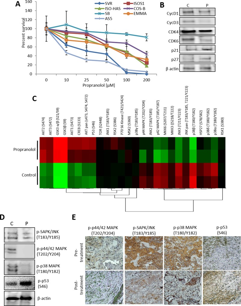
Figure 1. β-AR antagonism inhibits angiosarcoma viability and mitogenic/survival signaling.
(A) Angiosarcoma cell lines were treated with increasing concentrations of the non-selective beta blocker propranolol (0 to 200 µM). Cell viability was measured after 48 hours. (B) SVR cells were treated with a DMSO control or 25 µM propranolol for 24 hours. Protein lysates were collected and subjected to immunoblotting to detect levels of key cell cycle regulators. β-Actin was used as a loading control. (C) SVR cells were treated with 25 µM propranolol for 1 hour. Protein lysates were subjected to duplicate antibody arrays that simultaneously detected the relative phosphorylation of 24 kinases. The normalized levels of the phosphorylated proteins across the treatments are visualized via a heat map (red=increased phosphorylation, green=decreased phosphorylation). (D) Angiosarcoma xenografts were injected with an isotonic saline control or propranolol (15 mg/kg). Cell lysates were collected at 15 minutes post-treatment and subjected to immunoblotting for key mitogenic and survival signaling regulators. β-actin was used as a control to ensure equal loading of the samples. (E) Tumor sections from an angiosarcoma patient collected at diagnostic biopsy (before propranolol administration, pre-treatment) and at one week after administration of propranolol (120 mg/day) were analyzed by IHC to determine the levels of p-p44/42 (Thr202/Tyr204), p-SAPK/JNK (Thr183/Tyr185), p-p38 (T180/Y182), and p-p53 (S46). Brown coloration indicates positive antigen staining.

