Abstract
To update the melanoma staging system of the American Joint Committee on Cancer (AJCC) a large database was assembled comprising >46,000 patients from 10 centers worldwide with stages I, II, and III melanoma diagnosed since 1998. Based on analyses of this new database, the existing seventh edition AJCC stage IV database, and contemporary clinical trial data, the AJCC Melanoma Expert Panel introduced several important changes to the Tumor, Nodes, Metastasis (TNM) classification and stage grouping criteria. Key changes in the eighth edition AJCC Cancer Staging Manual include: 1) tumor thickness measurements to be recorded to the nearest 0.1 mm, not 0.01 mm; 2) definitions of T1a and T1b are revised (T1a, <0.8 mm without ulceration; T1b, 0.8–1.0 mm with or without ulceration or <0.8 mm with ulceration), with mitotic rate no longer a T category criterion; 3) pathological (but not clinical) stage IA is revised to include T1b N0 M0 (formerly pathologic stage IB); 4) the N category descriptors “microscopic” and “macroscopic” for regional node metastasis are redefined as “clinically occult” and “clinically apparent”; 5) prognostic stage III groupings are based on N category criteria and T category criteria (ie, primary tumor thickness and ulceration) and increased from 3 to 4 subgroups (stages IIIA–IIID); 6) definitions of N subcategories are revised, with the presence of microsatellites, satellites, or in-transit metastases now categorized as N1c, N2c, or N3c based on the number of tumor-involved regional lymph nodes, if any; 7) descriptors are added to each M1 subcategory designation for lactate dehydrogenase (LDH) level (LDH elevation no longer upstages to M1c); and 8) a new M1d designation is added for central nervous system metastases. This evidence-based revision of the AJCC melanoma staging system will guide patient treatment, provide better prognostic estimates, and refine stratification of patients entering clinical trials.
Keywords: American Joint Committee on Cancer (AJCC), melanoma, metastasis, pathology, prognosis, staging, survival, TNM classification
Introduction
To improve the outcomes of patients with cutaneous melanoma, treatment based on accurate staging and patient stratification into clinically relevant stage groups is fundamental. Not only does staging inform prognostic assessment and clinical decision making, it also facilitates centralized cancer registry reporting and the design, conduct, and analysis of clinical trials.
Since the early 1990s, a major advance in the management of patients with cutaneous melanoma has involved the technique of lymphatic mapping and sentinel lymph node (SLN) biopsy1; this is now routinely used as a staging procedure2 for patients with T1b, T2, T3, and T4 (according to the eighth edition of the American Joint Committee on Cancer [AJCC] Cancer Staging Manual)3 primary cutaneous melanomas and clinically negative regional lymph nodes in most melanoma treatment centers throughout the world.4 The frequency of SLN metastasis increases with increasing tumor thickness and other adverse clinicopathological prognostic factors.2,5 Clinical imaging technologies have also advanced, having become more sophisticated and more widely available, facilitating the detection of distant meta-static disease when it is of low volume and asymptomatic.
More recently, based upon improved knowledge of both the molecular pathogenesis of melanoma and cancer immunology, there has been a revolution in the treatment of patients with advanced stage and unresectable melanoma.6–20 This has already resulted in major improvements in patient outcomes. Two major new classes of effective systemic therapeutic agents are now in widespread clinical use: immunotherapies (eg, checkpoint inhibitors against cytotoxic T lymphocyte antigen 4 [CTLA-4] and/or programmed death 1 [PD-1]), which enhance the natural host antitumor immune response; and molecularly targeted antitumor therapies (eg, B-Raf proto-oncogene, serine/threonine kinase [BRAF] inhibitors alone or in combination with mitogen-activated protein kinase-kinase [MEK] inhibitors for the approximately 40%–50% of patients with BRAF V600-mutant melanoma).21 Moreover, adjuvant therapy with new agents has shown impressive ability to improve clinical outcomes in patients with resected stage III melanoma.22–24 It is against this background that the AJCC appointed a Melanoma Expert Panel to undertake the task of revising the cutaneous melanoma staging system for the eighth edition of the AJCC Cancer Staging Manual.
The seventh edition AJCC melanoma staging system (hereafter referred to as the seventh edition) has been widely adopted since its publication in 2009 and implementation in 2010.25 For the eighth edition AJCC melanoma staging system (hereafter referred to as the eighth edition), a contemporary international database was assembled to provide an evidence-based rationale for revisions to the cutaneous melanoma staging system that would have more current applicability.4 The objective was to analyze detailed, multi-institutional clinicopathological data collected in a standardized fashion to empirically establish Tumor (T), Node (N), and Metastasis (M) categories and stage groupings for the eighth edition. Here, we report the results of analyses using this large melanoma database, supplemented by analyses from the seventh edition AJCC stage IV database and by data from contemporary clinical trials. These provided the evidence base for revisions of the eighth edition as well as the Union for International Cancer Control (UICC) eighth edition TNM Classification of Malignant Tumors.26 The revised T, N, and M categories and stage groupings are presented below. To ensure that the necessary infrastructure is in place across the cancer care community, the eighth edition, which was originally published in October 2016, will not be formally implemented in the United States until January 1, 2018.27
Database and Methods
To assist the eighth edition Melanoma Expert Panel in its review of T and N categories and stage I through III subgroupings, a protocol-based International Melanoma Database and Discovery Platform (IMDDP) was created at The University of Texas MD Anderson Cancer Center (MD Anderson) (Houston, TX). This protocol was approved by the MD Anderson Institutional Review Board (IRB), and formal data use agreements were implemented across all participating institutions, each also having obtained approval from their own IRB. This overall approach built upon collaborative efforts of the previous AJCC Melanoma Task Forces (renamed the AJCC Melanoma Expert Panel for the eighth edition) and an expanded network of national and international academic melanoma clinician-investigators representing institutions, cooperative groups, and tumor registries. The database included de-identified patient records from 10 institutions in the United States, Europe, and Australia with well annotated clinicopathological and follow-up data for patients who had stage I through III melanomas at initial diagnosis and had received treatment since 1998. Importantly, the database reflected a contemporary clinical practice era during which the use of lymphatic mapping and SLN biopsy was well established in nearly all academic medical centers worldwide for patients who were considered at significant risk for occult regional node metastasis. Patients who were treated in the pre-SLN era (ie, before the 1990s) and in the early SLN era (early through mid-1990s) were deliberately omitted. During the latter period, SLN biopsy surgical techniques had evolved and matured (with the development and implementation of a dual-modality, intraoperative approach using blue dye and a radiotracer with gamma probe detection) along with pathological assessment of the SLN (with the widespread implementation of “enhanced” pathological assessment using step or serial sectioning and immunohistochemistry).1,2,28–32
In the analyses undertaken for the eighth edition, the database platform included the records of more than 46,000 patients with melanoma (see Supporting Information Table 1), of whom 43,792 qualified for analysis. Only data from patients for whom relevant covariates were known (see Supporting Information Table 2) were included in each analysis.
Given the unprecedented changes in the still rapidly evolving landscape of the management of patients with stage IV melanoma, the Melanoma Expert Panel concluded that it was premature to embark on a broad-based analytic initiative involving data from patients with stage IV melanoma who were treated during the past 8 years. Instead, the legacy seventh edition AJCC stage IV International Melanoma Database containing details of approximately 10,000 patients who presented with or developed stage IV disease was used as the primary data source for the eighth edition and was supplemented by data from published contemporary clinical trials.6–20
Statistical Analyses
Melanoma-specific survival (MSS) was calculated from the date of initial melanoma diagnosis. MSS curves were computed using the Kaplan-Meier method. Multivariable analyses were conducted using Cox proportional hazards regression models and recursive partitioning analysis. Analyses were performed using S+ (Windows version 8.2; TIBCO Software, Inc.). Recursive partitioning analysis was performed using the S+ “tree” libraries on the MSS null martingale residuals.
Major Changes
Table 14 summarizes the major changes introduced for the T, N, and M categories and stage groupings in the eighth edition. The rationale for these changes is described below.
TABLE 1.
A Summary of the Major Changes Introduced and Highlights of the Eighth Edition of the AJCC Melanoma Staging Systema
| CHANGE | DETAILS OF CHANGE/HIGHLIGHT |
|---|---|
| Definition of primary tumor (T) | All principal T-category tumor thickness ranges are maintained, but T1 is now subcategorized by tumor thickness strata at 0.8-mm threshold |
| Tumor mitotic rate is removed as a staging criterion for T1 tumors: T1a melanomas are now defined as nonulcerated and <0.8 mm in thickness; T1b is now defined as melanomas 0.8–1.0 mm in thickness regardless of ulceration status OR ulcerated melanomas <0.8 mm in thickness | |
| T0 definition has been clarified: T0 should be used to designate when there is no evidence of a primary tumor or that the site of the primary tumor is unknown (eg, in a patient who presents with an axillary metastasis with no known primary tumor); staging may be based on the clinical suspicion of the primary tumor with the tumor categorized as T0 (Tis, not T0, designates melanoma in situ) | |
| Tumor thickness measurements are now recorded to the nearest 0.1 mm, not the nearest 0.01 mm, because of impracticality and imprecision of measurements, particularly for tumors >1 mm thick; tumors ≤1 mm may be measured to the nearest 0.01 mm when practical but should be reported rounded to the nearest 0.1 mm (eg, melanomas measured to be anywhere in the range from 0.75 mm to 0.84 mm are reported as 0.8 mm in thickness [and hence T1b]) | |
| Tis (melanoma in situ), T0 (no evidence of or unknown primary tumor), and TX (tumor thickness cannot be determined) may now be used as the T-category designation for stage groupings | |
| Definition of regional lymph node (N) | The number of metastasis-containing regional lymph nodes is retained |
| Previously empirically defined “microscopic” and “macroscopic” descriptors are redefined as “clinically occult” (ie, clinical stage I–II with nodal metastasis determined at sentinel node biopsy) and “clinically apparent” regional node disease (clinical stage III), respectively | |
| Sentinel node tumor burden is considered a regional disease prognostic factor that should be collected for all patents with positive sentinel nodes but is not used to determine N-category groupings | |
| Non-nodal regional disease, including microsatellites, satellites, and in-transit cutaneous and/or subcutaneous metastases, is more formally stratified by N category according to the number of tumor- involved lymph nodes (the presence of microsatellites, satellites, or in-transit metastases is now categorized as N1c, N2c, or N3c based on the number of tumor-involved, regional lymph nodes, if any) | |
| “Gross” extranodal extension no longer used as an N staging criterion (but the presence of “matted nodes” is retained) | |
| Definition of distant metastasis (M) | M1 is now defined by both anatomic site of distant metastatic disease and serum lactate dehydrogenase (LDH) value for all anatomic site subcategories |
| Descriptions of distant anatomic sites of disease are clarified in M subcategories | |
| Descriptors are now added to M1 subcategory designation that provides LDH values (designated as “0” for “not elevated” and “1” for “elevated”) for all sites of distant disease; eg, skin/soft tissue/nodal metastases with elevated LDH are now M1a(1), not M1c | |
| A new M1d designation is added to include distant metastasis to the central nervous system (CNS), with or without any other distant sites of disease; M1c no longer includes CNS metastasis | |
| Elevated LDH level no longer defines M1c | |
| AJCC prognostic stage groups | No overall change in T subcategories, but definitions of stages IA and IB are refined |
| N category is now composed of 4 substages rather than 3, and stage III subgroupings are based on multivariable models, including T-category (tumor thickness and ulceration) and N-category (number of lymph nodes, satellites/in-transits/microsatellites) elements that demonstrate a significant impact of primary tumor factors in assigning N substage | |
| Clarified that stage IV is not further substaged (ie, M1c is stage IV, not stage IVC) |
Used with permission of the American Joint Committee on Cancer (AJCC), Chicago, Illinois. The original and primary source for this information is the AJCC Cancer Staging Manual, eighth edition (2017) published by Springer International Publishing (Gershenwald JE, Scolyer RA, Hess KR, et al. Melanoma of the skin. In: Amin MB, Edge SB, Greene FL, et al, eds. AJCC Cancer Staging Manual. 8th ed. New York: Springer International Publishing; 2017:563–5854).
The T Category
Breslow tumor thickness
In prior editions of the AJCC Cancer Staging Manual,25,33 it was implied (but not explicitly stated) that primary melanoma tumor thickness should be recorded to the nearest 0.01 mm. This has been clarified in the eighth edition. On the basis of consensus recommendations by the International Collaboration on Cancer Reporting34 and the International Melanoma Pathology Study Group, already widely adopted in the pathology community,35 thickness measurements should be recorded to the nearest 0.1 mm, not the nearest 0.01 mm, because of the impracticality and imprecision of measurements,35 particularly for tumors >1 mm thick, and the reality that tumor thickness may vary by 0.1 mm or more between different histological tissue sections cut from the same paraffin tissue block of the tumor.36 Tumors ≤1 mm thick may initially be measured to the nearest 0.01 mm but should be rounded up or down to be recorded to the precision of a single digit after the decimal (ie, to the nearest 0.1 mm). The convention for rounding decimal values in the hundredth’s place is to round down those ending in 1 to 4 and to round up those ending in 5 to 9. For example, a melanoma measuring 0.75 mm in thickness would be recorded as 0.8 mm in thickness (ie, T1b), and those measuring from 0.95 to 1.04 mm would be rounded to 1.0 mm (ie, T1b). Primary tumor thickness should be measured using an ocular micrometer that has been calibrated to the magnification of the microscope used for the measurement. Microsatellites should not be included in the measurement of tumor thickness. Additional specific recommendations for the measurement of tumor thickness in particular clinical circumstances have been previously documented34 and will be further detailed in a planned separate publication on pathological aspects of melanoma staging from the International Melanoma Pathology Study Group.
In the eighth edition, the T-category thresholds of melanoma thickness continue to be defined at 1, 2, and 4 mm (Table 2).4 However, the T categories have been revised to promote consistency, with the recommendation that thickness be rounded to the nearest 0.1 mm, as described above. By using these rounding conventions, T2 melanomas include melanomas with a tumor thickness from 1.05 to 2.04 mm, because T2 is now presented as from >1.0 to 2.0 mm in thickness compared with 1.01 to 2.0 mm in the seventh edition.25,37,38
TABLE 2.
Definition of Primary Tumor (T)a
| T CATEGORY | THICKNESS | ULCERATION STATUS |
|---|---|---|
| TX: Primary tumor thickness cannot be assessed (eg, diagnosis by curettage) | Not applicable | Not applicable |
| T0: No evidence of primary tumor (eg, unknown primary or completely regressed melanoma) | Not applicable | Not applicable |
| Tis (melanoma in situ) | Not applicable | Not applicable |
| T1 | ≤1.0 mm | Unknown or unspecified |
| T1a | <0.8 mm | Without ulceration |
| T1b | <0.8 mm | With ulceration |
| 0.8–1.0 mm | With or without ulceration | |
| T2 | >1.0–2.0 mm | Unknown or unspecified |
| T2a | >1.0–2.0 mm | Without ulceration |
| T2b | >1.0–2.0 mm | With ulceration |
| T3 | >2.0–4.0 mm | Unknown or unspecified |
| T3a | >2.0–4.0 mm | Without ulceration |
| T3b | >2.0–4.0 mm | With ulceration |
| T4 | >4.0 mm | Unknown or unspecified |
| T4a | >4.0 mm | Without ulceration |
| T4b | >4.0 mm | With ulceration |
Adapted with permission of the American Joint Committee on Cancer (AJCC), Chicago, Illinois. The original and primary source for this information is the AJCC Cancer Staging Manual, Eighth Edition (2017) published by Springer International Publishing (modified from: Gershenwald JE, Scolyer RA, Hess KR, et al. Melanoma of the skin. In: Amin MB, Edge SB, Greene FL, et al, eds. AJCC Cancer Staging Manual. 8th ed. New York: Springer International Publishing; 2017:563–5854).
Several previously published reports have indicated that survival among patients with T1 melanomas is related to tumor thickness, with a possible clinically important “breakpoint” in the region of 0.7 to 0.8 mm.39–42 These observations were explored in the IMDDP database by seeking to identify a subgroup of patients who had exceptionally good outcomes compared with even the most favorable subcategory (T1a) in the seventh edition25 and hence in whom SLN biopsy would generally not be indicated. In the T1 cohort, the impact on outcome of a 0.8-mm tumor thickness threshold was evaluated as well as mitotic rate (as a dichotomous variable, <1 mitosis per mm2 vs ≥1 mitosis per mm2) and ulceration. In a multivariable analysis of factors predicting MSS (including tumor thickness, ulceration, and mitotic rate) among 7568 patients with T1 N0 melanoma, tumor thickness ≥0.8 mm had a hazard ratio (HR) of 1.7 versus tumor thickness <0.8 mm (P = .057), melanoma with ulceration had an HR of 2.6 versus nonulcerated melanoma (P = .035), and a mitotic rate ≥1 mitosis per mm2 had an HR of 0.85 versus a mitotic rate <1 mitosis per mm2 (P = .57). On the basis of these analyses of patients with T1 melanomas, tumor thickness (when dichotomized as <0.8 mm and 0.8–1.0 mm) and ulceration were stronger predictors of MSS than mitotic rate. Accordingly, because mitotic rate was not statistically significant in the model, T1 subcategory definitions have been revised: T1a is now defined as nonulcerated melanomas <0.8 mm in thickness, and T1b is defined as melanomas from 0.8 to 1.0 mm in thickness regardless of ulceration status and ulcerated melanomas less than 0.8 mm in thickness (Table 2). The eighth edition Melanoma Expert Panel also noted that the subcategorization of T1 melanomas at a 0.8-mm threshold has clinical relevance, particularly for the role of SLN biopsy in patients with T1 melanomas. Overall, SLN metastases are very infrequent (<5%) in melanomas <0.8 mm in thickness but occur in approximately 5% to 12% of patients with primary melanomas from 0.8 to 1.0 mm in thickness,43–46 and consensus guidelines have recommended that SLN biopsy be considered in this latter group of patients, particularly when other adverse prognostic parameters are also present.47–50
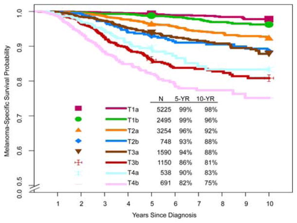
As in the seventh edition, patients with primary melanoma and no evidence of regional or distant metastasis are stratified into 8 T subcategories (T1a through T4b). MSS stratified by T subcategory for 23,001 patients with complete covariate data is illustrated in Figure 1. For these survival curves, patients with T1 melanomas were included if they had clinical (c) or pathological (p) T1 N0 melanomas, but patients with T2 through T4 melanomas were included only if they had pN0 melanoma (ie, no tumor-containing SLNs and no evidence of microsatellites, satellites, or in-transit metastases at diagnosis or after initial treatment). Overall, this approach aligns with the AJCC Principles of Cancer Staging (see Chapter 1 of the eighth edition AJCC Cancer Staging Manual).51 An implication of this approach is that patients with T2 through T4 melanomas who do not undergo SLN biopsy cannot be pathologically staged. Nonetheless, the Melanoma Expert Panel acknowledges that not all patients with T2 through T4 melanomas undergo SLN biopsy, and improved clinical prognostic models and tools (eg, clinical calculators, etc) may be developed to improve prognostic assessment among this cohort of patients in the future.
FIGURE 1.
Kaplan-Meier Melanoma-Specific Survival Curves According to T Subcategory for Patients With Stage I and II Melanoma From the Eighth Edition International Melanoma Database. Patients with N0 melanoma have been filtered, so that patients with T2 to T4 melanoma were included only if they had negative sentinel lymph nodes, whereas those with T1N0 melanoma were included regardless of whether they underwent sentinel lymph node biopsy.
In the eighth edition, the 5-year and 10-year MSS ranged from 99% and 98%, respectively, for patients with T1a N0 melanomas (ie, primary tumor thickness <0.8 mm, nonulcerated) to 82% and 75%, respectively, for patients with T4b N0 melanomas (ie, primary tumor thickness >4.0 mm, ulcerated). MSS for all T subcategories were notably higher than those reported in the seventh edition, in which the 10-year MSS rates were 93% and 39% for patients with T1a N0 and T4b N0 melanomas, respectively,25,37 or in the sixth edition.52 The higher survival of patients in the more contemporary cohort examined in this eighth edition effort is likely a consequence of the widespread use of SLN biopsy; the requirement of SLN biopsy for patients with T2 through T4 primary melanoma to be included in AJCC staging; and, to a lesser extent, newer imaging technologies that improve the detection of clinically occult metastatic disease, thereby defining more homogenous groups of patients and achieving more accurate staging.4,38 Some patients who, in the past, would have been classified as clinically node negative (cN0), would be expected to harbor clinically occult nodal metastasis identified on the basis of a positive SLN biopsy and are classified as pathologic N1 (pN1), pN2, etc, according to the overall number of tumor-involved lymph nodes. In a 2004 study using sixth edition criteria, for example, the risk of harboring a positive SLN ranged from 2% in patients with T1a melanoma (nonulcerated and ≤1.0 mm) to 53% in those with T4b melanoma.53
Other T-category definitions have been clarified in the eighth edition. Patients with melanoma in situ are properly categorized as Tis (not T0, which is reserved for an unknown or completely regressed primary site). Because tumor thickness can only be evaluated accurately in histological sections cut perpendicular to the epidermal surface, the T category should be recorded as TX if the thickness cannot be assessed (eg, in curettage specimens, when no tissue fragment shows a complete section of the tumor cut perpendicular to the surface). In some instances, if the tissue has been misembedded, then melting the paraffin block and re-embedding the tissue may enable perpendicular sections to be obtained. If there is evidence of regression of part of an invasive melanoma, then the thickness should be measured in the usual way to the deepest identifiable, viable tumor cell, and the tumor should be assigned to the appropriate T category. Partially regressed melanoma should not be designated TX or T0. T0 should be used if there is no evidence of a primary tumor (eg, in a patient who presents with nodal or visceral metastasis and no known primary tumor) or if a melanoma has regressed completely. If the invasive component of the melanoma has regressed but overlying in situ melanoma remains, then the tumor should be designated Tis.
Ulceration
Primary tumor ulceration is another T-category criterion. In the eighth edition, as in the seventh edition,4,25 the absence or presence of ulceration is designated “a” or “b,” respectively, in each T subcategory (eg, T2a and T2b correspond to nonulcerated and ulcerated T2 melanomas, respectively) (Table 2). Ulceration is defined as the full thickness absence of an intact epidermis above any portion of the primary tumor with an associated host reaction (characterized by a fibrinous and acute inflammatory exudate) above the primary tumor based on histopathological examination. If there is no host reaction, this likely represents artifactual loss of an intact epidermis overlying the primary melanoma, and the melanoma should not be recorded as ulcerated, because this may have resulted from sectioning artifact caused by the tissue sectioning techniques used in the laboratory. Epidermal loss caused by a prior biopsy should not be recorded as ulceration for staging purposes. If ulceration is present in either an initial partial biopsy or a re-excision specimen of a primary melanoma, then the tumor should be recorded as ulcerated for staging purposes. While the presence of “squared-off” edges of a scar can provide a clue to the presence of iatrogenic (prior biopsy-related) ulceration, at times, it may be difficult or impossible to distinguish between iatrogenic and noniatrogenic causes of ulceration on the basis of histopathologic assessment alone, and correlation with the clinical history is essential.54 If doubt remains as to whether ulceration is traumatic or iatrogenic in origin, then the tumor should be staged as an ulcerated primary tumor.
Ulceration is an adverse prognostic factor;4,25,37,41,55 the presence of an ulcerated primary was generally associated with an MSS similar to that of a patient with a nonulcerated primary in the next highest tumor thickness category (Fig. 1). For example, the 5-year and 10-year MSS rates are 93% and 88%, respectively, for patients with T2bpN0 primary cutaneous melanomas and 94% and 88%, respectively, for those with T3apN0 primary cutaneous melanomas.
Mitotic rate
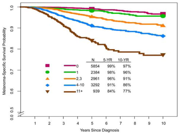
The mitotic rate, defined as the number of mitoses per square millimeter in the invasive portion of the tumor using the “hot-spot” method4 (ie, count beginning in a region where mitoses are more frequent and continue in immediately adjacent, nonoverlapping high-power fields), was a T1 category criterion in the seventh edition25 and was included as a dichotomous variable defined as <1 mitosis per mm2 versus ≥1 mitoses per mm2. In the eighth edition, the mitotic rate was not included as a T1 staging criterion (based on the T1 analysis described above; see Breslow tumor thickness). Nevertheless, among patients with clinically node-negative (cN0) primary melanoma in the eighth edition AJCC melanoma database, increasing mitotic rate was significantly associated with decreasing MSS in univariate analysis (Fig. 2). For example, in a univariate analysis of MSS for patients with T1 through T4 pN0 melanoma according to mitotic rate (mitoses per mm2), when categorized as <1, from 1 to 3, from 3 to 10, and >10 mitoses per mm2, the 5-year and 10-year MSS rates ranged from 99% and 97%, respectively, in patients who had primary tumors with <1 mitosis per mm2, to 84% and 77%, respectively, in those who had primary tumors with ≥11 mitoses per mm2 (P < .0001; log-rank test). As supported by this univariate analysis and previous reports,56,57 the mitotic rate is likely an important prognostic determinant when evaluated using its dynamic range across melanomas of all tumor thickness categories. Therefore, the AJCC Melanoma Expert Panel strongly recommends that mitotic rate be assessed and recorded for all primary melanomas,4 although it is not used for T1 staging in the eighth edition. The mitotic rate will likely be an important parameter for inclusion in the future development of prognostic models applicable to individual patients. Although it is not included in the T1 subcategory criteria, mitotic activity in T1 melanomas also has been associated with an increased risk of SLN metastasis.43,46,50,58
FIGURE 2.
Kaplan-Meier Melanoma-Specific Survival Curves According to Mitotic Rate (Mitoses per mm2) in Patients With Stage I and II Melanoma From the Eighth Edition International Melanoma Database.
The N Category
The N category documents metastatic disease both in regional lymph nodes and in non-nodal locoregional sites (ie, microsatellites, satellites, and in-transit metastases). For the eighth edition, the Melanoma Expert Panel sought to add further granularity throughout the N category by providing clarity of definitions.
Regional lymph node metastasis
In the eighth edition, N category criteria continue to include both the extent of regional node tumor involvement and the number of tumor-involved regional nodes. “Clinically occult” nodal metastasis describes patients with microscopically identified regional node metastasis detected by SLN biopsy and without clinical or radiographic evidence of regional node metastasis (termed “microscopic” nodal metastasis in the seventh edition). In contrast, “clinically detected” nodal metastasis describes patients with regional node metastasis identified by clinical, radiographic, or ultrasound examination (termed “macroscopic” nodal metastasis in the seventh edition) and usually (but not necessarily) confirmed by biopsy.51
Clinically occult (N1a, N2a, N3a) and clinically detected (N1b, N2b, N3b) N subcategories define patients with regional lymph node disease based on extent of regional node involvement and the number of tumor-involved regional nodes among patients without satellites, microsatellites, or in-transit metastases (Table 3).4 If at least one node is clinically detected and there are additional involved nodes detected only on microscopic examination, then the total number of involved nodes (ie, both those clinically detected and those identified only on microscopic examination of a complete lymphadenectomy specimen) should be recorded for N subcategory based on the total number of tumor-involved regional nodes. If microsatellites, satellites, or in-transit metastases are present, then patients are assigned to an N “c” subcategory according to the number of tumor-involved regional nodes, regardless of whether they are clinically occult or clinically detected: N1c, N2c or N3c if 0, 1 or ≥2 regional nodes contain tumor, respectively (Table 3).
TABLE 3.
Definition of Regional Lymph Node (N)a
| N CATEGORY | EXTENT OF REGIONAL LYMPH NODE AND/OR LYMPHATIC METASTASIS | |
|---|---|---|
|
| ||
| NO. OF TUMOR-INVOLVED REGIONAL LYMPH NODES | PRESENCE OF IN-TRANSIT,SATELLITE, AND/OR MICROSATELLITE METASTASES | |
| NX | Regional nodes not assessed (eg, sentinel lymph node [SLN] biopsy not performed, regional nodes previously removed for another reason); Exception: pathological N category is not required for T1 melanomas, use clinical N information | No |
| N0 | No regional metastases detected | No |
| N1 | One tumor-involved node or any number of in-transit, satellite, and/or microsatellite metastases with no tumor-involved nodes | |
| N1a | One clinically occult (ie, detected by SLN biopsy) | No |
| N1b | One clinically detected | No |
| N1c | No regional lymph node disease | Yes |
| N2 | Two or 3 tumor-involved nodes or any number of in-transit, satellite, and/or micro- satellite metastases with one tumor-involved node | |
| N2a | Two or 3 clinically occult (ie, detected by SLN biopsy) | No |
| N2b | Two or 3, at least one of which was clinically detected | No |
| N2c | One clinically occult or clinically detected | Yes |
| N3 | Four or more tumor-involved nodes or any number of in-transit, satellite, and/or microsatellite metastases with 2 or more tumor-involved nodes, or any number of matted nodes without or with in-transit, satellite, and/or microsatellite metastases | |
| N3a | Four or more clinically occult (ie, detected by SLN biopsy) | No |
| N3b | Four or more, at least one of which was clinically detected, or the presence of any number of matted nodes | No |
| N3c | Two or more clinically occult or clinically detected and/or presence of any number of matted nodes | Yes |
Adapted with permission of the American Joint Committee on Cancer (AJCC), Chicago, Illinois. The original and primary source for this information is the AJCC Cancer Staging Manual, Eighth Edition (2017) published by Springer International Publishing (modified from: Gershenwald JE, Scolyer RA, Hess KR, et al. Melanoma of the skin. In: Amin MB, Edge SB, Greene FL, et al, eds. AJCC Cancer Staging Manual. 8th ed. New York: Springer International Publishing; 2017:563–5854).
As noted in the seventh edition, there is no unequivocal evidence that there is a lower threshold for the size of a clinically occult melanoma regional lymph node tumor deposit that defines node-positive disease for staging purposes. Thus, a lymph node in which any metastatic tumor cells have been identified, irrespective of how small the tumor deposit or whether it has been identified on hematoxylin and eosin-stained or immunostained sections, should be designated as a tumor-involved lymph node. In the eighth edition, it has been clarified that, if melanoma cells are found in a lymphatic channel within or immediately adjacent to a lymph node, that node is regarded as tumor-involved for staging purposes.
In the eighth edition, the term “gross extranodal extension” is no longer used as an N category criterion, but the presence of matted nodes (defined as 2 or more nodes adherent to one another through involvement by metastatic disease, identified at the time the specimen is examined macroscopically in the pathology laboratory) is retained as an N3 criterion. Although it is not formally included as an eighth edition N category criterion, the definition of extra-nodal extension (ENE) (also termed extranodal spread or extracapsular extension) has been clarified. In the eighth edition, ENE is defined as the presence of a nodal metastasis extending through the lymph node capsule and into adjacent tissue, which may be macroscopically apparent but must be microscopically confirmed. It is recommended that this factor be recorded, as it may be useful for future analyses.59
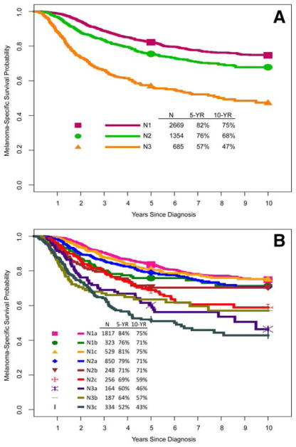
Several large series have demonstrated that patients with clinically occult regional node disease have better survival than those with clinically evident disease.52,60–62 This was also evident in the AJCC MSS curves according to N category and N subcategory, as shown in Figure 3. Overall, consistent with our observations in the seventh edition,25,37,62 there is marked heterogeneity in prognosis among patients with stage III regional node disease by N-category designation.
FIGURE 3.
Kaplan-Meier Melanoma-Specific Survival Curves According to (A) N Categories and (B) Subcategories From the Eighth Edition International Melanoma Database.
Non-nodal locoregional metastases (microsatellite, satellite, and in-transit metastases)
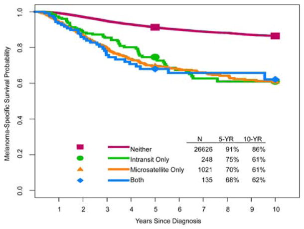
The presence and absence of microsatellite, satellite, or intransit metastases, regardless of the number of such lesions, are components of the N category in the eighth edition (Table 3).4 They are all thought to represent metastases that are a consequence of intralymphatic or possibly angiotrophic tumor spread. Satellite metastases have classically and somewhat arbitrarily been defined as clinically evident cutaneous and/or subcutaneous metastases occurring within 2 cm of the primary melanoma.33,51 Microsatellites have classically been defined as microscopic cutaneous and/or subcutaneous metastases found adjacent or deep to a primary melanoma on pathological examination (see discussion below). In-transit metastases have classically and somewhat arbitrarily been defined as clinically evident cutaneous and/or subcutaneous metastases identified at a distance more than 2 cm from the primary melanoma in the region between the primary and the first echelon of regional lymph nodes.33 Beginning with the sixth edition AJCC melanoma staging system, satellite and in-transit metastases were merged into a single staging entity reflective of intralymphatic regional metastases.33 Occasionally, satellite or in-transit metastases may occur distal to the primary site. An N “c” subcategory has been added into each of the N1, N2 and N3 categories (ie, N1c, N2c, N3c) (Table 3) in the eighth edition to incorporate contemporary knowledge of the prognostic importance of non-nodal locoregional metastases and to simplify the application of staging rules for patients who have them. Microsatellites, satellites, and in-transit metastases have been shown to portend a relatively poor prognosis.63–69 In univariate analysis of the eighth edition database that included patients with or without synchronous regional node involvement, there was no significant difference in survival outcome for these anatomically defined entities (Fig. 4); hence, they were grouped together for staging purposes (Table 3). Planned IMDDP multivariable analyses will further explore the prognostic impact of non-nodal regional disease on MSS.
FIGURE 4.
Kaplan-Meier Melanoma-Specific Survival Curves According to the Presence or Absence of Microsatellites, Satellites, and/or In-Transit Metastases From the Eighth Edition International Melanoma Database. Note that in-transit in the figure means in-transit and/or satellite metastasis and both means microsatellites and in-transit and/or satellite metastasis.
In the seventh edition, a microsatellite was defined as “any tumor nest >0.05 mm in diameter that was separated by normal dermis from the main invasive component of a melanoma by distance of >0.5 mm.”25 The definition of microsatellite has been clarified and refined, so that, in the eighth edition, there is no minimum size threshold or distance from the primary tumor that defines a microsatellite; it is simply defined as a microscopic cutaneous and/or subcutaneous metastasis adjacent to or deep to and completely discontinuous from a primary melanoma with unaffected stroma occupying the space between, identified on pathological examination of the primary tumor site. Fibrous scarring and/or inflammation noted between an apparently separate nodule and the primary tumor (rather than normal stroma) may represent regression of the intervening tumor; if these findings are present, then the nodule is considered to be an extension of the primary tumor and not a microsatellite. Although occasionally seen in the primary melanoma diagnostic biopsy specimen, microsatellites, when present, are more commonly identified in the wide excision specimen.
Metastatic melanoma in lymph nodes without a known primary tumor
Patients who presented with melanoma in one or more lymph nodes without a known primary tumor were not included in the International Melanoma Database constructed for the analyses informing the eighth edition. However, based on data from the published literature (including from patients who were diagnosed before 199870–72) and analyses of patients who presented to Melanoma Institute Australia since 1998,72 such patients had an equivalent or slightly better survival than patients with a known primary tumor who presented with a similar number of clinically detected, tumor-involved nodes. The AJCC Melanoma Expert Panel recommended that such patients be assigned to the corresponding N category based on the number of lymph nodes containing metastatic disease and the presence or absence of satellite, microsatellite, or intransit metastases. Until additional data are available, patients who have melanoma with an unknown primary and metastatic disease in a lymph node or nodes should be staged as in Table 6.
TABLE 6.
AJCC Pathological (pTNM) Prognostic Stage Groupsa
| WHEN T IS… | AND N IS… | AND M IS… | THEN THE PATHOLOGICAL STAGE GROUP IS… |
|---|---|---|---|
| Tis | N0b | M0 | 0 |
| T1a | N0 | M0 | IA |
| T1b | N0 | M0 | IA |
| T2a | N0 | M0 | IB |
| T2b | N0 | M0 | IIA |
| T3a | N0 | M0 | IIA |
| T3b | N0 | M0 | IIB |
| T4a | N0 | M0 | IIB |
| T4b | N0 | M0 | IIC |
| T0 | N1b, N1c | M0 | IIIB |
| T0 | N2b, N2c, N3b or N3c | M0 | IIIC |
| T1a/b–T2a | N1a or N2a | M0 | IIIA |
| T1a/b–T2a | N1b/c or N2b | M0 | IIIB |
| T2b/T3a | N1a–N2b | M0 | IIIB |
| T1a–T3a | N2c or N3a/b/c | M0 | IIIC |
| T3b/T4a | Any N ≥N1 | M0 | IIIC |
| T4b | N1a–N2c | M0 | IIIC |
| T4b | N3a/b/c | M0 | IIID |
| Any T, Tis | Any N | M1 | IV |
Used with permission of the American Joint Committee on Cancer (AJCC), Chicago, Illinois. The original and primary source for this information is the AJCC Cancer Staging Manual, eighth edition (2017) published by Springer International Publishing (Gershenwald JE, Scolyer RA, Hess KR, et al. Melanoma of the skin. In: Amin MB, Edge SB, Greene FL, et al, eds. AJCC Cancer Staging Manual. 8th ed. New York: Springer International Publishing; 2017:563–5854).
Pathological stage 0 (melanoma in situ) and T1 do not require pathological evaluation of lymph nodes to complete pathological staging; use clinical N information to assign their pathological stage.
The M Category
For the eighth edition, the Melanoma Expert Panel concluded that, because of the rapidly changing and still evolving landscape for the management of patients with stage IV melanoma, it was premature to embark on a broad-based, analytic initiative based on new data from patients who were treated in recent years. Instead, the legacy seventh edition AJCC stage IV International Melanoma Database was used for the eighth edition as the primary data source (and no new analyses were conducted), supplemented by published contemporary clinical trial data.6–20 In the eighth edition, M-category definitions were clarified and refined, and a new category for patients with central nervous system (CNS) metastases was added (M1d). For patients with distant metastases, M1 is defined by both anatomic site of distant metastatic disease and serum lactate dehydrogenase (LDH) level for all anatomic site subcategories.
Anatomic site(s) of distant metastatic disease
The anatomic site(s) of metastasis is used to assign patients to 1 of 4 (previously 3) M subcategories: M1a, M1b, M1c, and—new to the eighth edition—M1d (Table 4).4 The definition of each M1 anatomic site subcategory was also clarified. Patients with distant metastasis to skin, subcutaneous tissue, muscle, or distant lymph nodes, regardless of serum LDH level, are categorized as M1a. Patients with metastasis to lung (with or without concurrent metastasis to skin, subcutaneous tissue, muscle, or distant lymph nodes and regardless of serum LDH level) are categorized as M1b. Patients with metastases to any other visceral site(s) (exclusive of the CNS) are designated as M1c. New to the eighth edition, patients with metastases to the CNS (ie, involving the brain, spinal cord, leptomeninges, or other components of the CNS)4 are designated as M1d (irrespective of the presence of metastatic disease at other sites); these patients were previously designated as M1c in the seventh edition. This revision to include an M1d category reflects the expert panel’s assessment that, in addition to the historically poor overall survival outcome for patients with CNS metastases, contemporary clinical trial eligibility and exclusion criteria, as well as stratification and analysis, are often based on the presence/absence of CNS disease.6–20,73,74 Therefore, this additional level of granularity in the M category “maps” better to contemporary clinical practice and clinical trial decision making and analysis.
TABLE 4.
Definition of Distant Metastasis (M)a
| M CATEGORYb | M CRITERIA | |
|---|---|---|
|
| ||
| ANATOMIC SITE | LDH LEVEL | |
| M0 | No evidence of distant metastasis | Not applicable |
| M1 | Evidence of distant metastasis | See below |
| M1a | Distant metastasis to skin, soft tissue including muscle, and/or nonregional lymph node | Not recorded or unspecified |
| M1a(0) | Not elevated | |
| M1a(1) | Elevated | |
| M1b | Distant metastasis to lung with or without M1a sites of disease | Not recorded or unspecified |
| M1b(0) | Not elevated | |
| M1b(1) | Elevated | |
| M1c | Distant metastasis to non-CNS visceral sites with or without M1a or M1b sites of disease | Not recorded or unspecified |
| M1c(0) | Not elevated | |
| M1c(1) | Elevated | |
| M1d | Distant metastasis to CNS with or without M1a, M1b, or M1c sites of disease | Not recorded or unspecified |
| M1d(0) | Not elevated | |
| M1d(1) | Elevated | |
CNS indicates central nervous system; LDH, lactate dehydrogenase.
Used with permission of the American Joint Committee on Cancer (AJCC), Chicago, Illinois. The original and primary source for this information is the AJCC Cancer Staging Manual, eighth edition (2017) published by Springer International Publishing (Gershenwald JE, Scolyer RA, Hess KR, et al. Melanoma of the skin. In: Amin MB, Edge SB, Greene FL, et al, eds. AJCC Cancer Staging Manual. 8th ed. New York: Springer International Publishing; 2017:563–5854).
Suffixes for M category: (0) LDH not elevated, (1) LDH elevated. No suffix is used if LDH is not recorded or is unspecified.
Serum LDH level
In the seventh edition, an elevated LDH level was used to categorize a patient as M1c, regardless of anatomic site(s) of metastatic disease, given its significance as an independent, adverse predictor of survival among patients with stage IV disease. LDH remains a clinically significant factor associated with response, progression-free survival, MSS, and overall survival in the contemporary treatment era of targeted and immune therapies.75–77 In the eighth edition, an elevated LDH level no longer independently defines M1c disease. Instead, to better codify the impact of anatomic site and LDH level, descriptors were added to the M1 subcategory designation to indicate LDH status (designated as “[0]” for not elevated and “[1]” for elevated) for each M1 subcategory (Table 4).
The Stage Groups
As in prior editions of the AJCC Cancer Staging Manual, both clinical and pathological classifications are used in melanoma staging. In the eighth edition, clinical staging includes microstaging of the primary melanoma—as a standard practice, after biopsy of the primary melanoma—and clinical/radiologic assessment for regional and distant metastases, as well as biopsies performed to assess for regional and distant metastases, as appropriate (Table 5).4 There are no substages for clinical stage III melanoma. Pathological staging includes all clinical staging information, as well as any additional staging information derived from the wide excision (surgical) specimen that constitutes primary tumor surgical treatment, and pathological information about the clinically node-negative regional lymph nodes after SLN biopsy, with or without completion lymph node dissection (CLND), or therapeutic lymph node dissection for clinically evident regional lymph node disease (Table 6).4 In patients who undergo SLN biopsy and have a clinically occult regional lymph node metastasis identified by SLN biopsy but do not undergo additional surgery in the form of CLND, according to the eighth edition Principles of Cancer Staging (Chapter 1 of the eighth edition AJCC Cancer Staging Manual51) and the eighth edition melanoma chapter,4 category pN1a(sn) is assigned to specify that CLND was not performed. If a CLND is performed, then such patients would be assigned to subcategory pN1a (or another pN >0 subcategory, depending on the total number of tumor-involved lymph nodes) to distinguish these 2 clinical scenarios and to improve granularity in coding for clinical and analytic purposes.4,51
TABLE 5.
AJCC Clinical Prognostic Stage Groups (cTNM)a
| WHEN T IS… | AND N IS… | AND M IS… | THEN THE CLINICAL STAGE GROUP IS… |
|---|---|---|---|
| Tis | N0 | M0 | 0 |
| T1a | N0 | M0 | IA |
| T1b | N0 | M0 | IB |
| T2a | N0 | M0 | IB |
| T2b | N0 | M0 | IIA |
| T3a | N0 | M0 | IIA |
| T3b | N0 | M0 | IIB |
| T4a | N0 | M0 | IIB |
| T4b | N0 | M0 | IIC |
| Any T, Tis | ≥N1 | M0 | III |
| Any T | Any N | M1 | IV |
Used with permission of the American Joint Committee on Cancer (AJCC), Chicago, Illinois. The original and primary source for this information is the AJCC Cancer Staging Manual, eighth edition (2017) published by Springer International Publishing (Gershenwald JE, Scolyer RA, Hess KR, et al. Melanoma of the skin. In: Amin MB, Edge SB, Greene FL, et al, eds. AJCC Cancer Staging Manual. 8th ed. New York: Springer International Publishing; 2017:563–5854).
In part because of the low overall likelihood of nodal metastasis and lack of uniformly accepted criteria for SLN biopsy in T1 melanoma, neither pathological stage 0 (melanoma in situ [Tis]) nor T1 melanoma requires SLN biopsy to complete pathological staging among patients with clinically node-negative melanomas. Instead, cN information is used to assign the pathological stage for T1 melanomas if an SLN biopsy is not performed.
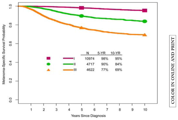
The MSS rates for all patients stratified by pathological stage groups I through III are shown in Figure 5. Patients with stage I, II, and III disease had 5-year and 10-year MSS rates of 98% and 95%, 90% and 84%, and 77% and 69%, respectively, and were overall slightly improved compared with patients who had similar stages of melanoma in the seventh edition analyses.25,37
FIGURE 5.
Kaplan-Meier Melanoma-Specific Survival Curves According to Stage in Patients With Stage I to III Melanoma From the Eighth Edition International Melanoma Database.
Stage I and II subgroupings
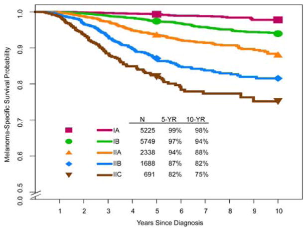
For pT-category stage groups, 5-year and 10-year MSS rates ranged from 99% and 98%, respectively, in patients with stage IA melanoma, to 82% and 75%, respectively, in those with stage IIC disease (Fig. 6). As in the seventh edition, patients with clinical T1b N0 melanoma are included in clinical stage IB. In contrast, patients with pathological T1b N0 melanoma are included in pathological stage IA (and not stage IB as in the seventh edition) (Table 6). This stage grouping reflects the better survival of patients who have T1b melanoma with pathologically negative nodes because, if SLN biopsy was performed, it only includes those with a tumor-negative SLN (ie, T1b pN1 patients would be stage III), compared with a group of patients with T1b melanoma who were only clinically staged. The 5-year and 10-year MSS rates were 97% and 93%, respectively, for patients with clinical T1b N0 melanoma, compared with 99% and 96%, respectively, for those with pathological T1b N0 melanoma.
FIGURE 6.
Kaplan-Meier Melanoma-Specific Survival Curves According to T Category Stage Group for Patients With Stage I and II Melanoma From the Eighth Edition International Melanoma Database. Patients with N0 melanoma were filtered, so that patients with T2+ melanoma were included only if they had negative sentinel lymph nodes, whereas those with T1N0 melanoma were included regardless of whether they underwent sentinel lymph node biopsy.
Stage III subgroupings
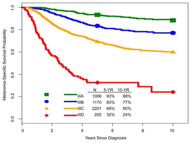
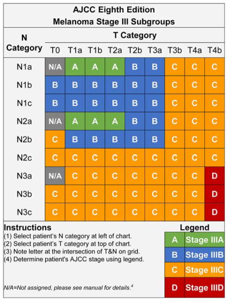
In the seventh edition, both regional lymph node factors (the number of nodes involved, microscopic vs macroscopic node involvement) as well as primary tumor ulceration determined stage III groups. Although the N category alone predicts MSS in the eighth edition analysis (Fig. 3), the Melanoma Expert Panel hypothesized that more accurate prognostic estimates could be obtained by including both T-category factors, tumor thickness and ulceration status, along with the number of tumor-involved lymph nodes and whether they were detected clinically or were clinically occult (ie, positive SLN), and the presence of microsatellite, satellite, and/or in-transit metastases (ie, 9 N categories) (Table 3). This was evaluated using recursive partitioning analysis. Initially, 8 pathological stage III subgroups were created, including 3 “pairs” of subgroups that had similar 5-year MSS (data not shown). On the basis of discussions by the Melanoma Expert Panel that explored the relative merits of “grouping” versus “splitting” and the observation that the adoption of 5 N-stage groups would result in a total of 11 overall stage groups across T, N, and M (5 + 5 + 1 = 11), which would not conform to the total number of stage groups across the broad AJCC cancer disease site landscape, the 8 subgroups were combined to create 4 stage III subgroups that maintained the overall prognostic heterogeneity of the base model (Fig. 7). As such, these 4 subgroups stratify patients with stage III melanoma in the eighth edition, compared with the 3 subgroups that were used to stratify stage III patients in the seventh edition.25,37 A clinic workstation guide to combining T and N categories into stage III subgroups is provided in Figure 8 (see also Supporting Information Fig. 1 for a black-and-white version and Supporting Information Fig. 2 for a full-page color version). The 5-year MSS rate according to stage III subgroups ranges from 93% in patients with stage IIIA disease (1–3 clinically occult, tumor-involved SLNs [N1a or N2a] and T1a, T1b, or T2a primaries) to 32% for those with stage IIID disease (patients with a thick and ulcerated primary [T4b] and either ≥4 tumor-involved regional nodes [N3a or N3b] or ≥2 tumor-involved nodes and evidence of microsatellite, satellite, or in-transit metastases [N3c]) (Fig. 7). In the seventh edition, the 5-year MSS rates for patients with stage IIIA, IIIB, and IIIC disease were 78%, 59%, and 40%, respectively.37 These differences, particularly for patients with stage IIIA disease, have implications for clinical decision making and counseling as well as the design, eligibility, stratification, and analysis of adjuvant therapy clinical trials.
FIGURE 7.
Kaplan-Meier Melanoma-Specific Survival Curves According to Stage III Subgroups From the Eighth Edition International Melanoma Database.
FIGURE 8.
American Joint Committee on Cancer (AJCC) Eighth Edition Stage III Subgroups Based on T and N Categories.
Distant metastases (stage IV)
Although revisions to the M category have been implemented in the eighth edition, as described in detail above (Tables 4, 5, and 6), no M-stage subgroups were proposed, and no new data have been analyzed to date. This is because the availability of contemporary data is limited and because survival differences among patients with stage IV melanoma historically were small (before the recent revolution in treatment options for patients with advanced melanoma). It is anticipated that, as recently introduced systemic therapies gain a foothold in the treatment repertoire of patients with advanced disease and even better treatment modalities become available, stage IV survival outcomes will continue to improve. An international stage IV melanoma database is planned in the future to explore this new and still evolving treatment landscape for patients with advanced disease.
Additional Recommendations
Multiple Primary Melanomas
It is well established that patients may be diagnosed with synchronous or metachronous primary melanomas. In general, according to the eighth edition AJCC Principles of Cancer Staging,51 when patients present with multiple primary cutaneous melanomas, each is considered a different primary site, and each is separately categorized. In the uncommon clinical scenario where patients who harbor regional node metastases have multiple primary melanomas draining to the same regional node basin, the primary tumor with the highest T category should be assigned as the originating primary tumor with respect to the nodal metastases; if distant metastases are present, then the primary tumor with the highest N category (or the highest T category if N0) should be assigned as the origin of the distant metastases.51 Moreover, in patients with multiple primary melanomas, the recorded stage should map to the highest stage group of any of the primary tumors. According to the Principles of Cancer Staging chapter,51 if there are multiple synchronous melanomas with no evidence of metastatic disease, then the assigned category is based on the tumor with the highest T category, and, by convention, the m suffix is used. For example, T2a(m) would be used to describe a 1.4-mm, nonulcerated melanoma diagnosed synchronously with a 0.7-mm, nonulcerated melanoma. Alternatively, another acceptable approach is to designate the number of primary tumors instead of the m suffix (ie, T2a(2) in the above example).51 To the extent possible, if the number of synchronous multiple primary melanomas at presentation is known, then this latter approach is preferred by the Melanoma Expert Panel.
Other Important Primary Tumor Factors
Although detailed discussion is beyond the scope of this article, in addition to the variables discussed (eg, tumor thickness, ulceration, mitotic rate), the Melanoma Expert Panel recommends the routine collection of multiple other known or putative primary tumor factors: level of invasion, tumor-infiltrating lymphocytes, lymphovascular invasion, and neurotropism. The interested reader is referred to a comprehensive description and discussion of these and other factors in the melanoma chapter of the eighth edition AJCC Cancer Staging Manual.4
SLN Microscopic Tumor Burden
There is significant and growing evidence that microscopic tumor burden in the SLN is prognostically important.78–90 SLN tumor burden can be assessed by a variety of micro-morphometric parameters, including the maximum size of the largest metastasis, the maximum subcapsular depth (also known as tumor penetrative depth88 of the deposits and measured from the inner surface of the lymph node capsule to the deepest intranodal tumor cell), the microanatomic location of SLN tumor deposits, the percentage cross-sectional area of the SLN that is involved, and the presence of extranodal extension. In various studies, one or more of these parameters has predicted survival in SLN-positive patients.78–90
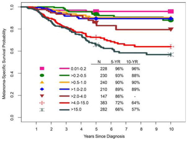
The impact of extent of SLN tumor burden (based on the greatest maximum dimension of the largest discrete, metastatic melanoma deposit) was assessed for the subset of patients with known SLN tumor burden in the IMDDP. In univariate analysis, increasing SLN tumor burden was associated with reduced MSS (Fig. 9). Although this histopathological parameter is not a formal staging criterion for the N category in the eighth edition, documentation of SLN tumor burden is an important prognostic factor that will be included in and likely will guide the development of future prognostic models and ultimately validated clinical tools (eg, calculators, nomograms, etc) for patients with regional metastatic disease.
FIGURE 9.
Kaplan-Meier Melanoma-Specific Survival Curves According to Maximum Dimension of Sentinel Lymph Node Metastatic Focus (mm) From the Eighth Edition International Melanoma Database. Note that there were insufficient data (<10 cases) to estimate 10-year melanoma-specific survival for patients who had a maximum sentinel lymph node metastatic focus of 2 to 4 mm.
Microscopic SLN tumor burden has already been implemented as an inclusion criterion in some clinical trials (eg, European Organization for Research and Treatment of Cancer [EORTC] trial 18071, adjuvant ipilimumab in stage III melanoma;23 and COMBI-AD, adjuvant dabrafenib plus trametinib in stage III mela-noma24). In these trials, patients with a single positive SLN must have a microscopic tumor burden >1 mm in diameter, based on the relatively worse prognosis of this patient subgroup.
On the basis of the currently available evidence, the AJCC Melanoma Expert Panel recommends that, at a minimum, the single largest maximum dimension (measured in millimeters to the nearest 0.1 mm using an ocular micrometer) of the largest discrete, metastatic melanoma deposit in SLNs be recorded in pathology reports.4 To further advance this field, the AJCC Melanoma Expert Panel and the International Melanoma Pathology Study Group plan to continue efforts to harmonize and standardize the assessment and reporting of SLN tumor burden. Planned IMDDP analyses will also further explore the prognostic impact of SLN tumor burden.
The Number of Distant Metastatic Sites and the Extent of Distant Metastatic Disease Burden
The number of metastases at distant sites has previously been documented as an important prognostic factor.76,91–93 This was also confirmed in previous preliminary multivariable analyses using the seventh edition AJCC stage IV melanoma database. However, this feature was not incorporated into the eighth edition as a formal staging criterion due in part to significant variability in the deployment of diagnostic imaging to comprehensively search for distant metastases (ranging from a chest x-ray in some centers to high-resolution, double-contrast computed tomography, positron emission tomography/computed tomography, and magnetic resonance imaging in others) as well as the heterogeneity with which extent of disease results are codified across databases. Until recording of the indications for and types of investigations used and the extent of distant metastatic disease are better standardized, the Melanoma Expert Panel concluded that the number of metastases cannot reproducibly be used for staging purposes.
Approach to Staging Patients After Neoadjuvant (“Up-Front”) Therapy
Historically, surgery represented the mainstay of treatment for patients with cutaneous melanoma. For several solid tumors, neoadjuvant therapy (systemic therapy before surgical resection) is often used as part of multidisciplinary treatment approaches for patients with locally advanced and/or regional disease and, for others, an “up-front” approach with systemic therapy (without a definitive plan for surgery to follow) is used.94 The availability of effective systemic therapies has greatly expanded potential treatment approaches for patients with unresectable and regionally advanced melanoma over the past several years and has led to tremendous interest in leveraging these clinical advances to develop neoadjuvant strategies for patients who have melanoma with locally advanced or metastatic disease. To stage such patients after treatment, the eighth edition Principles of Cancer Staging chapter includes a post-therapy or postneoadjuvant therapy classification, yTNM, which includes T, N, and M categorization after systemic or radiation treatment intended as definitive therapy (ycTNM) or after neoadjuvant therapy followed by planned surgery (ypTNM).51 Although this classification has been used infrequently in melanoma to date, because a robust portfolio of neoadjuvant clinical trials in patients with melanoma are currently under way and still more are planned, the “y” classification schema may prove useful in characterizing such patients, and the information can be compared with clinical stages assigned to patients before the start of neoadjuvant therapy. Future analyses will likely allow refinement of this not yet widely used classification schema.
Approach to Staging Patients After Recurrence/Retreatment
By definition, clinical and pathological classification according to the AJCC staging system occurs at the time of initial melanoma presentation. Thus, those who have regional node or non-nodal regional metastases at the time of initial presentation are characterized as having stage III disease, and those who present with distant metastases at the time of initial presentation are characterized as having stage IV disease. To accommodate staging for patients who have recurred, the eighth edition Principles of Cancer Staging chapter also includes an additional classification schema for patients who recur, rTNM, which is further divided into “r-clinical” (rcTNM) and “r-pathological” (rpTNM) stages. Such an approach may be useful to better characterize the extent of disease along the disease continuum in an individual patient with melanoma.51 Because, to date, this staging classification is relatively unknown and infrequently used by the global melanoma community, future analyses will likely inform revisions of this classification schema for patients with recurrent melanoma.
Conclusions
In the eighth edition AJCC staging system for cutaneous melanoma, particular attention was directed to clarifying major themes and terminology, introducing clinically relevant revisions, and creating a new, contemporary international database. The Melanoma Expert Panel focused most of its attention on evidence-based revisions of stage I to III melanoma for the eighth edition AJCC Cancer Staging Manual and established a framework for the development of robust and iteratively refined clinical prognostic models that will assist in the development of clinical tools to ultimately enhance clinical decision making. Importantly, based on analyses of this contemporary melanoma database, survival outcomes for equivalent stage groupings were substantially higher than those for similar stage groups of patients in prior editions, including the seventh edition, with implications for clinical decision making and clinical trial design, eligibility, stratification, and analysis.
Given the rapidly evolving landscape of treatment for stage IV melanoma in recent years, which already has resulted in significantly improved progression-free and overall survival for patients, the Melanoma Expert Panel strategically paused and did not establish a stage IV database or perform analyses of patients with stage IV disease. Instead new, clinically relevant M-category criteria were introduced into the eighth edition that will facilitate the refined collection of stage IV data, including more precise data collection for patients with CNS metastases. These new criteria will be essential to support future assessment of prognosis, as well as clinical trial design, eligibility, stratification, and analysis, for patients with advanced melanoma. Strategic development of analytic efforts for the population of patients with stage IV melanoma in the current new era of effective targeted therapies and immunotherapy is now under way as part of the IMDDP. These analyses are expected not only to improve prognostic assessment for patients with advanced disease but also to inform further revisions of the staging system and facilitate the development of clinical tools in the foreseeable future.
Additional enhancements to the eighth edition melanoma staging system, including yTNM and rTNM classifications, will enable contemporary patients with melanoma to be accurately risk stratified across the disease continuum. This will assist clinicians and patients in clinical management planning and enhance the design, conduct, and analysis of clinical trials that should ultimately lead to improved patient outcomes. Undoubtedly, melanoma staging will continue to evolve as new prognostic factors and evidence-based approaches—including the integration of clinical, pathological, molecular, and immunological endpoints— are developed, refined, and validated.
Supplementary Material
Practical Implications for Continuing Education.
The eighth edition of the American Joint Committee on Cancer melanoma staging system provides an updated framework for the classification and staging of patients with cutaneous melanoma.
Accurate melanoma staging is essential for reliable assessment of prognosis, rational treatment planning, and meaningful selection and stratification of patients entering clinical trials.
Because clinical care providers, pathologists, radiologists, translational researchers, cancer registrars, and others need to understand and effectively integrate the information included in this revised melanoma staging system into their clinical practice and registry-related activities, broad-based educational initiatives are necessary.
Acknowledgments
Eighth Edition AJCC Melanoma Expert Panel: Jeffrey E. Gershenwald, MD, FACS (Professor of Surgery and Cancer Biology; Medical Director, Melanoma and Skin Center, The University of Texas MD Anderson Cancer Center [Chair]); Richard A. Scolyer, MD, FRCPA, FRCPath (Conjoint Medical Director, Melanoma Institute Australia; Clinical Professor, The University of Sydney; Senior Staff Pathologist, Royal Prince Alfred Hospital [Vice Chair]); Michael B. Atkins, MD (Deputy Director, Georgetown-Lombardi Cancer Center); Charles M. Balch, MD, FACS (Professor of Surgery, The University of Texas MD Anderson Cancer Center); Raymond L. Barnhill, MD, MSc (Professor of Pathology, Institut Curie); Karl Y. Bilimoria, MD, MS (Director, Surgical Outcomes and Quality-Improvement Center, Vice Chair for Quality, Department of Surgery, Northwestern University); James D. Brierley, MS, MB, FRCR, FRCPC (Professor, University of Toronto; Staff Physician, Princess Margaret Hospital/University Health Network); Antonio C. Buzaid, MD (General Director, Centro Oncologico Antonio Ermirio de Moras, Hospital Sao Jose); David R. Byrd, MD (Professor of Surgery, University of Washington); Paul B. Chapman, MD (Medical Oncologist, Memorial Sloan Kettering Cancer Center); Alistair J. Cochran, MD (Ronald Reagan UCLA Medical Center); Daniel G. Coit, MD, FACS (Memorial Sloan Kettering Cancer Center); Alexander M. M. Eggermont, MD, PhD (Director General, Gustave Roussy Cancer Institute); David E. Elder, MD, MBChB, FRCPA (Hospital of the University of Pennsylvania); Mark B. Faries, MD (Codirector, Melanoma Program; Head, Surgical Oncology, The Angeles Clinic and Research Institute); Keith T. Flaherty, MD (Director, Termeer Center for Targeted Therapy, Massachusetts General Hospital Cancer Center); Claus Garbe, MD (Professor, University of Tubingen); Julie M. Gardner, MHA, BS (Manager, Clinical Protocol Administration, The University of Texas MD Anderson Cancer Center); Phyllis A. Gimotty, PhD (Professor of Biostatistics, University of Pennsylvania Perelman School of Medicine); Allan C. Halpern, MD (Chief, Dermatology Service, Memorial Sloan Kettering Cancer Center); Lauren E. Haydu, PhD (Manager, Clinical Data Management Systems, The University of Texas MD Anderson Cancer Center); Kenneth R. Hess, PhD (Professor, Department of Biostatistics, The University of Texas MD Anderson Cancer Center); Timothy M. Johnson, MD (Senior Associate Dean of Clinical Affairs, University of Michigan); John M. Kirkwood, MD (Professor of Medicine, Dermatology, and Translational Science, University of Pittsburgh); Alexander J. Lazar, MD, PhD, FCAP (Professor of Pathology, Dermatology, and Translational Molecular Pathology; Director, Melanoma Molecular Diagnostics, The University of Texas MD Anderson Cancer Center; and College of American Pathologists [CAP] AJCC Melanoma Representative); Anne W. M. Lee, MBBS, FRCR, FHKCR, FHKAM (Head, Department of Clinical Oncology, The University of Hong Kong and the University of Hong Kong-Shenzhen Hospital); Georgina V. Long, BSc, MBBS, PhD, FRACP (Conjoint Medical Director of Melanoma Institute Australia, Professor of Melanoma Medical Oncology and Translational Research, Melanoma Institute Australia and Royal North Shore Hospital, The University of Sydney); Grant A. McArthur, MD, BS, PhD, FRACP, FAHMS (Executive Director, Victorian Comprehensive Cancer Centre); Martin C. Mihm, Jr, MD, FACP (Professor of Dermatology, Harvard Medical School); Victor G. Prieto, MD, PhD (Chair, Professor of Pathology, The University of Texas MD Anderson Cancer Center); Merrick I. Ross, MD (Professor of Surgery, The University of Texas MD Anderson Cancer Center); Arthur J. Sober, MD (Professor of Dermatology, Harvard Medical School, Massachusetts General Hospital); Vernon K. Sondak, MD (Department Chair, Cutaneous Oncology, Moffitt Cancer Center); John F. Thompson, MD (Professor of Melanoma and Surgical Oncology, The University of Sydney, Melanoma Institute Australia); Richard L. Wahl, MD (Chairman, Department of Radiology, Washington University in St. Louis); and Sandra L. Wong, MD, MS (Professor and Chair, Department of Surgery, Geisel School of Medicine at Dartmouth, Dartmouth-Hitchcock Medical Center).
The AJCC Eighth Edition Melanoma Expert Panel acknowledges the following institutions and associated individuals for their data contributions to the eighth edition International Melanoma Database of the International Melanoma Database and Discovery Platform to perform analyses that informed the revisions incorporated into the eighth edition AJCC melanoma staging system (listed in alphabetical order): Fondazione Instituto di Recovero e Cura a Carattere Scientifico (IRCCS) Istituto Nazionale dei Tumori (Mario Santinami, MD and Andrea Maurichi, MD); Instituto Valenciano de Oncologia (Eduardo Nagore, MD); John Wayne Cancer Institute (Mark Faries, MD); Melanoma Institute Australia, The University of Sydney (John F. Thompson, MD, FRACS, FACS; Richard A. Scolyer, MD, FRCPA, FRCPath; Serigne Lo, PhD; Jonathan R. Stretch, MBBS, DPhil[Oxon], FRACS; Robyn R. P. Saw, MB MS, FRACS; and Andrew J. Spillane, MD, FRACS); Melbourne Melanoma Project: Peter MacCallum Cancer Centre (Grant McArthur, MBBS, PhD, FRACP; David Gyorki, MD, FRACS; Michael Henderson, MD, FRACS; and Sonia Mailer, BBc), Alfred Hospital Melbourne (John Kelly, MBBS, FACD), and Austin Hospital Melbourne (Johnathon Cebon, MBBS, PhD, FRACP); Department of Dermatology, National and Kapodistrian University of Athens School of Medicine, Andreas Sygros Hospital (Alexander Stratigos, MD); National and Kapodistrian University of Athens School of Medicine, General Hospital of Athens, Laiko (Helen Gogas, MD); The University of Texas MD Anderson Cancer Center (Jeffrey E. Gershenwald, MD; Lauren E. Haydu, MIPH; and Julie M. Gardner, MHA); Veneto Institute of Oncology-IOV (Carlo Riccardo Rossi, MD; Antonio Sommariva, MD; and Paolo Del Fiore); and Winship Cancer Institute of Emory University (Keith Delman, MD). The AJCC also acknowledges the Southwest Oncology Group for their contributions to the eighth edition melanoma effort (Antoni Ribas, MD, PhD; Lawrence Flaherty, MD; and James Moon, MS).
DISCLOSURES
This work was supported in part by the National Institutes of Health Specialized Program of Research Excellence (SPORE) Melanoma Grant P50 CA93459 (to The University of Texas MD Anderson Cancer Center); the National Institutes of Health/National Cancer Institute through The University of Texas MD Anderson Cancer Center Support Grant P30CA016672; a Melanoma Research Alliance Team Science Award; the generous philanthropic contributions to The University of Texas MD Anderson Melanoma Moon Shots Program; the Robert and Lynne Grossman Family Foundation; the Michael and Patricia Booker Melanoma Research Endowment; and the National Health and Medical Research Council of Australia, Melanoma Institute Australia, and the Medical Foundation of The University of Sydney. Jeffrey E. Gershenwald reports personal fees from Merck and Castle Biosciences outside the submitted work. Vernon K. Sondak reports personal fees from Merck, Genentech/Roche, Provectus Biopharmaceuticals, Bristol-Myers Squibb, Novartis, Array, Polynoma, and Pfizer outside the submitted work. Georgina V. Long reports personal fees from Amgen, Bristol-Myers Squibb, Novartis, Roche, Pierre Fabre, and Array Biopharma outside the submitted work. Merrick I. Ross reports personal fees and travel expenses/honorarium from Amgen and Merck and travel expenses/honorarium from Castle Biosciences, GlaxoSmithKline, and Provectus Biopharmaceuticals outside the submitted work. Mark B. Faries reports grants from the National Institutes of Health and personal fees from Novartis, Castle Biosciences, and Immune Design outside the submitted work. John M. Kirkwood reports grants from Prometheus; grants and personal fees from Merck; and personal fees from Bristol-Myers Squibb, Novartis, Roche, Genentech, EMD Serono, and Array Biopharma outside the submitted work. Alexander M. M. Eggermont reports personal fees from Acte-lion, Agenus, Bristol-Myers Squibb, GlaxoSmithKline, HalioDX, Incyte, ISA Pharmaceuticals, Merck Sharp & Dohme, Nektar, Novartis, Pfizer, and Sanofi outside the submitted work. John F. Thompson reports advisory board membership and honoraria from GlaxoSmithKline, Bristol-Myers Squibb, and Provectus Pharmaceuticals outside the submitted work. Allan Halpern reports consulting fees from Canfield Scientific, Inc, DermTech, and SciBase; is on the Data and Saftey Monitoring Board for Janssen Research & Development, LLC and Quintiles; and is part of the Scientific Advisory Board of Caliber ID. Martin Mihm reports consulting fees from Novartis, Alnylam, and MelaSciences; and is part of the Advisory Board of Caliber ID and BioCoz. Michael Atkins reports consulting fees from Bristol Myers Squibb, Merck, Novartis, Genentech/Roche, Array, and Arduro. Antonio C. Buzaid reports contracted research and speakers bureau fees from Roche, Bristol-Myers Squibb, Merck Sharp & Dohme, Pfizer, and Novartis. Helen Gogas reports consulting fees from Bristol-Myers Squibb, Merck Sharp & Dohme, Roche, Amgen, and Novartis; and grants from Bristol-Myers Squibb, Roche, Novartis. Alexander J. Stratigos reports consulting fees from LEO Pharma, Novartis, and Roche; and grants from Novartis, Roche. The remaining authors made no disclosures.
Author Contributions
Jeffrey E. Gershenwald: Conceptualization, methodology, validation, formal analysis, investigation, resources, data curation, writing–original draft, writing–review and editing, visualization, supervision, project administration, and funding acquisition. Richard A. Scolyer: Conceptualization, methodology, investigation, resources, writing–original draft, writing–review and editing, supervision, and project administration. Kenneth R. Hess: Formal analysis, writing–review and editing, and visualization. Vernon K. Sondak: Conceptualization, formal analysis, writing–original draft, writing–review and editing, and visualization. Georgina V. Long: Conceptualization, methodology, investigation, resources, writing–original draft, supervision, and funding acquisition. Merrick I. Ross: Writing–review and editing. Alexander J. Lazar: Conceptualization, methodology, formal analysis, investigation, writing–original draft, and writing–review and editing. Mark B. Faries: Investigation, resources, and writing–review and editing. John M. Kirkwood: Conceptualization, methodology, investigation, and writing–review and editing. Grant A. McArthur: Methodology, investigation, resources, and writing–review and editing. Lauren E. Haydu: Conceptualization, methodology, software, validation, investigation, resources, data curation, writing–review and editing, visualization, and project administration. Alexander M. M. Eggermont: Conceptualization, resources, and writing–review and editing. Keith T. Flaherty: Conceptualization, writing-original draft, writing-review, and editing. Charles M. Balch: Conceptualization, methodology, investigation, and writing–review and editing. John F. Thompson: Conceptualization, methodology, investigation, resources, writing–original draft, writing–review and editing, and project administration.
Footnotes
The AJCC Melanoma Expert Panel (in alphabetical order): Michael B. Atkins, Charles M. Balch, Raymond L. Barnhill, Karl Y. Bilimoria, Antonio C. Buzaid, David R. Byrd, Alistair J. Cochran, Alexander M. M. Eggermont, David E. Elder, Mark B. Faries, Keith T. Flaherty, Claus Garbe, Julie M. Gardner, Jeffrey E. Gershenwald (Chair), Phyllis A. Gimotty, Allan C. Halpern, Lauren E. Haydu, Kenneth R. Hess, Timothy M. Johnson, John M. Kirkwood, Alexander J. Lazar, Anne W. M. Lee, Georgina V. Long, Grant A. McArthur, Martin C. Mihm, Victor G. Prieto, Merrick I. Ross, Richard A. Scolyer (Vice-Chair), Arthur J. Sober, Vernon K. Sondak, John F. Thompson, and Sandra L. Wong.
The International Melanoma Database and Discovery Platform (in alphabetical order): Keith A. Delman, Mark B. Faries, Jeffrey E. Gershenwald (PI), Helen Gogas, David E. Gyorki, Lauren E. Haydu, Michael Henderson, Andrea Maurichi, Grant A. McArthur, Eduardo Nagore, Carlo Riccardo Rossi, Mario Santinami, Richard A. Scolyer, Antonio Sommariva, Alexander J. Stratigos, and John F. Thompson.
Additional supporting information may be found in the online version of this article.
References
- 1.Morton DL, Wen DR, Wong JH, et al. Technical details of intraoperative lymphatic mapping for early stage melanoma. Arch Surg. 1992;127:392–399. doi: 10.1001/archsurg.1992.01420040034005. [DOI] [PubMed] [Google Scholar]
- 2.Gershenwald JE, Thompson W, Mansfield PF, et al. Multi-institutional melanoma lymphatic mapping experience: the prognostic value of sentinel lymph node status in 612 stage I or II melanoma patients. J Clin Oncol. 1999;17:976–983. doi: 10.1200/JCO.1999.17.3.976. [DOI] [PubMed] [Google Scholar]
- 3.Amin MB, Edge SB, Greene FL, et al., editors. AJCC Cancer Staging Manual. 8. New York: Springer International Publishing; 2017. [Google Scholar]
- 4.Gershenwald JE, Scolyer RA, Hess KR, et al. Melanoma of the skin. In: Amin MB, Edge SB, Greene FL, et al., editors. AJCC Cancer Staging Manual. 8. New York: Springer International Publishing; 2017. pp. 563–585. [Google Scholar]
- 5.Morton DL, Thompson JF, Cochran AJ, et al. Final trial report of sentinel-node biopsy versus nodal observation in melanoma. N Engl J Med. 2014;370:599–609. doi: 10.1056/NEJMoa1310460. [DOI] [PMC free article] [PubMed] [Google Scholar]
- 6.Chapman PB, Hauschild A, Robert C, et al. Improved survival with vemurafenib in melanoma with BRAF V600E mutation. N Engl J Med. 2011;364:2507–2516. doi: 10.1056/NEJMoa1103782. [DOI] [PMC free article] [PubMed] [Google Scholar]
- 7.Falchook GS, Long GV, Kurzrock R, et al. Dabrafenib in patients with melanoma, untreated brain metastases, and other solid tumours: a phase 1 dose-escalation trial. Lancet. 2012;379:1893–1901. doi: 10.1016/S0140-6736(12)60398-5. [DOI] [PMC free article] [PubMed] [Google Scholar]
- 8.Flaherty KT, Infante JR, Daud A, et al. Combined BRAF and MEK inhibition in melanoma with BRAF V600 mutations. N Engl J Med. 2012;367:1694–1703. doi: 10.1056/NEJMoa1210093. [DOI] [PMC free article] [PubMed] [Google Scholar]
- 9.Flaherty KT, Puzanov I, Kim KB, et al. Inhibition of mutated, activated BRAF in meta-static melanoma. N Engl J Med. 2010;363:809–819. doi: 10.1056/NEJMoa1002011. [DOI] [PMC free article] [PubMed] [Google Scholar]
- 10.Hauschild A, Grob JJ, Demidov LV, et al. Dabrafenib in BRAF-mutated metastatic melanoma: a multicentre, open-label, phase 3 randomised controlled trial. Lancet. 2012;380:358–365. doi: 10.1016/S0140-6736(12)60868-X. [DOI] [PubMed] [Google Scholar]
- 11.Hodi FS, O’Day SJ, McDermott DF, et al. Improved survival with ipilimumab in patients with metastatic melanoma. N Engl J Med. 2010;363:711–723. doi: 10.1056/NEJMoa1003466. [DOI] [PMC free article] [PubMed] [Google Scholar]
- 12.Larkin J, Ascierto PA, Dreno B, et al. Combined vemurafenib and cobimetinib in BRAF-mutated melanoma. N Engl J Med. 2014;371:1867–1876. doi: 10.1056/NEJMoa1408868. [DOI] [PubMed] [Google Scholar]
- 13.Larkin J, Chiarion-Sileni V, Gonzalez R, et al. Combined nivolumab and ipilimumab or monotherapy in untreated melanoma. N Engl J Med. 2015;373:23–34. doi: 10.1056/NEJMoa1504030. [DOI] [PMC free article] [PubMed] [Google Scholar]
- 14.Long GV, Stroyakovskiy D, Gogas H, et al. Combined BRAF and MEK inhibition versus BRAF inhibition alone in melanoma. N Engl J Med. 2014;371:1877–1888. doi: 10.1056/NEJMoa1406037. [DOI] [PubMed] [Google Scholar]
- 15.Long GV, Stroyakovskiy D, Gogas H, et al. Dabrafenib and trametinib versus dabrafenib and placebo for Val600 BRAF-mutant melanoma: a multicentre, double-blind, phase 3 randomised controlled trial. Lancet. 2015;386:444–451. doi: 10.1016/S0140-6736(15)60898-4. [DOI] [PubMed] [Google Scholar]
- 16.Long GV, Trefzer U, Davies MA, et al. Dab-rafenib in patients with Val600Glu or Val600Lys BRAF-mutant melanoma meta-static to the brain (BREAK-MB): a multi-centre, open-label, phase 2 trial. Lancet Oncol. 2012;13:1087–1095. doi: 10.1016/S1470-2045(12)70431-X. [DOI] [PubMed] [Google Scholar]
- 17.Robert C, Karaszewska B, Schachter J, et al. Improved overall survival in melanoma with combined dabrafenib and trametinib. N Engl J Med. 2015;372:30–39. doi: 10.1056/NEJMoa1412690. [DOI] [PubMed] [Google Scholar]
- 18.Robert C, Long GV, Brady B, et al. Nivolumab in previously untreated melanoma without BRAF mutation. N Engl J Med. 2015;372:320–330. doi: 10.1056/NEJMoa1412082. [DOI] [PubMed] [Google Scholar]
- 19.Robert C, Schachter J, Long GV, et al. Pembrolizumab versus ipilimumab in advanced melanoma. N Engl J Med. 2015;372:2521–2532. doi: 10.1056/NEJMoa1503093. [DOI] [PubMed] [Google Scholar]
- 20.Andtbacka RH, Kaufman HL, Collichio F, et al. Talimogene laherparepvec improves durable response rate in patients with advanced melanoma. J Clin Oncol. 2015;33:2780–2788. doi: 10.1200/JCO.2014.58.3377. [DOI] [PubMed] [Google Scholar]
- 21.Long GV, Menzies AM, Nagrial AM, et al. Prognostic and clinicopathologic associations of oncogenic BRAF in metastatic melanoma. J Clin Oncol. 2011;29:1239–1246. doi: 10.1200/JCO.2010.32.4327. [DOI] [PubMed] [Google Scholar]
- 22.Weber J, Mandala M, Del Vecchio M, et al. Adjuvant nivolumab versus ipilimumab in resected stage III or IV melanoma [published online ahead of print September 10, 2017] N Engl J Med. 2017 doi: 10.1056/NEJMoa1709030. [DOI] [PubMed] [Google Scholar]
- 23.Eggermont AM, Chiarion-Sileni V, Grob JJ, et al. Prolonged survival in stage III melanoma with ipilimumab adjuvant therapy. N Engl J Med. 2016;375:1845–1855. doi: 10.1056/NEJMoa1611299. [DOI] [PMC free article] [PubMed] [Google Scholar]
- 24.Long GV, Hauschild A, Santinami M, et al. Adjuvant dabrafenib plus trametinib in stage III BRAF-mutated melanoma [published online ahead of print September 10, 2017] N Engl J Med. 2017 doi: 10.1056/NEJMoa1708539. [DOI] [PubMed] [Google Scholar]
- 25.Balch CM, Gershenwald JE, Atkins MB, Buzaid AC, Cascinelli N, Cochran AJ. Melanoma of the skin. In: Edge SB, Byrd DR, Compton CC, Fritz AG, Greene FL, Trotti A, editors. AJCC Cancer Staging Manual. 7. New York: Springer International Publishing; 2010. pp. 325–346. [Google Scholar]
- 26.Brierley JD, Gospodarowicz MK, Wittekind C, editors. TNM Classification of Malignant Tumours. 8. Hoboken, NJ: Wiley-Blackwell; 2017. [Google Scholar]
- 27.American Joint Committee on Cancer (AJCC) Implementation of AJCC 8th Edition Cancer Staging System. Chicago, IL: AJCC; 2017. [Accessed May 28, 2017]. cancerstaging.org/About/news/Pages/Implementation-of-AJCC-8th-Edition-Cancer-Staging-System.aspx. [Google Scholar]
- 28.Gershenwald JE, Colome MI, Lee JE, et al. Patterns of recurrence following a negative sentinel lymph node biopsy in 243 patients with stage I or II melanoma. J Clin Oncol. 1998;16:2253–2260. doi: 10.1200/JCO.1998.16.6.2253. [DOI] [PubMed] [Google Scholar]
- 29.Scolyer RA, Murali R, McCarthy SW, Thompson JF. Pathologic examination of sentinel lymph nodes from melanoma patients. Semin Diagn Pathol. 2008;25:100–111. doi: 10.1053/j.semdp.2008.04.002. [DOI] [PubMed] [Google Scholar]
- 30.Li LX, Scolyer RA, Ka VS, et al. Pathologic review of negative sentinel lymph nodes in melanoma patients with regional recurrence: a clinicopathologic study of 1152 patients undergoing sentinel lymph node biopsy. Am J Surg Pathol. 2003;27:1197–1202. doi: 10.1097/00000478-200309000-00002. [DOI] [PubMed] [Google Scholar]
- 31.Morton DL, Thompson JF, Essner R, et al. Validation of the accuracy of intraoperative lymphatic mapping and sentinel lymphadenectomy for early-stage melanoma: a multicenter trial. Multicenter Selective Lymphadenectomy Trial Group. Ann Surg. 1999;230:453–463. doi: 10.1097/00000658-199910000-00001. discussion 463–465. [DOI] [PMC free article] [PubMed] [Google Scholar]
- 32.Prieto VG, Clark SH. Processing of sentinel lymph nodes for detection of metastatic melanoma. Ann Diagn Pathol. 2002;6:257–64. doi: 10.1053/adpa.2002.35400. [DOI] [PubMed] [Google Scholar]
- 33.Greene FL, Compton CC, Fritz AG, Shah JP, Winchester DP, editors. AJCC Cancer Staging Atlas. 6. New York: Springer; 2006. Melanoma of the skin; pp. 207–216. [Google Scholar]
- 34.Scolyer RA, Judge MJ, Evans A, et al. Data set for pathology reporting of cutaneous invasive melanoma: recommendations from the International Collaboration on Cancer Reporting (ICCR) Am J Surg Pathol. 2013;37:1797–1814. doi: 10.1097/PAS.0b013e31829d7f35. [DOI] [PMC free article] [PubMed] [Google Scholar]
- 35.Ge L, Vilain RE, Lo S, Aivazian K, Scolyer RA, Thompson JF. Breslow thickness measurements of melanomas around American Joint Committee on Cancer staging cut-off points: imprecision and terminal digit bias have important implications for staging and patient management. Ann Surg Oncol. 2016;23:2658–2663. doi: 10.1245/s10434-016-5196-1. [DOI] [PubMed] [Google Scholar]
- 36.Patrick RJ, Corey S, Glass LF. The use of sequential serial sectioning of thin melanomas in determining maximum Breslow depth. J Am Acad Dermatol. 2007;57:S127–S128. doi: 10.1016/j.jaad.2006.02.007. [DOI] [PubMed] [Google Scholar]
- 37.Balch CM, Gershenwald JE, Soong SJ, et al. Final version of 2009 AJCC melanoma staging and classification. J Clin Oncol. 2009;27:6199–6206. doi: 10.1200/JCO.2009.23.4799. [DOI] [PMC free article] [PubMed] [Google Scholar]
- 38.Edge SB, Byrd DR, Compton CC, Fritz AG, Greene FL, Trotti A, editors. AJCC Cancer Staging Manual. 7. New York: Springer; 2010. [Google Scholar]
- 39.Breslow A. Thickness, cross-sectional areas and depth of invasion in the prognosis of cutaneous melanoma. Ann Surg. 1970;172:902–908. doi: 10.1097/00000658-197011000-00017. [DOI] [PMC free article] [PubMed] [Google Scholar]
- 40.Gimotty PA, Elder DE, Fraker DL, et al. Identification of high-risk patients among those diagnosed with thin cutaneous melanomas. J Clin Oncol. 2007;25:1129–1134. doi: 10.1200/JCO.2006.08.1463. [DOI] [PubMed] [Google Scholar]
- 41.Balch CM, Murad TM, Soong SJ, Ingalls AL, Halpern NB, Maddox WA. A multifactorial analysis of melanoma: prognostic histopathological features comparing Clark’s and Breslow’s staging methods. Ann Surg. 1978;188:732–742. doi: 10.1097/00000658-197812000-00004. [DOI] [PMC free article] [PubMed] [Google Scholar]
- 42.Green AC, Baade P, Coory M, Aitken JF, Smithers M. Population-based 20-year survival among people diagnosed with thin melanomas in Queensland, Australia. J Clin Oncol. 2012;30:1462–1346. doi: 10.1200/JCO.2011.38.8561. [DOI] [PubMed] [Google Scholar]
- 43.Andtbacka RH, Gershenwald JE. Role of sentinel lymph node biopsy in patients with thin melanoma. J Natl Compr Canc Netw. 2009;7:308–317. doi: 10.6004/jnccn.2009.0023. [DOI] [PubMed] [Google Scholar]
- 44.Cordeiro E, Gervais MK, Shah PS, Look Hong NJ, Wright FC. Sentinel lymph node biopsy in thin cutaneous melanoma: a systematic review and meta-analysis. Ann Surg Oncol. 2016;23:4178–4188. doi: 10.1245/s10434-016-5137-z. [DOI] [PubMed] [Google Scholar]
- 45.Han D, Zager JS, Shyr Y, et al. Clinicopathologic predictors of sentinel lymph node metastasis in thin melanoma. J Clin Oncol. 2013;31:4387–4393. doi: 10.1200/JCO.2013.50.1114. [DOI] [PubMed] [Google Scholar]
- 46.Murali R, Haydu LE, Quinn MJ, et al. Sentinel lymph node biopsy in patients with thin primary cutaneous melanoma. Ann Surg. 2012;255:128–133. doi: 10.1097/SLA.0b013e3182306c72. [DOI] [PubMed] [Google Scholar]
- 47.Gershenwald JE, Coit DG, Sondak VK, Thompson JF. The challenge of defining guidelines for sentinel lymph node biopsy in patients with thin primary cutaneous melanomas. Ann Surg Oncol. 2012;19:3301–3303. doi: 10.1245/s10434-012-2562-5. [DOI] [PubMed] [Google Scholar]
- 48.Sondak VK, Wong SL, Gershenwald JE, Thompson JF. Evidence-based clinical practice guidelines on the use of sentinel lymph node biopsy in melanoma. Am Soc Clin Oncol Educ Book. 2013 doi: 10.1200/EdBook_AM.2013.33.e320. [DOI] [PubMed] [Google Scholar]
- 49.Wong SL, Balch CM, Hurley P, et al. Sentinel lymph node biopsy for melanoma: American Society of Clinical Oncology and Society of Surgical Oncology joint clinical practice guideline. J Clin Oncol. 2012;30:2912–2918. doi: 10.1200/JCO.2011.40.3519. [DOI] [PMC free article] [PubMed] [Google Scholar]
- 50.Mandala M, Galli F, Cattaneo L, et al. Mitotic rate correlates with sentinel lymph node status and outcome in cutaneous melanoma greater than 1 millimeter in thickness: a multi-institutional study of 1524 cases. J Am Acad Dermatol. 2017;76:264–273. e2. doi: 10.1016/j.jaad.2016.08.066. [DOI] [PubMed] [Google Scholar]
- 51.Gress DM, Edge SB, Greene FL, et al. Principles of cancer staging. In: Amin MB, Edge SB, Greene FL, et al., editors. AJCC Cancer Staging Manual. 8. New York: Springer International Publishing; 2017. pp. 3–30. [Google Scholar]
- 52.Balch CM, Buzaid AC, Soong SJ, et al. Final version of the American Joint Committee on Cancer staging system for cutaneous melanoma. J Clin Oncol. 2001;19:3635–3648. doi: 10.1200/JCO.2001.19.16.3635. [DOI] [PubMed] [Google Scholar]
- 53.Rousseau DL, Jr, Ross MI, Johnson MM, et al. Revised American Joint Committee on Cancer staging criteria accurately predict sentinel lymph node positivity in clinically node-negative melanoma patients. Ann Surg Oncol. 2003;10:569–574. doi: 10.1245/aso.2003.09.016. [DOI] [PubMed] [Google Scholar]
- 54.Scolyer RA, Shaw HM, Thompson JF, et al. Interobserver reproducibility of histopathologic prognostic variables in primary cutaneous melanomas. Am J Surg Pathol. 2003;27:1571–1576. doi: 10.1097/00000478-200312000-00011. [DOI] [PubMed] [Google Scholar]
- 55.In ’t Hout FE, Haydu LE, Murali R, Bonenkamp JJ, Thompson JF, Scolyer RA. Prognostic importance of the extent of ulceration in patients with clinically localized cutaneous melanoma. Ann Surg. 2012;255:1165–1170. doi: 10.1097/SLA.0b013e31824c4b0b. [DOI] [PubMed] [Google Scholar]
- 56.Azzola MF, Shaw HM, Thompson JF, et al. Tumor mitotic rate is a more powerful prognostic indicator than ulceration in patients with primary cutaneous melanoma: an analysis of 3661 patients from a single center. Cancer. 2003;97:1488–1498. doi: 10.1002/cncr.11196. [DOI] [PubMed] [Google Scholar]
- 57.Thompson JF, Soong SJ, Balch CM, et al. Prognostic significance of mitotic rate in localized primary cutaneous melanoma: an analysis of patients in the multi-institutional American Joint Committee on Cancer melanoma staging database. J Clin Oncol. 2011;29:2199–2205. doi: 10.1200/JCO.2010.31.5812. [DOI] [PMC free article] [PubMed] [Google Scholar]
- 58.Wat H, Senthilselvan A, Salopek TG. A retrospective, multicenter analysis of the predictive value of mitotic rate for sentinel lymph node (SLN) positivity in thin melanomas. J Am Acad Dermatol. 2016;74:94–101. doi: 10.1016/j.jaad.2015.09.014. [DOI] [PubMed] [Google Scholar]
- 59.Crookes TR, Scolyer RA, Lo S, et al. Extra-nodal spread is associated with recurrence and poor survival in stage III cutaneous melanoma patients. Ann Surg Oncol. 2017;4:1378–1385. doi: 10.1245/s10434-016-5723-0. [DOI] [PubMed] [Google Scholar]
- 60.Balch CM, Soong S, Ross MI, et al. Long-term results of a multi-institutional randomized trial comparing prognostic factors and surgical results for intermediate thickness melanomas (1.0 to 4.0 mm). Intergroup Melanoma Surgical Trial. Ann Surg Oncol. 2000;7:87–97. doi: 10.1007/s10434-000-0087-9. [DOI] [PubMed] [Google Scholar]
- 61.Cascinelli N, Belli F, Santinami M, et al. Sentinel lymph node biopsy in cutaneous melanoma: the WHO Melanoma Program experience. Ann Surg Oncol. 2000;7:469–474. doi: 10.1007/s10434-000-0469-z. [DOI] [PubMed] [Google Scholar]
- 62.Balch CM, Gershenwald JE, Soong SJ, et al. Multivariate analysis of prognostic factors among 2,313 patients with stage III melanoma: comparison of nodal micrometastases versus macrometastases. J Clin Oncol. 2010;28:2452–2459. doi: 10.1200/JCO.2009.27.1627. [DOI] [PMC free article] [PubMed] [Google Scholar]
- 63.Buzaid AC, Ross MI, Balch CM, et al. Critical analysis of the current American Joint Committee on Cancer staging system for cutaneous melanoma and proposal of a new staging system. J Clin Oncol. 1997;15:1039–1051. doi: 10.1200/JCO.1997.15.3.1039. [DOI] [PubMed] [Google Scholar]
- 64.Cascinelli N, Bufalino R, Marolda R, et al. Regional non-nodal metastases of cutaneous melanoma. Eur J Surg Oncol. 1986;12:175–180. [PubMed] [Google Scholar]
- 65.Day CL, Jr, Harrist TJ, Gorstein F, et al. Malignant melanoma. Prognostic significance of “microscopic satellites” in the reticular dermis and subcutaneous fat. Ann Surg. 1981;194:108–112. doi: 10.1097/00000658-198107000-00019. [DOI] [PMC free article] [PubMed] [Google Scholar]
- 66.Harrist TJ, Rigel DS, Day CL, Jr, et al. “Microscopic satellites” are more highly associated with regional lymph node metastases than is primary melanoma thickness. Cancer. 1984;53:2183–2187. doi: 10.1002/1097-0142(19840515)53:10<2183::aid-cncr2820531029>3.0.co;2-9. [DOI] [PubMed] [Google Scholar]
- 67.Leon P, Daly JM, Synnestvedt M, Schultz DJ, Elder DE, Clark WH., Jr The prognostic implications of microscopic satellites in patients with clinical stage I melanoma. Arch Surg. 1991;126:1461–1468. doi: 10.1001/archsurg.1991.01410360031006. [DOI] [PubMed] [Google Scholar]
- 68.Read RL, Haydu L, Saw RP, et al. In-transit melanoma metastases: incidence, prognosis, and the role of lymphadenectomy. Ann Surg Oncol. 2015;22:475–481. doi: 10.1245/s10434-014-4100-0. [DOI] [PubMed] [Google Scholar]
- 69.Rao UN, Ibrahim J, Flaherty LE, Richards J, Kirkwood JM. Implications of microscopic satellites of the primary and extracapsular lymph node spread in patients with high-risk melanoma: pathologic corollary of Eastern Cooperative Oncology Group Trial E1690. J Clin Oncol. 2002;20:2053–2057. doi: 10.1200/JCO.2002.08.024. [DOI] [PubMed] [Google Scholar]
- 70.Cormier JN, Xing Y, Feng L, et al. Meta-static melanoma to lymph nodes in patients with unknown primary sites. Cancer. 2006;106:2012–2020. doi: 10.1002/cncr.21835. [DOI] [PubMed] [Google Scholar]
- 71.Lee CC, Faries MB, Wanek LA, Morton DL. Improved survival after lymphadenectomy for nodal metastasis from an unknown primary melanoma. J Clin Oncol. 2008;26:535–541. doi: 10.1200/JCO.2007.14.0285. [DOI] [PubMed] [Google Scholar]
- 72.van der Ploeg AP, Haydu LE, Spillane AJ, et al. Melanoma patients with an unknown primary tumor site have a better outcome than those with a known primary following therapeutic lymph node dissection for macroscopic (clinically palpable) nodal disease. Ann Surg Oncol. 2014;21:3108–3116. doi: 10.1245/s10434-014-3679-5. [DOI] [PubMed] [Google Scholar]
- 73.Goldberg SB, Gettinger SN, Mahajan A, et al. Pembrolizumab for patients with melanoma or non-small-cell lung cancer and untreated brain metastases: early analysis of a non-randomised, open-label, phase 2 trial. Lancet Oncol. 2016;17:976–983. doi: 10.1016/S1470-2045(16)30053-5. [DOI] [PMC free article] [PubMed] [Google Scholar]
- 74.Margolin K, Ernstoff MS, Hamid O, et al. Ipilimumab in patients with melanoma and brain metastases: an open-label, phase 2 trial. Lancet Oncol. 2012;13:459–465. doi: 10.1016/S1470-2045(12)70090-6. [DOI] [PubMed] [Google Scholar]
- 75.Kelderman S, Heemskerk B, van Tinteren H, et al. Lactate dehydrogenase as a selection criterion for ipilimumab treatment in metastatic melanoma. Cancer Immunol Immunother. 2014;63:449–458. doi: 10.1007/s00262-014-1528-9. [DOI] [PMC free article] [PubMed] [Google Scholar]
- 76.Long GV, Grob JJ, Nathan P, et al. Factors predictive of response, disease progression, and overall survival after dabrafenib and trametinib combination treatment: a pooled analysis of individual patient data from randomised trials. Lancet Oncol. 2016;17:1743–1754. doi: 10.1016/S1470-2045(16)30578-2. [DOI] [PubMed] [Google Scholar]
- 77.Nosrati A, Tsai KK, Goldinger SM, et al. Evaluation of clinicopathological factors in PD-1 response: derivation and validation of a prediction scale for response to PD-1 monotherapy. Br J Cancer. 2017;116:1141–1147. doi: 10.1038/bjc.2017.70. [DOI] [PMC free article] [PubMed] [Google Scholar]
- 78.Cochran AJ, Wen DR, Huang RR, Wang HJ, Elashoff R, Morton DL. Prediction of metastatic melanoma in nonsentinel nodes and clinical outcome based on the primary melanoma and the sentinel node. Mod Pathol. 2004;17:747–755. doi: 10.1038/modpathol.3800117. [DOI] [PubMed] [Google Scholar]
- 79.Dewar DJ, Newell B, Green MA, Topping AP, Powell BW, Cook MG. The microanatomic location of metastatic melanoma in sentinel lymph nodes predicts nonsentinel lymph node involvement. J Clin Oncol. 2004;22:3345–3349. doi: 10.1200/JCO.2004.12.177. [DOI] [PubMed] [Google Scholar]
- 80.Egger ME, Bower MR, Czyszczon IA, et al. Comparison of sentinel lymph node micrometastatic tumor burden measurements in melanoma. J Am Coll Surg. 2014;218:519–528. doi: 10.1016/j.jamcollsurg.2013.12.014. [DOI] [PubMed] [Google Scholar]
- 81.Fink AM, Weihsengruber F, Duschek N, et al. Value of micromorphometric criteria of sentinel lymph node metastases in predicting further nonsentinel lymph node metastases in patients with melanoma. Melanoma Res. 2011;21:139–143. doi: 10.1097/CMR.0b013e328343ecf4. [DOI] [PubMed] [Google Scholar]
- 82.Francischetto T, Spector N, Neto Rezende JF, et al. Influence of sentinel lymph node tumor burden on survival in melanoma. Ann Surg Oncol. 2010;17:1152–1158. doi: 10.1245/s10434-009-0884-8. [DOI] [PubMed] [Google Scholar]
- 83.Frankel TL, Griffith KA, Lowe L, et al. Do micromorphometric features of metastatic deposits within sentinel nodes predict non-sentinel lymph node involvement in melanoma? Ann Surg Oncol. 2008;15:2403–2411. doi: 10.1245/s10434-008-0024-x. [DOI] [PubMed] [Google Scholar]
- 84.Gershenwald JE, Andtbacka RH, Prieto VG, et al. Microscopic tumor burden in sentinel lymph nodes predicts synchronous nonsentinel lymph node involvement in patients with melanoma. J Clin Oncol. 2008;26:4296–4303. doi: 10.1200/JCO.2007.15.4179. [DOI] [PMC free article] [PubMed] [Google Scholar]
- 85.Ranieri JM, Wagner JD, Azuaje R, et al. Prognostic importance of lymph node tumor burden in melanoma patients staged by sentinel node biopsy. Ann Surg Oncol. 2002;9:975–981. doi: 10.1007/BF02574515. [DOI] [PubMed] [Google Scholar]
- 86.Scolyer RA, Li LX, McCarthy SW, et al. Micromorphometric features of positive sentinel lymph nodes predict involvement of nonsentinel nodes in patients with melanoma. Am J Clin Pathol. 2004;122:532–539. doi: 10.1309/TDWJ-TR15-TDM1-TG7Q. [DOI] [PubMed] [Google Scholar]
- 87.Starz H, Balda BR, Kramer KU, et al. A micromorphometry-based concept for routine classification of sentinel lymph node metastases and its clinical relevance for patients with melanoma. Cancer. 2001;91:2110–2121. [PubMed] [Google Scholar]
- 88.van Akkooi AC, Nowecki ZI, Voit C, et al. Sentinel node tumor burden according to the Rotterdam criteria is the most important prognostic factor for survival in melanoma patients: a multicenter study in 388 patients with positive sentinel nodes. Ann Surg. 2008;248:949–955. doi: 10.1097/SLA.0b013e31818fefe0. [DOI] [PubMed] [Google Scholar]
- 89.van der Ploeg AP, van Akkooi AC, Haydu LE, et al. The prognostic significance of sentinel node tumour burden in melanoma patients: an international, multicenter study of 1539 sentinel node-positive melanoma patients. Eur J Cancer. 2014;50:111–120. doi: 10.1016/j.ejca.2013.08.023. [DOI] [PubMed] [Google Scholar]
- 90.van der Ploeg AP, van Akkooi AC, Rutkowski P, et al. Prognosis in patients with sentinel node-positive melanoma is accurately defined by the combined Rotterdam tumor load and Dewar topography criteria. J Clin Oncol. 2011;29:2206–2214. doi: 10.1200/JCO.2010.31.6760. [DOI] [PubMed] [Google Scholar]
- 91.Balch CM, Soong SJ, Murad TM, Ingalls AL, Maddox WA. A multifactorial analysis of melanoma: III. Prognostic factors in melanoma patients with lymph node metastases (stage II) Ann Surg. 1981;193:377–388. doi: 10.1097/00000658-198103000-00023. [DOI] [PMC free article] [PubMed] [Google Scholar]
- 92.Barth A, Wanek LA, Morton DL. Prognostic factors in 1,521 melanoma patients with distant metastases. J Am Coll Surg. 1995;181:193–201. [PubMed] [Google Scholar]
- 93.Brand CU, Ellwanger U, Stroebel W, et al. Prolonged survival of 2 years or longer for patients with disseminated melanoma. An analysis of related prognostic factors. Cancer. 1997;79:2345–2353. [PubMed] [Google Scholar]
- 94.Sondak VK, Khushalani NI. Adjuvant and neoadjuvant therapy in high-risk stage III cutaneous melanoma. Int J Radiat Oncol Biol Phys. 2017;98:16–17. doi: 10.1016/j.ijrobp.2017.02.224. [DOI] [PubMed] [Google Scholar]
Associated Data
This section collects any data citations, data availability statements, or supplementary materials included in this article.


