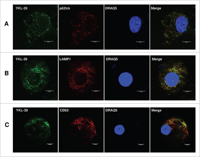
Figure 5.

Intracellular distribution of YKL-39 in human macrophages. Human CD14+ monocytes were stimulated with IL4+TGF-beta for 12 days. YKL-39 was detected with rat mAb 18H10 and anti-rat Alexa488-conjugated secondary antibody (shown in green). Other proteins are visualized in red and nuclear are visualized in blue. Merge of green and red is shown in yellow. (A) Co-localization of YKL-39 and p62lck (late endosomes); (B) Co-localization of YKL-39 and LAMP-1 (lysosomes); (C) Co-localization of YKL-39 and CD63 (secretory lysosomes). Scale bars: 5 µm.
