

Abstract
We report on an incidental detection of a meningioma on [18F]-2′-fluoro-5-methyl-1-beta-D-arabinofuranosyluracil (18F-FMAU) PET/CT scan that was performed during a prospective investigation of 18F-FMAU PET/CT for targeted biopsy of potential sites of tumor in men with suspected prostate cancer based on elevated prostate specific antigen level. Neither prostate multiparamteric MRI nor 18F-FMAU PET/CT localized small volume Gleason 3+3 tumor deposits. However, an incidental focal high accumulation of 18F-FMAU was observed in high right parietal lobe that displayed characteristics of a meningioma on a subsequent brain MRI.
Keywords: Prostate, Cancer, FMAU, PET, CT, meningioma
Figure 1.
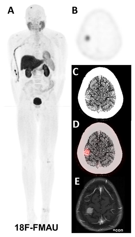
A 69-year-old man with elevated serum prostate-specific antigen (PSA) level of 6.9 ng/mL and suspected prostate cancer underwent a clinical 3T multi-parametric magnetic resonance imaging (MRI) of the prostate gland and an Internal Review Board approved whole body research protocol PET/CT with 18F-FMAU. The maximum intensity projection PET/CT showed normal biodistribution of 18F-FMAU except for a focal intense activity in the right head region (A). Axial 18F-FMAU PET (B), axial CT at brain window level (C) and axial fused PET and CT at brain window level (D) localized the focal tracer accumulation to high right parietal lobe (SUVmax 6.2, mediastinal blood pool SUVmean 0.4) without definite localizing sign on CT alone. A follow-up 3T brain MRI (post-contrast T1-weighted sequence is shown; T1 pre-contrast, T2/FLAIR, GRE, and DTI sequences are not shown) demonstrated homogeneously enhancing, round, 1.5 × 1.4 × 0.9 cm extra-axial mass compatible with a meningioma (E). Meningiomas are the most common nonglial primary brain tumors (1). The mainstay imaging study for diagnosis of meningioma is contrast-enhanced MRI that demonstrates isointense to slightly hypointense signal on T1-weighted, hyperintense signal on T2-weighted and homogeneous post-contrast T1 sequence enhancement relative to normal brain cortex (2). PET/CT is not routinely used in the imaging evaluation of meningiomas. However, meningioma can accumulate a variety of PET radiotracers (e.g. 18F-fluoroodeoxyglucose, 11C- or 18F-labeled amino acids 11C-or 18F-labeled choline, 11C-acetate, 13N-NH3, 68Ga-DOTA-peptides, and radiolabeled prostate-specific membrane antigen agents) (3). 18F-FMAU is a thymidine analog that tracks the thymidine salvage pathway of DNA synthesis and after phosphorylation by thymidine kinase (TK, mitochondrial TK2 > cytoplasmic TK1) is incorporated into the DNA and has been investigated for tumor imaging (4–7). The high accumulation of 18F-FMAU in meningioma may be related to the enhanced TK enzymatic activity in meningiomas (8). With regards to the prostate gland in this case, multi-parametric MRI showed diffusely heterogeneous gland and PET demonstrated diffuse mild tracer localization (not shown). A subsequent prostate biopsy demonstrated low volume tumor deposits in 3 of 15 biopsy specimens (0.5–1.5 mm length, 5–10% tumor, Gleason 3+3). Neither multi-parametric MRI nor 18F-FMAU PET/CT localized these small tumor deposits.
Acknowledgments
Supported by the National Institute of Biomedical Imaging Bioengineering, National Institutes of Health, grant number R21-EB017568 (H. Jadvar).
Footnotes
Conflict of Interest: The authors declare no conflicts of interest.
References
- 1.Kunimatsu A, Kunimatsu N, Kamiya K, et al. Variants of meningiomas: a review of imaging findings and clinical features. Jpn J Radiol. 2016;34:459–469. doi: 10.1007/s11604-016-0550-6. [DOI] [PubMed] [Google Scholar]
- 2.Tamrazi B, Shiroishi MS, Liu C-SJ. Advanced imaging of intracranial meningiomas. Neurosurg Clin N Am. 2016;27:137–143. doi: 10.1016/j.nec.2015.11.004. [DOI] [PMC free article] [PubMed] [Google Scholar]
- 3.Comelius JF, Langen KJ, Stoffels G, et al. Positron emission tomography imaging of meningioma in clinical practice: review of literature and future direction. Neurosurgery. 2012;70:1033–1041. doi: 10.1227/NEU.0b013e31823bcd87. [DOI] [PubMed] [Google Scholar]
- 4.Alauddin MM, Conti PS, Fissekis JD. Synthesis of [18F]-labeled 2′-deoxy-2′-fluoro-5-methyl-1-beta-D-arabinofuranosyluracil ([18F]FMAU) J Labeled Comp Radiopharm. 2002;45:583–590. [Google Scholar]
- 5.Tehrani OS, Muzik O, Heibrun LK, et al. Tumor imaging using 1-(2′-deoxy-2′-18F-fluoro-beta-D-arabinofuranosyl)thymin and PET. J Nucl Med. 2007;48:1436–1441. doi: 10.2967/jnumed.107.042762. [DOI] [PubMed] [Google Scholar]
- 6.Alauddin MM. Journey of 2′-deoxy-2′-fluoro-methyl-1-beta-D-arabinofuranosyluracil (FMAU): from antiviral drug to PET imaging agent. Curr Med Chem. 2018;25:1–12. doi: 10.2174/0929867325666171129125217. [DOI] [PubMed] [Google Scholar]
- 7.Sun H, Mangner TJ, Collins JM, et al. Imaging DNA synthesis in vivo with 18F-FMAU and PET. J Nucl Med. 2005;46:292–296. [PubMed] [Google Scholar]
- 8.Persson L, Gronowitz SJ, Kallander CF. Thymidine kinase in extracts of human brain tumors. Acta Neurochir (Wien) 1986;80:123–127. doi: 10.1007/BF01812286. [DOI] [PubMed] [Google Scholar]