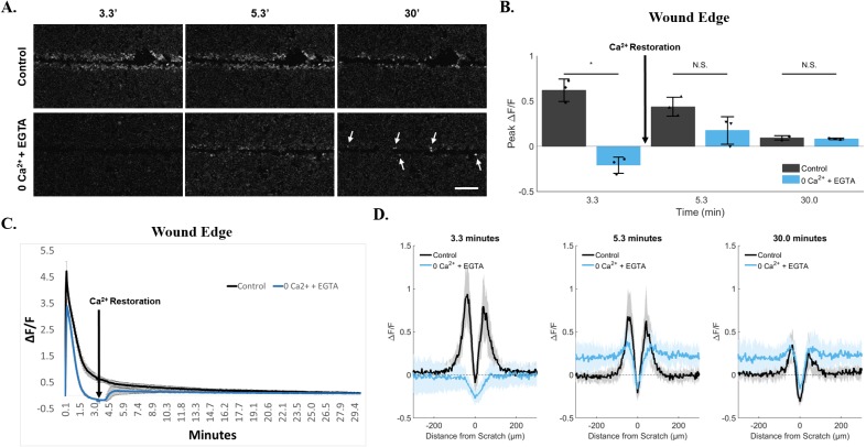
Figure 6. Persistence at the wound edge is regenerated by calcium restoration.
(A) Cells were incubated in 0Ca2+ + EGTA media to deplete external calcium and imaged for 30 minutes. Compared with control, persistent calcium was clearly blocked by 200 seconds (3.3 minutes). Calcium was then replenished in the external media via media exchange initiated at 200 seconds (3.3 minutes). This resulted in a sudden and lasting rescue of persistent calcium in cells at the wound edge (arrows). Data was quantified using both ROI-dependent (B, C) and ROI-independent approaches (D). Importantly, quantification shows a differential peak ΔF/F between cells at the wound edge vs. neighboring cells (D, 0Ca2+ + EGTA at 30 minutes). Data presented as mean ± standard deviation. *indicates significance from control, P < 0.05 via paired t-test. For ROI-based analysis (B, C), data represent N=3 [control: 0.6 ± 0.12 at 3.3min, 0.4 ± 0.11 at 5.3min, 0.09 ± 0.02 at 30min. 0Ca2+ + EGTA: −0.2 ± 0.09 at 3.3min, 0.18 ± 0.15 at 5.3min, 0.08 ± 0.01 at 30min]. For ROI-independent analysis (D), data represent N=9 in total from 3 independent experiments. Scale bar equals 200μm.

