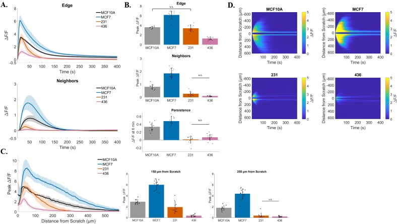
Figure 8. Signaling in response to scratch is disrupted in multiple breast cancer cell lines.
(A) Traces show ΔF/F plotted through time for wound edge cells (upper traces) vs. neighboring cells (lower traces) in human breast MCF10A epithelial cells and human breast MCF-7, MDA-MB-231, and MDA-MB-436 cancer cells. (B) Summary of peak ΔF/F analysis from wound edge cells and neighboring cells, and persistent calcium signaling at 6 minutes, for each cell line tested. Compared with non-tumorigenic breast epithelial cells (MCF10A), the three breast cancer cell lines showed significantly different signaling in response to scratch. MCF-7 cells showed an increased scratch-induced intracellular calcium in both wound edge and neighboring cells, as well as increased persistent calcium signaling. All of these parameters were significantly decreased in MDA-MB-231 and MDA-MB-436, compared with MCF10A (with exception of Edge peak ΔF/F MCF10A vs. 231). (C) Total distance of signal propagation away from the wound edge was plotted for each cell line tested. Traces and bar graphs show that signaling to neighbors is differentially propagated between cell lines. Compared with MCF10A, MDA-MB-231 and MDA-MB-436 cells show a diminished intercellular signaling between neighboring and wound edge cells, while this is enhanced in MCF-7 cells. (D) Kymographs (ROI-independent analysis) reveal similar mechanisms Kymographs (ROI-independent analysis) reveal similar mechanisms. Signaling across the cell monolayer in response to scratch is enhanced in MCF-7 cells compared with MCF10A, while it is severely reduced in MDA-MB-231 and MDA-MB-436 cells. Kymographs also show a reduced long term persistent signaling for MDA-MB-231 and MDA-MB-436 cells. Data presented as mean ± standard deviation. Unless noted by N.S., all values are significantly different with significance set at P < 0.05 via one-way ANOVA with a post-hoc Tukey's honest difference criterion. N.S. indicates no significant difference. Data represent N=10 (MCF10A), N=14 (MCF-7), N=10 (231), N=15 (436) in total from 2 independent experiments for each group.

