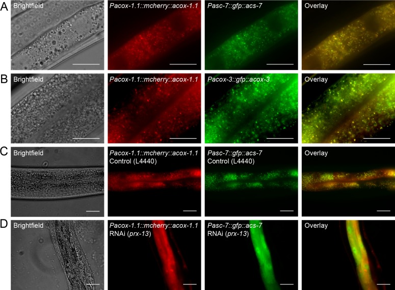
Figure 6. Peroxisomal co-localization of ACS-7, ACOX-1.1, and ACOX-3.
(A) Co-injection of plasmids containing Pacox-1.1::mcherry::acox-1.1 and Pacs-7::gfp::acs-7 into wild-type worms shows that ACOX-1.1 and ACS-7 co-localize in a punctate pattern in the intestine. (B) Co-injection of plasmids containing Pacox-1.1::mcherry::acox-1.1 and Pacox-3::gfp::acox-3 into wild-type worms shows that ACOX-1.1 and ACOX-3 co-localize in a punctate pattern in the intestine. (C) RNAi using the control plasmid L4440 in Pacox-1.1::mcherry::acox-1.1;Pacs-7::gfp::acs-7 worms gives a punctate pattern of expression in the intestine. (D) RNAi against prx-13 in Pacox-1.1::mcherry::acox-1.1; Pacs-7::gfp::acs-7 worms gives a diffuse pattern of expression in the intestine. In (A–D), L4 to young adult stage worms were imaged. Scale bar = 20 μm.


