. 2018 May 9;7:e35439. doi: 10.7554/eLife.35439

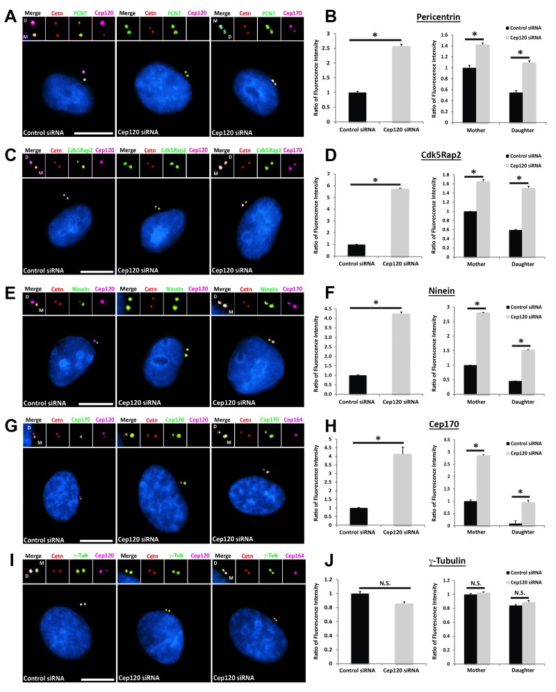
Figure 2. Loss of Cep120 in quiescent cells causes accumulation of PCM components on the daughter centriole.

MEF cells transfected with control or Cep120-targeting siRNA were immunostained for centrin (centrioles), Cep120, Cep164 or Cep170 (to identify the mother centriole), along with the indicated PCM components. Graphs show quantification of the fluorescence intensity for each PCM protein, normalized to control cells. We noted an overall increase in the abundance of PCM proteins at the centrosome in Cep120-depleted cells, with a large increase observed on the daughter centriole. (A–B) Pericentrin: N = 320 (control) and 320 (Cep120) siRNA. (C–D) Cdk5Rap2: N = 400 (control) and 400 (Cep120) siRNA. (E–F) Ninein: N = 430 (control) and 430 (Cep120) siRNA. (G–H) Cep170: N = 480 (control) and 480 (Cep120) siRNA. (I–J) In contrast, there was no significant change in γ-tubulin levels in Cep120-depleted cells. N = 400 (control) and 400 (Cep120) siRNA. Results are averages from three independent experiments; *p<0.05. N.S. = not significant. Scale bars = 10 μm.

Figure 2.

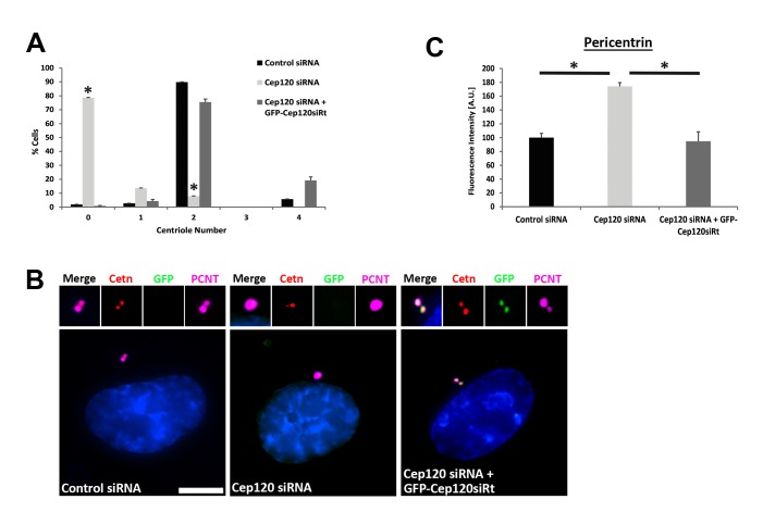
Figure 2—figure supplement 1. – Rescue of centriole duplication and PCM levels following Cep120 depletion.

Figure 2—figure supplement 1.

(A) Quantification of centriole number during the cycling stage of cells transfected with control siRNA, Cep120 siRNA, or Cep120-depleted cells transfected with siRNA-resistant GFPCep120 (GFP-Cep120siRt). Cells were transfected with control or Cep120-siRNA, incubated for 24 hr, then transfected with GFPCep120siRt. Centrioles were counted at 48 hr post-transfection. Loss of Cep120 in dividing cells causes defective centriole duplication, resulting in an increase in the fraction of cells with 0 and 1 centriole. Expression of GFP-Cep120siRt rescues centriole number. N = 420 (control siRNA), 553 (Cep120 siRNA) and 124 (Cep120 siRNA +GFP-Cep120siRt). (B–C) Expression of GFP-Cep120siRt in Cep120-depleted cells restores pericentrin levels at the centrosome. Cells were stained for GFP (to visualize exogenous GFP-Cep120siRt), centrin (centrioles), and pericentrin. Graph shows the relative fluorescence intensity of centrosomal pericentrin. N = 102 (control siRNA), 84 (Cep120 siRNA) and 86 (Cep120 siRNA +GFP-Cep120siRt). Results are averages of two independent experiments; *p<0.05. Scale bar = 10 μm.
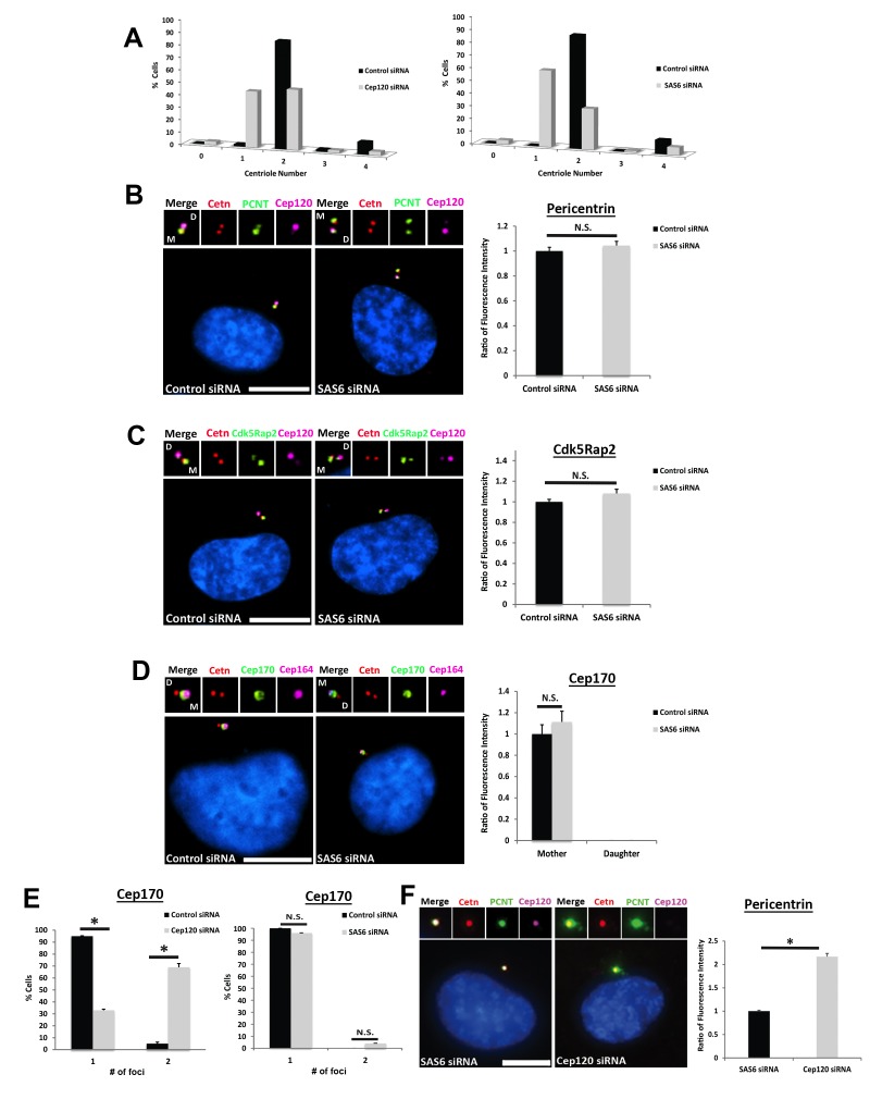
Figure 2—figure supplement 2. – Depletion of Sas6 does not affect PCM levels at the centrosome.

Figure 2—figure supplement 2.

(A) Quantification of centriole number during the cycling stage of control, Cep120- or Sas6-depleted cells. Centrioles were counted at 48 hr post-transfection. Similar to Cep120, loss of Sas6 in dividing cells causes defective centriole duplication, resulting in an increase in the fraction of cells with one centriole. N = 500 (control), 500 (Cep120) and 500 (Sas6) siRNA. (B–C) In contrast to Cep120, depletion of Sas6 did not result in significant change in Pericentrin (B) or Cdk5Rap2 (C) levels at the centrosome. N = 280 (control) and 280 (Sas6) siRNA. (D) Depletion of Sas6 did not alter Cep170 localization or abundance at the centrosome. N = 210 (control) and 210 (Sas6) siRNA. (E) Loss of Cep120 caused accumulation of Cep170 on the daughter centriole, resulting in an increase in the percentage of cells with 2 foci of Cep170. In contrast, depletion of Sas6 did not alter the number of Cep170 foci at centrioles. N = 300 (control), 300 (Cep120) and 300 (Sas6) siRNA. Results are averages of three independent experiments; *p<0.05. N.S. = not significant. Scale bars = 10 μm. (F) Quantification of pericentrin levels in G1 cells containing a single parental daughter centriole. Cells were transfected with siRNA targeting Sas6 or Cep120, incubated for 24 hr, then fixed and stained with antibodies against Cep120, centrin and pericentrin. Graph shows quantification of pericentrin levels in cells containing the single parental centriole with Cep120 (Sas6 siRNA) versus cells lacking Cep120 (Cep120 siRNA). N = 214 (control) and 244 (Sas6) siRNA. Results are averages of two independent experiments; *p<0.05.