

Yuk-Ming Dennis Lo is Professor and Chair of Chemical Pathology at the Chinese University of Hong Kong.
Dennis has transformed the practice of Obstetrics as the father of noninvasive prenatal testing. Having discovered placental/fetal nucleic acids in the maternal circulation in 1997, he worked systematically and artfully to develop a method to assess the fetal genome from maternal blood. This breakthrough, and others, will transform the care of pregnant women in the 21st century. For this, among many other reasons, he is being recognized as a “Giant.”
Early Years: Hong Kong, Cambridge, and Oxford
Dennis was born in Hong Kong when it was still a British colony. His mother, a music teacher, was born in the territory, and his father’s family emigrated to Hong Kong from Chaozhou, a city in the Canton (Guangdong) province on the coast of the South China Sea. For 13 years, Dennis attended St. Joseph’s College, one of the oldest schools in Hong Kong, which was founded by the Jesuits in 1875. Dennis recalled that St. Joseph’s College allowed students great academic freedom to pursue their subjects of interest, and considered this to have been an ideal environment for him to learn and grow.
Dennis was accepted at Stanford University in the United States to study electrical engineering and at the Universities of Hong Kong and Cambridge, United Kingdom, to study medicine. He chose medicine and the next decision was whether to stay in Hong Kong or study abroad – he decided to go to the United Kingdom. Three years later, he moved to the University of Oxford for his medical education, a decision that he said was made, in part, because of the architecture of Sir Christopher Wren, one of the most highly acclaimed English architects in history, whose work includes the Oxford’s Christ Church College, where Dennis became a student.
He graduated with Bachelor of Medicine and Bachelor of Chirurgiae (Latin for “surgery”) degrees (BM, BCh), followed by a DM degree, the latter being equivalent to the MD degree at Oxford. Dennis stayed at Oxford for another seven years after he qualified as a medical doctor. He met his wife Alice, a physicist, at Oxford. In the Nuffield Department of Pathology and Bacteriology at Oxford University, Dennis was a Wellcome Trust Medical Graduate Fellow for four years, and then a University Lecturer in Clinical Biochemistry for three years. It was there in 1996 that Dennis began the groundbreaking research that would change prenatal testing in obstetrical practice. In 1997, he returned to Hong Kong as a Senior Lecturer in Chemical Pathology at The Chinese University of Hong Kong. This was the year in which Hong Kong ceased to be a British colony and was returned to China.
Placental/fetal DNA is present in the maternal circulation: how it all began
Initially, Dennis had used nucleated fetal cells in maternal blood for DNA-based prenatal diagnosis1 and reported bidirectional cell traffic between mother and fetus.2 However, prenatal diagnosis based on DNA amplification from fetal cells in the maternal circulation was hampered by the extremely low concentration of fetal cells and by the fact that some fetal cell populations persist from one pregnancy to the next.3
Dennis decided to look for fetal cell-free DNA in the maternal circulation. What prompted him to undertake this research were two reports indicating that circulating tumor DNA could be detected in the plasma or serum of patients with cancer.4, 5 Reasoning that a pregnancy resembled a very large and rapidly growing “tumor,” Dennis postulated that it should be possible to detect placental/fetal DNA in the mother’s circulation. He used Y-chromosomal DNA as a marker for DNA of a male fetus and demonstrated that fetal cell-free DNA could be detected in maternal plasma. His scientific instincts were right, and depending on whether plasma or serum was used, he was able to detect fetal cell-free DNA in 70–80% of pregnant women; he published his findings in the Lancet in July 1997 (Figure 1).6
FIGURE 1.

The discovery of cell-free fetal DNA in the maternal circulation
Lancet. 1997 Aug 16;350:485–487.6 Reproduced with permission.
A polymerase chain reaction unit for Christmas
Dennis then pursued the question of how early in pregnancy, and at which concentrations, fetal cell-free DNA could be detected in the maternal circulation. To answer these questions, he needed a special polymerase chain reaction (PCR) machine, which at the time had just been developed7 and was very expensive. Still, at a Christmas party near London, Dennis asked Professor Magnus Hjelm, his future Department Head, to purchase a PCR machine, and without hesitation, Professor Hjelm agreed. It was to be the best Christmas present Dennis ever received.
Return to Hong Kong and the systematic study of the kinetics of fetal DNA in maternal blood
Immediately upon his return to Hong Kong, Dennis started to measure the concentrations of cell-free fetal DNA in maternal plasma and serum in a cohort of pregnant women. He found that fetal DNA could be detected as early as seven weeks of gestation and in much higher concentrations than fetal nucleated cells: 3% in early pregnancy and 6% in late pregnancy.8 Dennis then conducted clearance studies to determine if cell-free fetal DNA persisted in the maternal circulation from one pregnancy to the next, as fetal nucleated cells can.9
He first tested blood drawn six weeks after delivery, and finding no fetal cell-free DNA, he progressively shortened the interval from delivery to sample collection to one week and then to one day. Still having found no fetal cell-free DNA, he analyzed blood collected at progressively shorter intervals after cesarean deliveries, and determined that the half-life of cell-free fetal DNA in the maternal circulation was only 16 minutes.9
Given such a short half-life, Dennis calculated that the fetus was delivering about 30,000 genomes per minute into the maternal circulation to maintain the levels of fetal cell-free DNA found during the third trimester of pregnancy. He wondered whether the fetus was sending a signal to the mother by delivering the DNA into her circulation.
Placental/fetal DNA in preeclampsia and preterm labor
Chris Redman, Professor of Obstetric Medicine at Oxford, had shown that trophoblast microparticles could be proinflammatory to the mother,10, 11 which made Dennis wonder if placental fetal DNA might also be “toxic.” So, Dennis measured the concentration of fetal cell-free DNA in mothers with preeclampsia and found that it was 5–10 times higher than in normal pregnancy12 (Figure 2). He also discovered that in women with an episode of preterm labor who went on to have a preterm delivery, the fetal cell-free DNA concentration was twice that of normal pregnancies13 (Figure 3). Both findings were subsequently confirmed by other investigators.14–16 In 2000, Dennis participated in a conference focusing on the maternal-fetal dialogue, hosted by the Perinatology Research Branch of the Eunice Kennedy Shriver National Institute of Child Health and Human Development (NICHD) and chaired by Dr. Diana Bianchi, where he presented what was known about nucleic acids in the maternal circulation and the potential of this line of investigation (Figures 4 and 5).
FIGURE 2.

Preeclampsia is associated with increased concentrations of circulating cell-free fetal DNA
Clin Chem. 1999 Feb; 45:184–188.12 Reproduced with permission.
FIGURE 3.
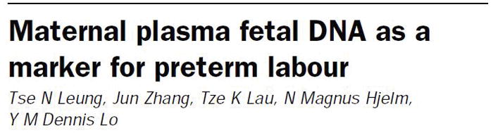
Cell-free fetal DNA concentrations are increased in the maternal circulation in spontaneous preterm labor
Lancet. 1998 Dec 12;352(9144):1904–5. Erratum in: Lancet 1999 Aug 28;354(9180):780.13 Reproduced with permission.
FIGURE 4.
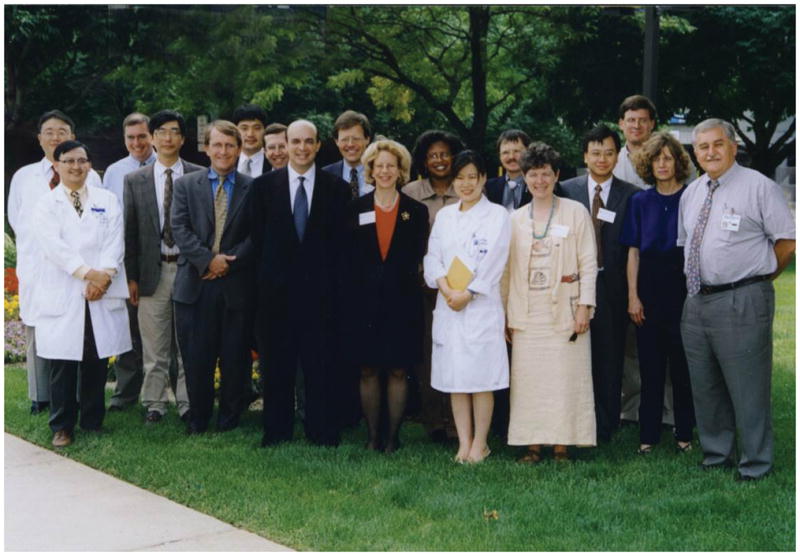
Maternal-Fetal Cell Trafficking Conference at the Perinatology Research Branch, Detroit, MI, in July 2000
Photo courtesy of Dr. Roberto Romero.
FIGURE 5.

Dr Dennis Lo and Dr Diana Bianchi in Hong Kong
Dr Diana Bianchi, Director of the Eunice Kennedy Shriver National Institute of Child Health and Human Development and Editor-in-Chief of Prenatal Diagnosis, with Dr. Dennis Lo at the International Commercial Centre, the tallest building in Hong Kong. Photo courtesy of Dr. Dennis Lo.
Progress toward development of a non-invasive test for fetal aneuploidy
The next step in developing non-invasive prenatal testing (NIPT) was to determine if the concentration of cell-free fetal DNA in the maternal circulation was higher in cases of aneuploidy. In collaboration with Dr. Bianchi (then Professor of Pediatrics and Obstetrics and Gynecology at Tufts University, now Director of NICHD), they found that the concentration of fetal DNA was twice as high in aneuploidy as in euploidy, but the overlap was too high to be practically useful as a screening test.17 This led him look for fetal RNA in the maternal circulation, as fetal cells might express different genes from maternal ones, and gene expression patterns might be different in cases of aneuploidy compared to euploidy.18
Non-invasive determination of fetal rhesus blood group status
An early success in developing NIPT was the determination of fetal rhesus D (RhD) blood group status in mothers who were RhD-negative. Using maternal plasma and real-time PCR, Dennis and his group were able to identify fetal rhesus D blood group status with high accuracy.19, 20 This was the first clinical application of NIPT: it has been available in Europe since 2001, and was adopted in the United States a few years later. This success changed obstetrical practice, as patients who had an RhD-negative fetus no longer needed to undergo serial invasive procedures, such as amniocentesis.
Noninvasive diagnosis of fetal trisomy 21
Subsequently, Dennis sought to develop a noninvasive method for the prenatal diagnosis of fetal chromosomal aneuploidies, starting with trisomy 21. He spent the next ten years using a variety of approaches to try to distinguish fetal from maternal DNA in plasma to accurately measure the increase in the dose of DNA in the circulation of women with a fetus having trisomy 21.
Because DNA from different tissues can be biochemically modified—that is, to undergo epigenetic changes—Dennis reasoned that it might be possible to identify such changes in DNA that were fetus-specific. The most widely studied epigenetic changes involve DNA methylation, and, in 2002, Dennis described a method for detecting fetal DNA in maternal plasma based on DNA methylation.21 Subsequent experiments showed that the technique could be used to detect not only trisomy 21 but also trisomy 18.22
Because different genes are also “switched on and off” in different tissues, Dennis also tried to find genes that were switched “off” in the mother but switched “on” in the fetus. He looked for the products of gene expression in maternal plasma – specifically, messenger RNA (mRNA). Yet, Dennis was surprised to find mRNA in the maternal circulation,23 as mRNA was believed to be fragile; however, it is actually protected because it is packaged within extracellular vesicles and, therefore, could be used in NIPT for trisomy 21.
Although the techniques based on DNA methylation and mRNA were feasible methods for fetal NIPT, they were complex and required additional analysis of the variations in genetic markers that might not work for all fetus-mother combinations. Therefore, Dennis developed yet another precise method to quantify the change in fetal DNA dosage by counting DNA molecules in maternal plasma one at a time.24 He first utilized digital PCR,25 and the following year, employed a then-new technique called massively parallel sequencing, or next-generation sequencing26 (Figure 6). Large-scale validation studies showed that the technology could achieve 100% sensitivity and 98% specificity.27 In 2011, the technology was introduced into clinical practice, and over the next three years, the test was performed in more than 700,000 plasma samples in over 50 countries.
FIGURE 6.

A classic
Proc Natl Acad Sci U S A. 2008 Dec 23; 105:20458–20463.26 Reproduced with permission.
The first human fetal genome characterized from a sample of maternal blood
Dennis and his team continued to push the technological envelope. In 2010, they reported the sequence of an entire fetal genome derived from maternal plasma samples, which opened up the possibility of detecting fetal de novo mutations on a genome-wide scale 28 (Figure 7). After three years, they reported that a fetal epigenome could also be determined from maternal plasma.29 This provides a potentially valuable tool with which to investigate how environmental factors can modify the fetal genome.
FIGURE 7.
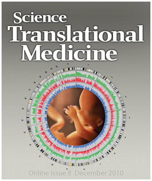
A fetal genome in the maternal circulation
Cover article: Lo YM, Chan KC, Sun H, Chen EZ, Jiang P, Lun FM, Zheng YW, Leung TY, Lau TK, Cantor CR, Chiu RW. Maternal plasma DNA sequencing reveals the genome-wide genetic and mutational profile of the fetus. Sci Transl Med. 2010 Dec 8; 2(61):61ra91.28 Reproduced with permission.
Equally (if not more) exciting is what might be learned using transcriptomics, made possible by the ability to study fetal RNA in the maternal circulation. Thus, Dennis developed a noninvasive approach to profile the transcriptome of the human fetus. This methodology is beset with technical challenges, as plasma RNA is unstable, and “the techniques used to stabilize it add considerable complexity to the pre-analytic side of the issue”, but the technique allows examination of fetal gene expression. This has the potential of allowing characterization of the maternal-fetal dialogue.
Single-cell transcriptomics of the placenta and in patients with preeclampsia
An important step forward is the use of large-scale microfluidic single-cell transcriptomic technology to comprehensively characterize cellular heterogeneity of the human placenta. This technology allowed Dennis’s team to identify the cellular composition of human placentas based on their transcriptional signatures. Their publication in the Proceedings of the National Academy of Science in 2017 reported the delineation of the cellular dynamics during normal pregnancy and the dysfunction identified in the placentas of patients with preeclampsia.30 Moreover, this work demonstrated that the changes observed in the human placenta at delivery could be detected in cell-free plasma RNA of the mother. This enables noninvasive elucidation of the cellular dynamics of pregnancy complications.30
Screening for Epstein-Barr virus and nasopharyngeal carcinoma
Besides prenatal diagnosis, the biology of cancer is of major interest to Dennis—specifically, the use of cell-free DNA detected in the plasma of patients with cancer for guiding treatment selection, monitoring response to treatment, and detecting residual disease. Most tumor markers are proteins, such as prostate-specific antigen for prostate cancer or alpha feto-protein for hepatocellular carcinoma. However, tumors release tumor DNA into the circulation, making detection of subclinical cancers possible.
Dennis was inducted as a member of the National Academy of Sciences in the United States in 2013, and his inaugural article in the Proceedings was entitled “Non-invasive detection of cancer-associated genome-wide hypomethylation and copy number aberrations by plasma DNA bisulfite sequencing.”31 The key concept was that DNA changes observed in cancer cells could be detected in plasma by analysis of cell-free DNA. This approach has been used in many types of cancer, including breast, lung, sarcomas, and neuroendocrine tumors. Dennis is optimistic that this may lead to a screening method to detect cancers with high sensitivity and high specificity.
A recent contribution of Dennis’s team to this story was published in August 2017.32 Nasopharyngeal carcinoma is prevalent in Southeast Asia: 35 cases per 100,000 individuals among middle-aged men in the highest incidence areas of south China. The Epstein-Barr virus has been implicated in the pathogenesis of this cancer. The Epstein-Barr virus DNA in plasma is released by nasopharyngeal carcinoma cells as cell-free DNA fragments, rather than as intact viral genomes contained inside viral particles. Thus, Dennis and his team tested whether cell-free Epstein-Barr virus DNA could be used as a screening test for nasopharyngeal carcinoma in asymptomatic individuals. They screened more than 20,000 subjects, of whom 309 had persistently positive tests. The sensitivity and specificity of Epstein-Barr virus cell-free DNA in plasma samples for the detection of asymptomatic nasopharyngeal carcinoma were 97% and 98%, respectively. Importantly, this allowed early detection of these cancers, and the patients with cancer identified following screening had an improved progression-free survival.
Mentors
Dennis recalled that Dr. Yuet Wai Kan, a scientist from Hong Kong and a medical school classmate of Dennis’s father, was an early and lasting influence. Dr. Kan, a Lasker Award winner, is now Professor of Hematology at University of California, San Francisco, and he established that a mutation could be responsible for human disease (thalassemia and sickle cell anemia).33 During medical school, Dennis attended a lecture by Sir John Bell, now the Regius Professor of Medicine at Oxford, who was one of the first to use PCR. Dennis asked Professor Bell to teach him how to do PCR and he graciously agreed. Dennis said that learning this technique changed his life. Another mentor was Dr. Kenneth Fleming, the former Head of the Medical Sciences Division at Oxford, who gave Dennis his first opportunity to do laboratory-based research (Figure 8).
FIGURE 8.
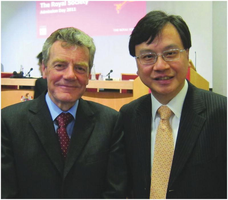
Dr Kenneth Fleming and Dr Dennis Lo
Photograph: http://clinchem.aaccjnls.org/content/61/1/32. Clinical Chemistry. 2015; 61(1):32–37. Reproduced with permission.
A career in academic medicine
Dennis first became interested in an academic career through his father, who was a psychiatrist in charge of Hong Kong’s public psychiatric service. He often traveled to international conferences, and sometimes rehearsed his talks in front of Dennis and his brother. “His career inspired me to become an academician,” Dennis said.
I asked Dennis how he managed to combine research with his responsibilities as chair of a department, associate dean of research, and head of a research institute. “Leading a group, and being an inventor or even an entrepreneur, are all, in a way, the same thing, in that they involve transforming a vision in my mind into something real,” he said. “My responsibility as the chair of my department, institution, and [as] associate dean of research, is to build an environment where one can innovate, and that is also my interest: to make sure the university is as conducive to innovation as possible.”
Dennis explained that The Chinese University of Hong Kong, which opened in 1963 and has 20,000 students, is a very friendly place that has a collegiate system rather like Oxford and Cambridge. “Maybe that is one of the reasons why most of the Rhodes Scholars from Hong Kong are graduates of The Chinese University.”
Awards and recognition
Dennis has received numerous prestigious awards and recognitions, some of which are particularly noteworthy. In 2011, Dennis was inducted as a Fellow of the Royal Society, the world’s oldest scientific society, founded in 1660 with the motto Nullius in verba (“take nobody’s word for it”). Fellowship in the Royal Society is given to individuals deemed to have made substantial contributions to the improvement of natural knowledge, and Dennis’s fellowship in this society places him on the company of Christopher Wren (founding member), Isaac Newton, Charles Darwin, Francis Crick, and Stephen Hawking.34
In 2014, Dennis received the King Faisal International Prize for Medicine for his work in the noninvasive diagnosis of fetal diseases (Figure 9). In 2016, Dennis received the Thomson Reuters Citation Laureate Award in Chemistry. This honor is given to scientists who rank in the top 0.1% of citations in their research area and whose contributions are considered to have had “unusually strong influence in the scientific community.”35 Thomson Reuters takes pride in that this award is often a precursor to the Nobel Prize. Also in 2016, Dennis was awarded the first Future Science Prize in Life Science, the Chinese equivalent of the Nobel Prize, for developing NIPT. But all of this is only part of the story: Dennis has dozens of patents, many of which have been licensed and led to the application of his discoveries to improve the understanding of pregnancy, cancer, and monitoring organ transplantation.
FIGURE 9.
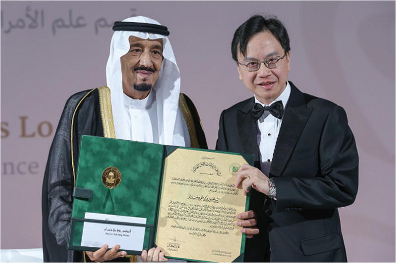
King Salman, then Crown Prince, of Saudi Arabia and Dr Dennis Lo in Riyadh, Saudi Arabia, on January 14, 2014
Dr Dennis Lo receives the King Faisal International Prize for Medicine for his work in the noninvasive diagnosis of fetal diseases. Photograph: https://kfip.org/professor-yuk-ming-dennis-lo/.
Hobbies
Photography, which has intrigued him since early childhood, is Dennis’s main hobby, and he remembers taking photographs and making slides for his father when he traveled to international conferences. Dennis had a dark room back then, and processing color photographs was a very complicated process that, to him, was like an experiment. Dennis considers himself a “technological geek” and is fascinated with digital cameras – he has moved on to a brand that takes 42-megapixel photographs and now prefers Adobe Photoshop to the chemicals of the darkroom.
Movies
Dennis also loves the movies, especially science fiction, as he feels transported into another world. “That’s where I can relax and let my imagination run wild,” he explained, noting that scientists often have their best ideas when they are completely relaxed. He found the Alien series very interesting, even “life-changing.” When he was considering becoming a pathologist, he saw Alien 3, which opens with an autopsy. Dennis told me that he was considering anatomical pathology at the time and thought, “do I really want to start my day with that…?”
Famously, there is another movie that influenced Dennis’s work. He had been immersed in thinking about how to approach the sequencing of the fetal genome when he and his wife, Alice, saw Harry Potter and the Half-Blood Prince in IMAX/3D. When the title flashed across the screen, the “H” looked to him like two chromosomes, and Dennis realized that for each pair of chromosomes, there is a maternal and a paternal copy. He thought he could develop separate algorithms to look for each copy, and said that it was quite incredible—the realization came within minutes after he saw the “H.” This was the basis for deducing the fetal genome using a combination of genotype data from the father and haplotype information from the mother.28
His favorite books are also about science. He recalls reading James Watson’s The Double Helix: A Personal Account of the Discovery of the Structure of DNA when he was quite young. The book advises junior scientists on how to compete with more established, renowned scientists, and suggests that “if your ideas are good enough, you might be able to win.”
This is a very good time to enter the field
Dennis considers the 21st century to be the “golden age of molecular medicine” – the tools and technology available are extraordinary. Dennis told me that “in the future, we will have noninvasive maternal and fetal genomes, a circulating epigenome, and a circulating transcriptome – but knowing how to leverage these breakthroughs to improve diagnosis and treatment will still be a challenge.” Dennis predicts that the discoveries in medicine that will be made in the next few decades will be more significant than all the discoveries in the last 2000 years.
Gratitude and loyalty
Dennis is particularly grateful to his longtime collaborators Rossa Chiu, Allen Chan, and others in his group for their long-standing partnership that have made so many breakthroughs possible (Figure 10). He has had a close relationship with the Department of Obstetrics and Gynecology of the Chinese University of Hong Kong, particularly with Professor T.Y. Leung, Chair of that Department, with whom he has published extensively on the applications of cell-free DNA for prenatal diagnosis and other conditions, such as the detection of fetomaternal hemorrhage,36 noninvasive assessment of the zygosity in twin gestations,37 and recently on the feasibility of gestational age determination by assessment of methylation and size profiling of maternal plasma DNA.38
FIGURE 10.
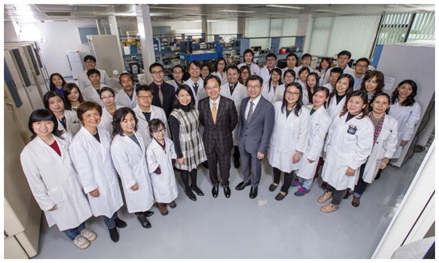
Dr Dennis Lo and his colleagues at the Chinese University of Hong Kong
Photograph:
Credit: Courtesy of Dr. Dennis Lo.
A Giant
Dennis Lo is a Professor of Medicine and Chemical Pathology – not an obstetrician or a gynecologist by training. He is a “Giant” in our discipline because his discoveries, inventions, and innovations have changed the practice of obstetrics, and promise to change the understanding of pregnancy, development, fetal growth, and the maternal-fetal dialogue, as well as improve the assessment of so many aspects of obstetrics. He has already done this by pioneering the noninvasive diagnosis of fetal Rh-D status and aneuploidy, the detection of fetomaternal hemorrhage, providing the first fetal human genome derived from a sample of maternal blood, a fetal methylome, and the fetal transcriptome. His contributions to the early diagnosis of cancer with cell-free DNA have far-reaching implications for obstetrics and gynecology, and medicine in general. We look forward to what else Dennis will discover in the years to come.
PORTRAIT.

Dr. Dennis Lo
Footnotes
The author reports no conflict of interest.
References
- 1.Lo YM, Patel P, Wainscoat JS, Sampietro M, Gillmer MD, Fleming KA. Prenatal sex determination by DNA amplification from maternal peripheral blood. Lancet. 1989;2:1363–5. doi: 10.1016/s0140-6736(89)91969-7. [DOI] [PubMed] [Google Scholar]
- 2.Lo YM, Lo ES, Watson N, Noakes L, Sargent IL, Thilaganathan B, et al. Two-way cell traffic between mother and fetus: biologic and clinical implications. Blood. 1996;88:4390–5. [PubMed] [Google Scholar]
- 3.Bianchi DW, Zickwolf GK, Weil GJ, Sylvester S, Demaria MA. Male fetal progenitor cells persist in maternal blood for as long as 27 years postpartum. Proc Natl Acad Sci U S A. 1996;93:705–8. doi: 10.1073/pnas.93.2.705. [DOI] [PMC free article] [PubMed] [Google Scholar]
- 4.Stroun M, Anker P, Maurice P, Lyautey J, Lederrey C, Beljanski M. Neoplastic characteristics of the DNA found in the plasma of cancer patients. Oncology. 1989;46:318–22. doi: 10.1159/000226740. [DOI] [PubMed] [Google Scholar]
- 5.Chen XQ, Stroun M, Magnenat JL, Nicod LP, Kurt AM, Lyautey J, et al. Microsatellite alterations in plasma DNA of small cell lung cancer patients. Nat Med. 1996;2:1033–5. doi: 10.1038/nm0996-1033. [DOI] [PubMed] [Google Scholar]
- 6.Lo YM, Corbetta N, Chamberlain PF, Rai V, Sargent IL, Redman CW, et al. Presence of fetal DNA in maternal plasma and serum. Lancet. 1997;350:485–7. doi: 10.1016/S0140-6736(97)02174-0. [DOI] [PubMed] [Google Scholar]
- 7.Heid CA, Stevens J, Livak KJ, Williams PM. Real time quantitative PCR. Genome Res. 1996;6:986–94. doi: 10.1101/gr.6.10.986. [DOI] [PubMed] [Google Scholar]
- 8.Lo YM, Tein MS, Lau TK, Haines CJ, Leung TN, Poon PM, et al. Quantitative analysis of fetal DNA in maternal plasma and serum: implications for noninvasive prenatal diagnosis. Am J Hum Genet. 1998;62:768–75. doi: 10.1086/301800. [DOI] [PMC free article] [PubMed] [Google Scholar]
- 9.Lo YM, Zhang J, Leung TN, Lau TK, Chang AM, Hjelm NM. Rapid clearance of fetal DNA from maternal plasma. Am J Hum Genet. 1999;64:218–24. doi: 10.1086/302205. [DOI] [PMC free article] [PubMed] [Google Scholar]
- 10.Smarason AK, Sargent IL, Starkey PM, Redman CW. The effect of placental syncytiotrophoblast microvillous membranes from normal and pre-eclamptic women on the growth of endothelial cells in vitro. Br J Obstet Gynaecol. 1993;100:943–9. doi: 10.1111/j.1471-0528.1993.tb15114.x. [DOI] [PubMed] [Google Scholar]
- 11.Smarason AK, Sargent IL, Redman CW. Endothelial cell proliferation is suppressed by plasma but not serum from women with preeclampsia. Am J Obstet Gynecol. 1996;174:787–93. doi: 10.1016/s0002-9378(96)70466-0. [DOI] [PubMed] [Google Scholar]
- 12.Lo YM, Leung TN, Tein MS, Sargent IL, Zhang J, Lau TK, et al. Quantitative abnormalities of fetal DNA in maternal serum in preeclampsia. Clin Chem. 1999;45:184–8. [PubMed] [Google Scholar]
- 13.Leung TN, Zhang J, Lau TK, Hjelm NM, Lo YM. Maternal plasma fetal DNA as a marker for preterm labour. Lancet. 1998;352:1904–5. doi: 10.1016/S0140-6736(05)60395-9. [DOI] [PubMed] [Google Scholar]
- 14.Farina A, Leshane ES, Romero R, Gomez R, Chaiworapongsa T, Rizzo N, et al. High levels of fetal cell-free DNA in maternal serum: a risk factor for spontaneous preterm delivery. Am J Obstet Gynecol. 2005;193:421–5. doi: 10.1016/j.ajog.2004.12.023. [DOI] [PubMed] [Google Scholar]
- 15.Levine RJ, Qian C, Leshane ES, Yu KF, England LJ, Schisterman EF, et al. Two-stage elevation of cell-free fetal DNA in maternal sera before onset of preeclampsia. Am J Obstet Gynecol. 2004;190:707–13. doi: 10.1016/j.ajog.2003.12.019. [DOI] [PubMed] [Google Scholar]
- 16.Bianchi DW, Romero R. Biological implications of bi-directional fetomaternal cell traffic: a summary of a National Institute of Child Health and Human Development-sponsored conference. J Matern Fetal Neonatal Med. 2003;14:123–9. doi: 10.1080/jmf.14.2.123.129. [DOI] [PubMed] [Google Scholar]
- 17.Lo YM, Lau TK, Zhang J, Leung TN, Chang AM, Hjelm NM, et al. Increased fetal DNA concentrations in the plasma of pregnant women carrying fetuses with trisomy 21. Clin Chem. 1999;45:1747–51. [PubMed] [Google Scholar]
- 18.Lo YM, Tsui NB, Chiu RW, Lau TK, Leung TN, Heung MM, et al. Plasma placental RNA allelic ratio permits noninvasive prenatal chromosomal aneuploidy detection. Nat Med. 2007;13:218–23. doi: 10.1038/nm1530. [DOI] [PubMed] [Google Scholar]
- 19.Lo YM, Bowell PJ, Selinger M, Mackenzie IZ, Chamberlain P, Gillmer MD, et al. Prenatal determination of fetal RhD status by analysis of peripheral blood of rhesus negative mothers. Lancet. 1993;341:1147–8. doi: 10.1016/0140-6736(93)93161-s. [DOI] [PubMed] [Google Scholar]
- 20.Lo YM, Hjelm NM, Fidler C, Sargent IL, Murphy MF, Chamberlain PF, et al. Prenatal diagnosis of fetal RhD status by molecular analysis of maternal plasma. N Engl J Med. 1998;339:1734–8. doi: 10.1056/NEJM199812103392402. [DOI] [PubMed] [Google Scholar]
- 21.Poon LL, Leung TN, Lau TK, Chow KC, Lo YM. Differential DNA methylation between fetus and mother as a strategy for detecting fetal DNA in maternal plasma. Clin Chem. 2002;48:35–41. [PubMed] [Google Scholar]
- 22.Tong YK, Ding C, Chiu RW, Gerovassili A, Chim SS, Leung TY, et al. Noninvasive prenatal detection of fetal trisomy 18 by epigenetic allelic ratio analysis in maternal plasma: Theoretical and empirical considerations. Clin Chem. 2006;52:2194–202. doi: 10.1373/clinchem.2006.076851. [DOI] [PubMed] [Google Scholar]
- 23.Poon LL, Leung TN, Lau TK, Lo YM. Presence of fetal RNA in maternal plasma. Clin Chem. 2000;46:1832–4. [PubMed] [Google Scholar]
- 24.Chiu RW, Cantor CR, Lo YM. Non-invasive prenatal diagnosis by single molecule counting technologies. Trends Genet. 2009;25:324–31. doi: 10.1016/j.tig.2009.05.004. [DOI] [PubMed] [Google Scholar]
- 25.Lo YM, Lun FM, Chan KC, Tsui NB, Chong KC, Lau TK, et al. Digital PCR for the molecular detection of fetal chromosomal aneuploidy. Proc Natl Acad Sci U S A. 2007;104:13116–21. doi: 10.1073/pnas.0705765104. [DOI] [PMC free article] [PubMed] [Google Scholar]
- 26.Chiu RW, Chan KC, Gao Y, Lau VY, Zheng W, Leung TY, et al. Noninvasive prenatal diagnosis of fetal chromosomal aneuploidy by massively parallel genomic sequencing of DNA in maternal plasma. Proc Natl Acad Sci U S A. 2008;105:20458–63. doi: 10.1073/pnas.0810641105. [DOI] [PMC free article] [PubMed] [Google Scholar]
- 27.Chiu RW, Akolekar R, Zheng YW, Leung TY, Sun H, Chan KC, et al. Non-invasive prenatal assessment of trisomy 21 by multiplexed maternal plasma DNA sequencing: large scale validity study. Bmj. 2011;342:c7401. doi: 10.1136/bmj.c7401. [DOI] [PMC free article] [PubMed] [Google Scholar]
- 28.Lo YM, Chan KC, Sun H, Chen EZ, Jiang P, Lun FM, et al. Maternal plasma DNA sequencing reveals the genome-wide genetic and mutational profile of the fetus. Sci Transl Med. 2010;2:61ra91. doi: 10.1126/scitranslmed.3001720. [DOI] [PubMed] [Google Scholar]
- 29.Lun FM, Chiu RW, Sun K, Leung TY, Jiang P, Chan KC, et al. Noninvasive prenatal methylomic analysis by genomewide bisulfite sequencing of maternal plasma DNA. Clin Chem. 2013;59:1583–94. doi: 10.1373/clinchem.2013.212274. [DOI] [PubMed] [Google Scholar]
- 30.Tsang JCH, Vong JSL, Ji L, Poon LCY, Jiang P, Lui KO, et al. Integrative single-cell and cell-free plasma RNA transcriptomics elucidates placental cellular dynamics. Proc Natl Acad Sci U S A. 2017;114:E7786–e95. doi: 10.1073/pnas.1710470114. [DOI] [PMC free article] [PubMed] [Google Scholar]
- 31.Chan KC, Jiang P, Chan CW, Sun K, Wong J, Hui EP, et al. Noninvasive detection of cancer-associated genome-wide hypomethylation and copy number aberrations by plasma DNA bisulfite sequencing. Proc Natl Acad Sci U S A. 2013;110:18761–8. doi: 10.1073/pnas.1313995110. [DOI] [PMC free article] [PubMed] [Google Scholar]
- 32.Chan KCA, Woo JKS, King A, Zee BCY, Lam WKJ, Chan SL, et al. Analysis of Plasma Epstein-Barr Virus DNA to Screen for Nasopharyngeal Cancer. N Engl J Med. 2017;377:513–22. doi: 10.1056/NEJMoa1701717. [DOI] [PubMed] [Google Scholar]
- 33.Kan YW. Development of DNA analysis for human diseases. Sickle cell anemia and thalassemia as a paradigm. Jama. 1992;267:1532–6. [PubMed] [Google Scholar]
- 34.Faye-Petersen OM, Heller DS, Joshi VV. Handbook of placental pathology. New York: Taylor and Francis group; Number of pages. [Google Scholar]
- 35.Regnault TR, Galan HL, Parker TA, Anthony RV. Placental development in normal and compromised pregnancies--a review. Placenta. 2002;23(Suppl A):S119–29. doi: 10.1053/plac.2002.0792. [DOI] [PubMed] [Google Scholar]
- 36.Lau TK, Lo KW, Chan LY, Leung TY, Lo YM. Cell-free fetal deoxyribonucleic acid in maternal circulation as a marker of fetal-maternal hemorrhage in patients undergoing external cephalic version near term. Am J Obstet Gynecol. 2000;183:712–6. doi: 10.1067/mob.2000.106582. [DOI] [PubMed] [Google Scholar]
- 37.Leung TY, Qu JZ, Liao GJ, Jiang P, Cheng YK, Chan KC, et al. Noninvasive twin zygosity assessment and aneuploidy detection by maternal plasma DNA sequencing. Prenat Diagn. 2013;33:675–81. doi: 10.1002/pd.4132. [DOI] [PubMed] [Google Scholar]
- 38.Jiang P, Tong YK, Sun K, Cheng SH, Leung TY, Chan KC, et al. Gestational Age Assessment by Methylation and Size Profiling of Maternal Plasma DNA: A Feasibility Study. Clin Chem. 2017;63:606–08. doi: 10.1373/clinchem.2016.265702. [DOI] [PubMed] [Google Scholar]