Abstract
Background
Sciatic nerve injuries following total hip arthroplasty are disabling complications. Although degrees of injury are variable from neuropraxia to neurotmesis, mechanical irritation of sciatic nerve might be occurred by protruding hardware. This case shows endoscopic decompression for protruded acetabular screw irritating sciatic nerve, the techniques described herein may permit broader arthroscopic/endoscopic applications for management of complications after reconstructive hip surgery.
Case presentation
An 80-year-old man complained of severe pain and paresthesias following acetabular component revision surgery. Physical findings included right buttock pain with radiating pain to lower extremity. Radiographs and computed tomography imaging showed that the sharp end of protruded screw invaded greater sciatic foramen anterior to posterior and distal to proximal direction at sciatic notch level. A protruding tip of the acetabular screw at the sciatic notch was decompressed by use of techniques gained from experience performing endoscopic sciatic nerve decompression. The pre-operative pain and paresthesias resolved post-operatively after recovering from anesthesia.
Conclusions
This case report describes the first documented endoscopic resection of the tip of the acetabular screw irritating sciatic nerve after total hip arthroplasty. If endoscopic resection of an offending acetabular screw can be performed in a safe and minimally invasive manner, one can envision a future expansion of the role of hip arthroscopic surgery in several complications management after total hip arthroplasty.
Electronic supplementary material
The online version of this article (10.1186/s12891-018-2091-x) contains supplementary material, which is available to authorized users.
Keywords: Endoscopic sciatic nerve decompression, Sciatic nerve neuropathy, Acetabular dome screw, Total hip arthroplasty
Background
Sciatic nerve irritation due to an acetabular screw following total hip arthroplasty (THA) is rare; the few reported were treated with revision procedure to remove an acetabular screw [1]. Recent reports of the orthopaedic literature document the use of endoscopic decompression of sciatic nerve entrapment syndrome/deep gluteal syndrome treatment [2]. Endoscopy allows for complete extrapelvic sciatic nerve visualization and safe nerve decompression in the deep gluteal space [3, 4]. This is the first documented case of completely endoscopic treatment of sciatic nerve irritation by a protruded acetabular dome screw following THA.
Case presentation
An 80-year-old man complained of severe pain and paresthesias following acetabular component revision surgery via the original posterolateral approach. The patient subsequently complained of right leg pain and paresthesias. The symptoms were exaggerated during initial hip flexion and internal rotation that limited ambulation. Physical findings included right buttock pain with radiating pain to the ipsilateral lower extremity. Radiographs and computed tomography imaging (Figs. 1, 2, 3, and 4) showed an acetabular screw tip protruding into the greater sciatic foramen in an anterior to posterior and distal to proximal direction at the level of the sciatic notch. The screw was 30 mm in length and positioned at posterior-inferior acetabular quadrant. Magnetic resonance imaging demonstrated tenting of the right piriformis muscle tented by the end of screw. Mechanical irritation of the sciatic nerve screw was suspected.
Fig. 1.
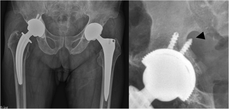
Preoperative hip radiographs (anteroposterior view; Left, iliac oblique view; Right) with protruded dome screw (arrowhead) into sciatic notch
Fig. 2.
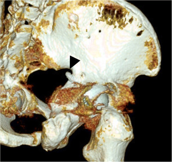
Oblique 3-dimensional computed tomography reconstruction showing an acetabular screw protruded into sciatic notch (arrowhead)
Fig. 3.
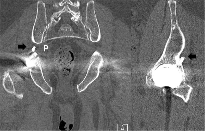
Coronal and computed tomography views of right hip showing sciatic notch area occupied by an acetabular screw (arrow). The protruded screw was placed superiorly than piriformis muscle (P)
Fig. 4.
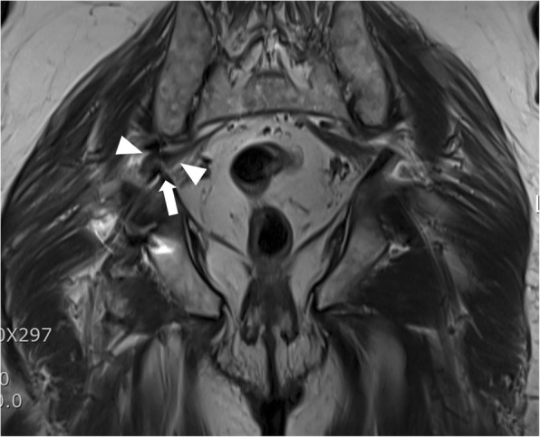
T2 turbo spin echo magnetic resonance image displaying a screw protrusion (arrowhead) and irritated piriformis muscle (arrow)
Eight months after the aforementioned surgery, the patient underwent supine hip arthroscopy without distraction on a fracture table (Hana®, Mizuho OSI). The operating table was tilted right side upward to increase accessibility of ipsilateral buttock. An anterolateral viewing portal and poster lateral working portal were developed without incident. For deep gluteal space visualization, a 70-degree high definition long arthroscope with adjustable length cannulas were utilized. The cannula was opened to maintain the fluid flow when utilizing the radiofrequency probe. Fluid pressure was set to 60 mmHg with intermittent pressure increases up 80 mmHg. After endoscopic greater trochanteric bursectomy, the repaired piriformis from previous revision surgery was well visualized (Fig. 5). Endoscopic visualization of the sciatic nerve revealed a hypo vascular appearance, with entrapped by repaired the piriformis muscle and adjacent fibrous tissue. Endoscopic dynamic testing demonstrated sciatic nerve hypomobility with limited excursion during hip flexion and extension in internal and external rotation. The repaired piriformis tendon was tenotomized and more adjacent scar tissue was observed. Endoscopic resection of fibrovascular bands and adhesions between the piriformis muscle and posterior acetabular wall was performed without incident, allowing visualization of the protruding screw tip penetrated through the piriformis muscle (Fig. 6). Endoscopic piriformis muscle dissection and adhesiolysis was performed, improving visualization of the screw tip adjacent to the supero-lateral aspect of the sciatic nerve. No sciatic nerve intra-substance splitting or tearing was observed, supporting the diagnosis of sciatic nerve irritation without direct nerve injury. Via endoscopic and intermittent multiplanar fluoroscopic visualization, further dissection was performed proximally and distally to prevent inadvertent injuries to the sciatic nerve and the superior gluteal neurovascular bundle. Partial osteoplasty of the sciatic notch with a motorized burr performed under endoscopic guidance. Endoscopic resection of the offending screw tip was performed with a 5.5 mm motorized round burr (Fig. 7). The adjustable cannula protected the adjacent superior gluteal vessels from iatrogenic harm. The screw was scored from the superolateral direction to minimize the risk of unexpected injury by the burr after decorticating the sciatic notch. The protruded screw tip was completely resected and subsequently recontoured to a smooth surface. The screw tip was removed enbloc with an arthroscopic grasper (Fig. 8). Generated metallic debris were removed via suction through the burr cannula. All visible metallic debris was removed. Dynamic arthroscopic and fluoroscopic examinations confirmed successful decompression of the acetabular screw with an immediate visible improvement in sciatic nerve vascularity and excursion. Key procedural steps of this arthroscopic procedure are shown as Additional file 1: Endoscopic technique.
Fig. 5.
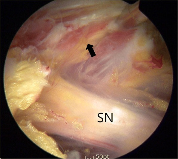
Endoscopic view from anterolateral portal of right hip (supine, upward tilt ipsilateral hip) showing repaired piriformis in previous surgery (arrow). Sciatic nerve (SN) is pale and loss of perineural fat. It is better visualized in the accompanying video
Fig. 6.
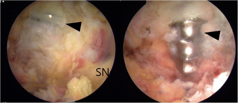
Endoscopic view from anterolateral portal of right hip (supine, ipsilateral upward tilt) showing screw tip (arrow) protruding from posterior column into sciatic notch being irritated sciatic nerve (SN)
Fig. 7.
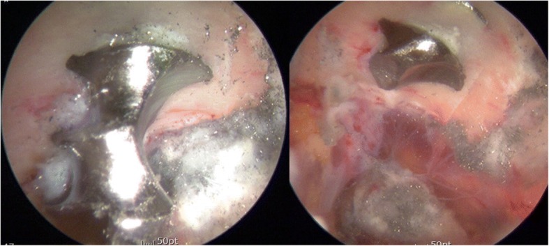
Endoscopic view from anterolateral portal of right hip (supine) showing an acetabular screw being cut proximally by arthroscopic burr
Fig. 8.
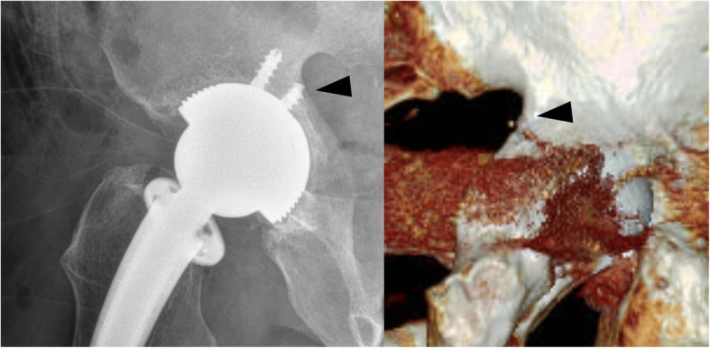
Postoperative radiography and 3-dimensional computed tomography reconstruction showing the resected acetabular screw at sciatic notch level
After this endoscopic surgery, immediate hip range of motion was begun with a continuous passive motion machine. The pre-operative pain and paresthesias resolved post-operatively after recovering from anesthesia. The patient advanced to household ambulation without upper extremity aids at 1 postoperative week despite recommendations for 2 weeks of ambulation using crutches. By 3 months postoperatively, he had returned to full activities. At 6 months postoperatively, he had a negative active and passive piriformis test and he remained pleased with the outcome. Postoperative imaging included radiographs and computed tomography scans with 3-dimensional reconstruction demonstrating complete resection of the offending portion of the acetabular screw.
Discussion
Nerve lesions following total hip arthroplasty (THA) are disabling complications occurring in 0.06 to 2.2% of arthroplasties [5, 6]. Hardware-induced sciatic nerve neuropathies are rarely reported [1, 6]. If placed in the posterior superior quadrant, the screws may be directed toward the sciatic nerve [7]. Mechanical irritation should be suspected in cases of any sign of sciatic neuropathy after THA. The treatment of nerve injuries is tailored its causation. In most cases, the cause is unknown and treatment is directed toward managing symptoms rather than reversing the nerve injury [8]. Open sciatic nerve exploration with dissection of nerve bundle and burr-decompression of the screw has been reported [9]. A protruding acetabular screw can tether the sciatic nerve, restricting its excursion. Endoscopic resection of an offending acetabular screw has now been described. Indeed, had this protruded screw not been resectable by endoscopic means, open exploration and screw removal or resection may have been required. Some advantages of deep gluteal space endoscopy as a minimally invasive procedure are demonstrated in this case report. Decreasing morbidity with minimal blood loss while avoiding re-revision surgery with hip dislocation was facilitated. Moreover, early joint mobilization, relatively rapid postoperative rehabilitation, and outstanding cosmesis were realized. Surgical skills gained from experience with endoscopic technique for deep gluteal syndrome exploration and comfort with the 70 degree arthroscope aided the performance of this surgery. Careful management of endoscopic fluid pressure minimized risk of iatrogenic fluid extravasation into the intra-abdominal and/or retroperitoneal spaces.
An expandable cannula facilitated safe resection of the protruding screw tip while avoiding injury to the adjacent superior gluteal nerve and sciatic nerve. Circulating endoscopy fluid might decrease thermogenesis during metal-on-metal burring of the offending screw. In addition, generated metallic debris is removed immediately through suction system attached burr and cannula to minimize bodily retention with possible adverse consequences.
The decision to remove rather than try to preserve the piriformis muscle was influenced by the surgeon’s experience with deep gluteal space exploration often performed for sciatic nerve decompression.
Relevant suggestions and pearls are summarized in Table 1.
Table 1.
Pearls for endoscopic resection of protruded acetabular screw irritating sciatic nerve
| Perform preoperative assessment of feasibility approaching the location of screw. Perform accurate preoperative self-assessment of surgical experience and arthroscopic skills. Consider patient position (supine or lateral) allowing for manual manipulation of the lower limb at the knee and hip joints for the full assessment of sciatic nerve excursion Lateral position may facilitate ease of conversion to open surgery Prepare for possible open resection of screw or revision total hip arthroplasty (rather than endoscopic resection) if endoscopic method fails. Consider fluoroscopic guidance to identify and confirm resection of the protruding screw. Mobilize and development of soft tissue around sciatic notch. Consider sciatic notch osteoplasty to expose proper cutting level of the screw. Pay careful attention to safe position of burr to prevent superior gluteal neurovascular bundle and sciatic nerve injury (may require several accessory portals). Circulating fluid during burr resection of screw may minimize thermogenesis and metallic debris retention. Confirm adequate resection and smooth recontour of the screw by arthroscopic dynamic testing while envisioning the sciatic nerve Allow early mobilization of hip commensurate with symptomatic improvement. |
For this patient, resection of offending screw tip using endoscopy was considered first, because the acetabular component implanted revision surgery had bony ingrowth without evidence of loosening. Although sciatic nerve irritation from a protruding acetabular screw is rare and its described treatment even rarer, the endoscopic techniques described herein may have broader applications. Recent interest in hip arthroscopy along with more advanced techniques gained from the endoscopic management of sciatic nerve entrapment syndrome/deep gluteal syndrome have allowed the application of minimally invasive hip surgery for conditions once thought treatable only by open sciatic nerve exploration, screw resection and/or revision hip arthroplasty. The endoscopic exploration and screw resection described herein enables less invasive surgery permitting early joint motion, accelerated rehabilitation, and potential outpatient management. Beyond iliopsoas tenotomy, endoscopic treatment may expand to other complication of THA including protruding screws causing adjacent neurovascular compromise.
Conclusion
A protruding acetabular screw at the sciatic notch was decompressed by use of techniques gained from experience performing endoscopic sciatic nerve decompression. Expandable cannula was used to protect superior gluteal neurovascular bundle proximally and sciatic nerve distally, followed by exposure of screw using an arthroscopic shaver.
Additional file
Endoscopic section of acetabular screw tip to decompress sciatic nerve. Via endoscopic and intermittent multiplanar fluoroscopic visualization, further dissection was performed proximally and distally to prevent inadvertent injuries to the sciatic nerve and the superior gluteal neurovascular bundle. Partial osteoplasty of the sciatic notch with a motorized burr performed under endoscopic guidance. Endoscopic resection of the offending screw tip was performed with a 5.5 mm motorized round burr. (MP4 4460 kb)
Acknowledgments
Funding
This paper was supported by Fund of Biomedical Research Institute, Chonbuk National University Hospital.
Availability of data and materials
Data are contained within the manuscript.
Abbreviation
- THA
Total hip arthroplasty
Authors’ contributions
SY wrote the manuscript including the literature review. MP is an experienced hip surgeon with an interest in the sciatic nerve injury, and had made substantial contributions to conception and design. DM had been involved in revising the manuscript critically. YC guided the literature search and anatomic considerations. All authors have read and approved the final manuscript.
Ethics approval and consent to participate
Not applicable.
Consent for publication
Written informed consent was obtained from the patient for publication of this Case report and any accompanying images. A copy of the written consent is available for review by the Editor of this journal.
Competing interests
The authors declare that they have no competing interests.
Publisher’s Note
Springer Nature remains neutral with regard to jurisdictional claims in published maps and institutional affiliations.
Footnotes
Electronic supplementary material
The online version of this article (10.1186/s12891-018-2091-x) contains supplementary material, which is available to authorized users.
Contributor Information
Sun-jung Yoon, Phone: 82-63-250-2798, Email: sunjungyoonkos@gmail.com.
Myung-sik Park, Email: 20355@cuh.co.kr.
Dean K. Matsuda, Email: saltandlight777@hotmail.com
Yun Ho Choi, Email: why76@jbnu.ac.kr.
References
- 1.Park JH, Hozack B, Kim P, Norton R, Mandel S, Restrepo C, et al. Common peroneal nerve palsy following total hip arthroplasty: prognostic factors for recovery. J Bone Joint Surg Am. 2013;95:e55. doi: 10.2106/JBJS.L.00160. [DOI] [PubMed] [Google Scholar]
- 2.Martin HD, Shears SA, Johnson JC, Smathers AM, Palmer IJ. The endoscopic treatment of sciatic nerve entrapment/deep gluteal syndrome. Arthrosc J Arthrosc Relat Surg Off Publ Arthrosc Assoc N Am Int Arthrosc Assoc. 2011;27:172–181. doi: 10.1016/j.arthro.2010.07.008. [DOI] [PubMed] [Google Scholar]
- 3.Martin HD, Reddy M, Gómez-Hoyos J. Deep gluteal syndrome. J Hip Preserv Surg. 2015;2:99–107. doi: 10.1093/jhps/hnv029. [DOI] [PMC free article] [PubMed] [Google Scholar]
- 4.Park M, Yoon S-J, Jung S, Kim S-H. Clinical results of endoscopic sciatic nerve decompression for deep gluteal syndrome: mean 2-year follow-up. BMC Musculoskelet Disord. 2016;17:218. doi: 10.1186/s12891-016-1062-3. [DOI] [PMC free article] [PubMed] [Google Scholar]
- 5.Brown GD, Swanson EA, Nercessian OA. Neurologic injuries after total hip arthroplasty. Am J Orthop Belle Mead NJ. 2008;37:191–197. [PubMed] [Google Scholar]
- 6.Vastamäki M, Ylinen P, Puusa A, Paavilainen T. Late hardware-induced sciatic nerve lesions after acetabular revision. Clin Orthop. 2008;466:1193–1197. doi: 10.1007/s11999-008-0176-9. [DOI] [PMC free article] [PubMed] [Google Scholar]
- 7.Wasielewski RC, Cooperstein LA, Kruger MP, Rubash HE. Acetabular anatomy and the transacetabular fixation of screws in total hip arthroplasty. J Bone Joint Surg Am. 1990;72:501–508. doi: 10.2106/00004623-199072040-00005. [DOI] [PubMed] [Google Scholar]
- 8.Schmalzried TP, Noordin S, Amstutz HC. Update on nerve palsy associated with total hip replacement. Clin Orthop. 1997;344:188–206. doi: 10.1097/00003086-199711000-00020. [DOI] [PubMed] [Google Scholar]
- 9.Xu LW, Veeravagu A, Azad TD, Harraher C, Ratliff JK. Delayed presentation of sciatic nerve injury after Total hip arthroplasty: neurosurgical considerations, diagnosis, and management. J Neurol Surg Rep. 2016;77(3):e134–e138. doi: 10.1055/s-0035-1568134. [DOI] [PMC free article] [PubMed] [Google Scholar]
Associated Data
This section collects any data citations, data availability statements, or supplementary materials included in this article.
Supplementary Materials
Endoscopic section of acetabular screw tip to decompress sciatic nerve. Via endoscopic and intermittent multiplanar fluoroscopic visualization, further dissection was performed proximally and distally to prevent inadvertent injuries to the sciatic nerve and the superior gluteal neurovascular bundle. Partial osteoplasty of the sciatic notch with a motorized burr performed under endoscopic guidance. Endoscopic resection of the offending screw tip was performed with a 5.5 mm motorized round burr. (MP4 4460 kb)
Data Availability Statement
Data are contained within the manuscript.


