Abstract
Objectives
The aims of this study were to determine whether the administration of anti-inflammatory and antifibrotic agents affect the proliferation, viability, and expression of markers involved in the fibrotic development of the fibroblasts obtained from arthrofibrotic tissue in vitro, and to evaluate the effect of the agents on arthrofibrosis prevention in vivo.
Methods
Dexamethasone, diclofenac, and decorin, in different concentrations, were employed to treat fibroblasts from arthrofibrotic tissue (AFib). Cell proliferation was measured by DNA quantitation, and viability was analyzed by Live/Dead staining. The levels of procollagen type I N-terminal propeptide (PINP) and procollagen type III N-terminal propeptide (PIIINP) were evaluated with enzyme-linked immunosorbent assay (ELISA) kits. In addition, the expressions of fibrotic markers were detected by real-time polymerase chain reaction (PCR). Fibroblasts isolated from healthy tissue (Fib) served as control. Further, a rabbit model of joint contracture was used to evaluate the antifibrotic effect of the three different agents.
Results
Dexamethasone maintained the viability and promoted the proliferation of AFib. Diclofenac decreased the viability and inhibited the cell proliferation during the first week of cultivation. However, decorin inhibited AFib proliferation and downregulated the expressions of fibrotic markers. Additionally, decorin could improve the flexion contracture angle and inhibit the deposition of interstitial matrix components in the rabbit joint model.
Conclusion
Decorin decreased the expression of myofibroblast markers in AFib, inhibited the proliferation of AFib, and prevented the initial procedure of arthrofibrosis in vivo, suggesting that decorin could be a promising treatment to inhibit the development of arthrofibrosis.
Cite this article: X. Tang, S. Teng, M. Petri, C. Krettek, C. Liu, M. Jagodzinski. The effect of anti-inflammatory and antifibrotic agents on fibroblasts obtained from arthrofibrotic tissue: An in vitro and in vivo study. Bone Joint Res 2018;7:213–222. DOI: 10.1302/2046-3758.73.BJR-2017-0219.R2.
Keywords: Arthrofibrosis, Anti-inflammation, Antifibrosis, Proliferation, Differentiation
Article Focus
The aims of this study were to determine whether the administration of anti-inflammatory and antifibrotic agents affected the proliferation, viability, and expression of markers involved in myofibroblasts obtained from arthrofibrotic tissue (AFib), to investigate the mechanism of their clinical antiarthrofibrotic effect at the cellular level, and to evaluate their effect on arthrofibrosis prevention in vivo.
Key Messages
Dexamethasone enhanced the proliferation of AFib, especially the concentration of 1 μM, while diclofenac with the concentration of 0.05 mg/ml and 0.5 mg/ml decreased the viability and proliferation of AFib.
The expressions of fibrotic markers were not downregulated by dexamethasone, while obvious downregulation was observed in the diclofenac group.
The proliferation of AFib was inhibited in the 200 nM decorin group after seven days of cultivation. In addition, both 100 nM and 200 nM of decorin decreased the expression of fibrotic markers.
Intra-articular administration of decorin could improve the flexion contracture angle and inhibit the deposition of interstitial matrix components in the joint.
Strengths and Limitations
The effects of two anti-inflammatory agents and one potentially antifibrotic agent on AFib were compared comprehensively.
The viability, proliferation, and differentiation of AFib cultured with different agents were investigated thoroughly.
A rabbit model of joint contracture was introduced to evaluate the antifibrotic effect of the three different agents.
No studies on optimal dosing and frequency of administration have been completed.
Introduction
Arthrofibrosis has been reported in most joints in the body.1 Early recognition and physical therapy are effective for the majority of patients.2 When these conservative measures fail, operative intervention is indicated, such as arthroscopic debridement.3 In addition to risks associated with further surgery, recurrent arthrofibrosis and stiffness are frequently reported. Thus, the prophylactic application of pharmacologic agents is desirable to prevent arthrofibrosis.4 Dexamethasone is a potent anti-inflammatory drug that is widely used in the treatment of a range of severe inflammatory and autoimmune diseases.5 Its anti-inflammatory and immunosuppressive effects lie in the binding of glucocorticoid receptors to glucocorticoid-responsive elements, the interaction of glucocorticoid receptors with other transcription factors, and the glucocorticoid receptor-mediated effects on second-messenger cascades.5 Diclofenac sodium is a non-steroidal anti-inflammatory drug (NSAID) that is commonly used in patients with musculoskeletal disorders. Non-steroidal anti-inflammatory drugs exert a therapeutic effect by inhibiting cyclooxygenase (COX)-2,6 which is a cytokine-induced isozyme producing prostaglandins that mediate inflammation.7 Decorin, a secreted 45 kDa proteoglycan with a core protein comprised primarily of leucine-rich repeats (LRR), has diverse functions.8 Decorin can suppress cell proliferation and has been found in the extracellular matrix (ECM) of several tissue types including skin, cartilage, and bone.9-11 Li et al12 demonstrated that decorin could prevent fibrosis in muscle tissue by inhibiting transforming growth factor beta 1 (TGF-β1).
In the present study, fibroblasts derived from arthrofibrotic knee joints were exposed to dexamethasone, diclofenac, and decorin, in varying concentrations. In addition, a rabbit model of joint contracture was introduced to assess the antifibrotic effect of the three different agents. The goal of this study was to investigate the effects of the three aforementioned medications on the development of arthrofibrosis and to explore the mechanism at the cellular level.
Materials and Methods
Cell isolation and culture
The Institutional Ethical Committee approved all procedures, and written informed consent was obtained from all subjects. The infrapatellar fat pads were obtained with ethical approval and informed consent from ten patients undergoing arthroscopic or open arthrolysis. Fibroblasts were isolated as previously described.13 Briefly, tissue was dissected and treated with 1 mg/ml collagenase IV (Roche, Mannheim, Germany), which was diluted in Dulbecco’s Modified Eagle’s Medium (DMEM), containing 10% fetal bovine serum (FBS), 1% L-Glutamine, and 1% Pen/Strep, for one hour at 37°C with constant agitation. After being vigorously vortexed to release cells, the supernatant was centrifuged at 1100 rpm for ten minutes at room temperature. The cell pellet was resuspended in DMEM and Ham’s F12 medium, supplemented with 10% FBS, 1% L-Glutamine, and 1% Pen/Strep. Then, 106 cells were seeded onto a 75 cm2 flask (Corning) and cultured in an incubator under standard conditions (37°C, 5% CO2, 95% humidity). Cells were raised until passage three, at which point they were used for experiments. To study the effect of different therapeutic medications on the viability, proliferation, and differentiation of the arthrofibrotic tissue (AFib), the cells were cultured in the expansion medium described above and supplemented with 10 nM or 1 μM dexamethasone (Sigma-Aldrich, St. Louis, Missouri), 0.05 mg/ml or 0.5 mg/ml diclofenac (Sigma-Aldrich), or 100 nM or 200 nM recombinant human decorin (R&D Systems, Inc., Minneapolis, Minnesota). Fibroblasts isolated from the healthy tissue (Fib) of patients suffering traumatic knee injury without arthrofibrosis and AFib, which were cultured with standard expansion medium, served as controls.
Cell viability assay
Fib and AFib were seeded into six-well plates at a density of 1 × 10⁵ cells/well and cultured with medium containing different medications. At time points three days and seven days, the LIVE/DEAD Viability/Cytotoxicity Kit (Invitrogen, Carlsbad, California) was utilized for cell viability analysis as previously described.14 Viable and non-viable cells were counted in each of five random fields of view for each well.
DNA quantitation
Cells were seeded in 25 cm2 tissue culture flasks at a density of 1 × 10⁵ cells/flask and cultured with 10 ml of a different medium. After one, three, five, and seven days, cell supernatants were collected and frozen at -70°C for later procollagen type I N-terminal propeptide (PINP) and procollagen type III N-terminal propeptide (PIIINP) analysis. Meanwhile, the DNA of the cells was extracted using the MasterPure Complete DNA Purification Kit (Epicentre, Madison, Wisconsin) as described previously.15 The DNA extracted from cells was quantified using a NanoDrop 8000 Spectrophotometer (ND Technologies, Wilmington, Delaware).
Enzyme-linked immunosorbent assay
As described above, the cell supernatant in each group was collected, centrifuged for 15 minutes at 3700 × rpm at 4°C, and stored at -70°C. The PINP and PIIINP concentration levels were determined by sandwich enzyme immunoassay kits (Human Collagen Type III (PIIINP) ELISA kit; Bio-Medical Assay, Peking, China) according to the manufacturer’s specifications. The content of DNA was used to standardize the expressions of PINP/PIIINP, the unit of which was ng/mg DNA.
Real-time polymerase chain reaction (PCR)
Cells were seeded in 25 cm2 tissue culture flasks at a density of 1 × 10⁵ cells/flask and cultured with 10 ml of a different medium. After three or seven days, the total cellular RNA was extracted with the NucleoSpin RNA II Kit (Macherey-Nagel, Düren, Germany) following the manufacturer’s instructions. Subsequently, RNA was reverse-transcribed into cDNA using the High-Capacity cDNA Reverse Transcriptase Kit (Applied Biosystems, Foster City, California). Real-time PCR was performed to measure transcript levels of three fibrotic marker genes: TGF-β1 (Hs00998133_m1); alpha-smooth muscle actin (α-SMA) (Hs00426835_g1); and Tenascin-C (TNC) (Hs01115665_m1) on the StepOnePlus Real Time PCR System using the TaqMan gene expression assay kit (Applied Biosystems). The cycle threshold (Ct) values of targeted genes were normalized to that of housekeeping gene glyceraldehyde-3-phosphate dehydrogenase (GAPDH). Data were expressed relative to control with the 2-ΔΔCt formula.
MTS assay
To study the effect of different medication on the proliferation of Fib, the cells were cultured in the expansion medium described above supplemented with 10 nM or 1 μM dexamethasone, 0.05 mg/ml or 0.5 mg/ml diclofenac, or 100 nM or 200 nM recombinant human decorin. Fib cultured with standard expansion medium served as controls. Fib was seeded in 96-well plates at density of 5 × 10⁴ cells/well. After one, three, five, and seven days, the proliferation of Fib was determined using 3-(4,5-dimethylthiazol-2-yl)-5-(3-carboxymethoxyphenyl)-2-(4-sulfophenyl)-2H-tetrazolium (MTS; Cell Titer 96 Aqueous Solution Cell Proliferation Assay, Promega, Madison, Wisconsin) following the manufacturer’s instructions. Briefly, culture medium was removed and the cells were incubated with 100 µl MTS, the concentration of which was 0.5 mg/ml. After two hours of incubation at 37 °C in 5% CO2, optical density (OD) was determined at 490 nm using a 96-well plate reader.
Western blot analysis
Fib were seeded in 25 cm2 tissue culture flasks at a density of 1 × 10⁵ cells/flask and cultured with 10 ml different medium. The lysate of Fib treated with different agents were used for Western blotting after three and seven days cultivation. Collected protein were separated by SDS gel polyacrylamide electrophoresis and transferred electrophoretically onto nitrocellulose membranes (0.2 mm). The membranes were then blocked using a Tris buffer containing 0.1% Tween-20 and 5% fat-free milk. Membranes were then incubated with primary antibody overnight at 4°C, washed with blocking buffer, and incubated with primary antibodies against platelet-derived growth factor receptor alpha (PDGFRα) (Upstate Biotechnology, Lake Placid, New York). Protein bands were determined by reacting the HRP-conjugated secondary antibodies (Thermo Fisher Scientific, Waltham, Massachusetts) before exposure. GAPDH was used as an internal control. Blots were read on a densitometer and normalized to GAPDH contents to quantify relative amounts of target proteins.
In vivo experiments
A total of 24 skeletally mature New Zealand white rabbits (12 male, 12 female) weighing 2.5 kg to 3.0 kg each were used for the study. All experimental procedures were approved by the Institutional Animal Review Committee. The model of joint contracture was performed as previously described.16,17 Briefly, 3 mm defects were created in the non-cartilaginous portion of the right femur above the medial and lateral femoral condyles. The anterior and posterior cruciate ligaments were transected. Additionally, the joint was hyperextended to 45° to disrupt the posterior capsule. The joint was then fully flexed and immobilized with a 1.6 mm Kirschner wire (K-wire) for eight weeks postoperatively. All rabbits were randomized into six groups. The four right limbs in different groups received four 1 ml intra-articular injections of different drugs (10 nM or 1 μM dexamethasone, 0.05 mg/ml or 0.5 mg/ml diclofenac, 100 nM or 200 nM decorin) over eight days (an injection every second day following surgery). The rabbits were euthanized with a pentobarbital overdose after eight weeks. After removing the wire, the joint angle was measured blinded at a torque of 20 NcM.16 Infrapatellar fat pads were sectioned to 7 μm and stained with haematoxylin and eosin (H&E). The deposition of interstitial matrix components in the joint was evaluated in a blinded manner. The left knee of each rabbit served as a nonoperative control within each group.
Statistical analysis
All values are expressed as mean values and standard deviations. Data were analyzed by one-way analysis of variance (ANOVA). Statistical analysis was performed using the Statistical Package for Social Sciences (SPSS 15.0 for Windows; IBM Corp., Armonk, New York). A significance level of 95% with a p-value of 0.05 was used in all statistical tests performed.
Results
Cell viability
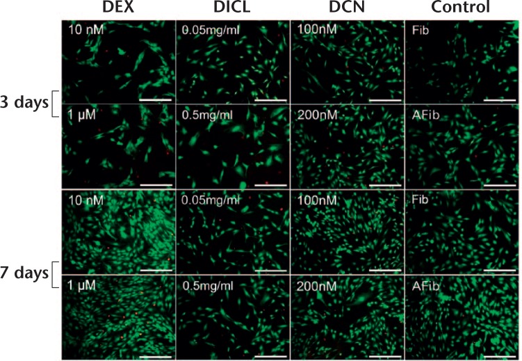
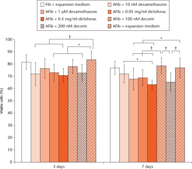
The percentage of viable AFib cells cultured with expansion medium was 83.35% (sd 6.11) and 76.84% (sd 5.43) after three and seven days, respectively, which was not significantly different from that of the Fib group (control groups in Figs 1 and 2). After three days, the cell viability was lower in the 10 nM dexamethasone group compared with the AFib in the expansion medium (p < 0.01). The same phenomenon was observed in both diclofenac groups (p < 0.05, p < 0.01). After seven days, the cell viability was further decreased to 67.78% (sd 8.91), 63.30% (sd 3.86), and 65.29% (sd 7.58) in 1 μM dexamethasone, 0.5 mg/ml diclofenac, and 200 nM decorin groups, respectively, statistically lower than the expansion medium group (p < 0.05, p < 0.01).
Fig. 1.
Viability of cells cultured in different groups was analyzed by Live/Dead staining after three and seven days. Viable and non-viable cells were counted in each of five random fields of view for each well. Scale bars represent 200 mm (red, nucleus of non-viable cells; green, viable cells).
Fig. 2.
Graph showing the cell viability of different groups. Data represent mean and standard deviation (n = 6, *p < 0.05, †p < 0.01; one-way analysis of variance). DEX, dexamethasone; DICL, diclofenac; DCN, decorin; AFib, arthrofibrotic tissue; Fib, fibroblasts isolated from healthy tissue.
Cell proliferation
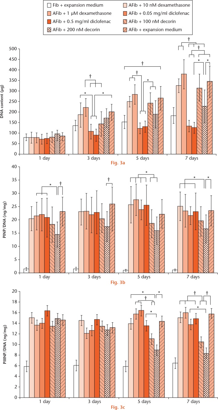
The DNA content in AFib cultured with expansion medium was, respectively, 1.08, 1.46, 1.79, and 1.94 times greater than the DNA in the Fib group after one, three, five, and seven days (Fig. 3a). The AFib DNA content in the diclofenac groups was significantly lower than that of AFib + expansion medium group after three, five, and seven days, respectively (p < 0.05, p < 0.01). After seven days, DNA content of AFib in 200 nM decorin was lower than that in the expansion medium group (p < 0.05).
Cells were seeded in 25 cm2 tissue culture flasks at a density of 1 × 10⁵ cells/flask and cultured with 10 ml of the experimental medium. a) After one, three, five, and seven days, the cells were trypsinized and the DNA of the cells was extracted and quantitated. b) and c) The procollagen type I N-terminal propeptide (PINP) and procollagen type III N-terminal propeptide (PIIINP) concentration levels of the cell supernatant obtained from different groups were determined by enzyme-linked immunosorbent assay (ELISA). The DNA content was used to standardize the expressions of PINP/PIIINP. Data represent mean and standard deviation (n = 6, *p < 0.05, †p < 0.01; one-way analysis of variance). AFib, arthrofibrotic tissue; Fib, fibroblasts isolated from healthy tissue.
To study the effect of different medication on the proliferation of fibroblast from normal joint, Fib was treated by dexamethasone, diclofenac, or decorin with different concentrations. As shown in Supplementary figure a, the date obtained from MTS assay demonstrated a time-dependent increase in all groups during the one-week cultivation. No inhabited effect on cell proliferation was observed in all groups. Additionally, no significant difference of optical density was detected at each timepoint.
Biochemical analysis
Following one week of culture, the PINP expression level in the Fib group was extremely low compared with the other groups. However, the PINP of AFib cultured with expansion medium remained at a high level throughout the whole experiment. Remarkably, decorin decreased the PINP, especially in the 200 nM group, 0.62-, 0.67-, 0.71-, 0.70-fold after one, three, five, and seven days, respectively, compared with the expression levels of AFib in expansion medium (Fig. 3b). Interestingly, the PIIINP was not affected by any of the interventions after one and three days’ culture. Inhibition was found in the decorin groups after five days, which is statistically different to the results of the other groups (p < 0.05, p < 0.01) (Fig. 3c). As a marker of normal fibroblasts, the expression of PDGFRα was quantified to evaluate the effect of different medication on Fib. As shown in Supplementary figure b, abundant expression of PDGFRα was detected in normal fibroblast cultured in expansion medium. No significant alteration was observed between different groups at each time point.
TGF-β1, α-SMA, and TNC mRNA expression in AFib
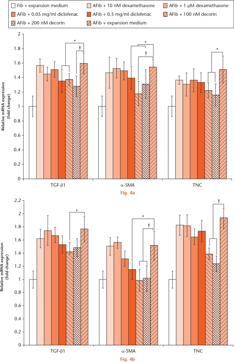
The gene expression in AFib cultured in expansion medium was significantly increased compared with that of the Fib group (p < 0.01). During one week of cultivation, no statistical difference was observed between the dexamethasone groups and the positive groups with respect to TGF-β1, α-SMA and TNC expression; 0.5 mg/ml diclofenac downregulated TGF-β1 and α-SMA expression after three and seven days. Notably, the expression of these genes in the decorin groups was significantly different to that of the positive group (p < 0.05, p < 0.01) (Fig. 4).
The expression of transforming growth factor-beta 1 (TGF-β1), alpha-smooth muscle actin (α-SMA) and Tenascin-C (TNC) genes was analyzed by real-time polymerase chain reaction (PCR) after a) three days and b) seven days of cultivation. Data represent mean and standard deviation (n = 6, *p < 0.05, †p < 0.01; one-way analysis of variance). AFib, arthrofibrotic tissue; Fib, fibroblasts isolated from healthy tissue.
Biomechanical analysis
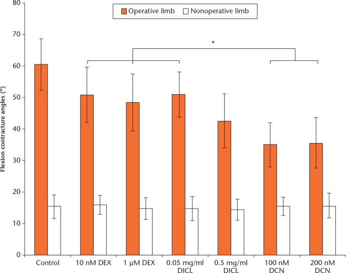
The flexion contracture angle of operative and non-operated limbs was analyzed at eight weeks. The extension loss angle of the operative limb was clearly visible in every group, compared with the non-operated limb. The flexion contracture angle was 60.55° (sd 8.15) in the control group, the highest among all groups. A slight decrease was observed in the 10 nM dexamethasone and 0.05 mg/ml diclofenac groups (p > 0.05 vs control group). Notably, significant decreases were found in 1 μM dexamethasone, 0.5 mg/ ml diclofenac (42.60° (sd 8.72)) and both decorin groups (35.01° (sd 7.02); 35.60° (sd 8.04)), compared with the control group (p < 0.05). Additionally, a significant difference was observed between the decorin group and the other groups (p < 0.05) (Fig. 5).
Fig. 5.
Graph depicting the flexion contracture angles of the knee joint in our rabbit model (n = 4, *p < 0.05; one-way analysis of variance). DEX, dexamethasone; DICL, diclofenac; DCN, decorin.
Histological analysis
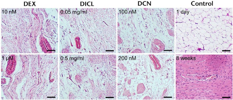
Histological analysis of the harvested tissues was performed blinded eight weeks following surgery. The number of vessels and the formation of fibroblastic tissue increased in all groups. Normal synovial intima and subsynovial adipose were replaced by fibroblastic tissue in the controls after eight weeks. A similar result also was observed in the dexamethasone groups. The spindled fibroblast was decreased slightly in the diclofenac groups, compared with the control group. Additionally, the distribution of fibroblastic tissue was not as extensive as that in the control group. In the decorin groups, a small amount of fibroblastic tissue was scattered in the subsynovial adipose tissue (Fig. 6).
Fig. 6.
Histological analysis of infrapatellar fat pad of the joints in different groups. The specimens in control group harvested after one day and eight weeks served as negative and positive controls, respectively (stain, hematoxylin, and eosin). DEX, dexamethasone; DICL, diclofenac; DCN, decorin. Scale bar = 200 μM.
Discussion
Arthrofibrosis is a debilitating complication after knee trauma and surgery. Stability and mobility are both essential for joint functions, and how to restore stability and preserve mobility remains a dilemma for orthopaedic surgeons.18 In this study, the proliferation of AFib was inhibited by diclofenac as determined by DNA quantitation. This negative effect of diclofenac was consistent with a previous study,19 although the cell source was different. An increase in non-viable cells was observed in the diclofenac groups, particularly at the higher concentrations of the drug. Bort et al19 reported that diclofenac-induced mitochondrial impairment, together with a futile consumption of nicotinamide adenine dinucleotide phosphate (NADPH), is the mechanism of cytotoxicity observed in hepatocytes. We postulate that this phenomenon was responsible for the observations in our experiments. In order to determine the specific effect of different agents on AFib, Fib from normal joints were treated with the same conditioned medium. No obvious difference was detected from MTS assay between different groups after one, three, five, or seven days (Supplementary fig. a). Sharon et al20 determined that platelet-derived growth factor receptor α (PDGFRα) was abundantly expressed by normal fibroblasts. We investigated the effect of dexamethasone, diclofenac, and decorin on the phenotype of Fib by detecting PDGFRα expression. No significant alteration was observed between different groups during one week of cultivation (Supplementary fig. b). According to these results, we concluded that dexamethasone, diclofenac, and decorin could not affect the activity of fibroblasts from a normal joint, which was essential to the clinical application of these agents. Contrasting effects of dexamethasone on myofibroblasts have been reported in the literature.21-23 In the present study, dexamethasone did not alter cell viability but it did enhance cell proliferation. Gu et al21 suggested that dexamethasone could not change the human fetal lung myofibroblast but El-Helou et al22 reported that dexamethasone has the capability to attenuate myofibroblast proliferation. Differences in drug concentrations and cell sources may account for these discrepancies.
According to previously published data, TGF-β1, α-SMA, and TNC have been widely investigated as myofibroblast markers.24,25 Pourjafar and Derakhshanfar26 reported that administration of diclofenac can induce renal fibrosis in rabbits. In contrast, we found that 0.5 mg/ml diclofenac did not downregulate TGF-β1 and α-SMA significantly in the present study. We presume that the differing effects of diclofenac in vivo and in vitro could explain the discrepancy between our findings and those of previously published studies. Decorin is a human proteoglycan that inactivates the effects of TGF-β1.27 TGF-β1 has long been known to induce matrix synthesis and contraction by fibroblasts, contributing to fibrotic disease.28 We found the expressions of TGF-β1, α-SMA, and TNC in AFib were downregulated by decorin, although the mechanism by which decorin acted on TGF-β1 was not investigated in detail in this study.
In total knee and hip arthroplasty literature, intraoperative and postoperative dexamethasone administration has been shown to quicken functional recovery and improve the range of joint movement.29,30 Desai et al31 reported that perioperative glucocorticoid administration improved elbow movement in terrible triad injuries. Intra-articular administration of diclofenac was demonstrated to be an effective treatment for arthritis due to its anti-inflammatory potential.32 However, we found no improvement in joint contractures in animals receiving 10 nM dexamethasone, while joint contracture angle was decreased by administration of 1 μM dexamethasone. Similar results were detected in the diclofenac groups. We postulate that only high concentrations of dexamethasone and diclofenac could inhibit the fibroblast proliferation and deposition of interstitial matrix components in the joint. Based on the flexion contracture angle and histological analysis, decorin was demonstrated to have antifibrotic activity in vivo, including the inhibition of collagen deposition. Murray et al33 established that an inhibitor of alpha V integrins protected mice from fibrosis in skeletal muscle by decreasing the activity of TGF-β1. We presumed that a similar mechanism might also exist in our decorin group. Abdel et al16 reported that decorin could not improve arthrofibrosis clinically when the contracture was already fully formed. We consider that the timing of administration is an essential factor. Decorin could be applied to prevent the initiation of arthrofibrosis, but may not reverse the established contractures.
In summary, dexamethasone could not downregulate the myofibroblast markers in AFib. Meanwhile, proliferation of AFib was not decreased by dexamethasone. Diclofenac could inhibit the metabolism and proliferation of AFib; however, such effects on the other cells in the joints should be investigated prior to clinical application. In addition, decorin benefited the downregulation of the myofibroblast markers in AFib, suggesting that decorin could be a promising approach to treat arthrofibrosis. These findings were further confirmed by intra-articular administration of the agents in the rabbit model of joint contracture. Since the mechanisms by which fibrosis develops are increasingly thought to be conserved throughout different organ systems,34 we plan to investigate the effect of decorin in other fibrotic disease settings, such as the formation of adhesions after flexor tendon injuries, which lead to compromised joint flexion or joint contracture.
Footnotes
Author Contributions: X. Tang: Experimental studies, Synthesizing the data, Drafting the manuscript, Reviewing the manuscript.
S. Teng: Study design, Experimental studies, Synthesizing the data, Reviewing the manuscript.
M. Petri: Experimental studies, Synthesizing the data, Reviewing the manuscript.
C. Krettek: Study design, Drafting the manuscript, Reviewing the manuscript.
C. Liu: Study design, Experimental studies, Synthesizing the data, Drafting the manuscript, Reviewing the manuscript.
M. Jagodzinski: Study design, Drafting the manuscript, Reviewing the manuscript.
Conflicts of Interest Statement: None declared
Follow us @BoneJointRes
Supplementary material
Graphs showing the MTS evaluation for, and the expression of PDGFRα in, fibroblasts isolated from healthy tissue treated with dexamethasone, diclofenac, and decorin are available alongside this article at www.bjr.boneandjoint.org.uk
Funding Statement
This work was financially supported by the AO Foundation’s Research Fund (AO 05-H74), the National Natural Sciences Foundation of China (31300799), and the Independent Innovative Research Funds of HUST (2014QN094).
References
- 1. Gillespie MJ, Friedland J, Dehaven KE. Arthrofibrosis: etiology, classification, histopathology, and treatment. Oper Tech Sports Med 1998;6:102-110. [Google Scholar]
- 2. Creighton RA, Bach BR., Jr Arthrofibrosis: evaluation, prevention, and treatment. Tech Knee Surg 2005;11:163-172. [Google Scholar]
- 3. Kim DH, Gill TJ, Millett PJ. Arthroscopic treatment of the arthrofibrotic knee. Arthroscopy 2004;20(Suppl 2):187-194. [DOI] [PubMed] [Google Scholar]
- 4. Barlow JD, Morrey ME, Hartzler RU, et al. Effectiveness of rosiglitazone in reducing flexion contracture in a rabbit model of arthrofibrosis with surgical capsular release: A biomechanical, histological, and genetic analysis. Bone Joint Res 2016;5:11-17. [DOI] [PMC free article] [PubMed] [Google Scholar]
- 5. Rhen T, Cidlowski JA. Antiinflammatory action of glucocorticoids–new mechanisms for old drugs. N Engl J Med 2005;353:1711-1723. [DOI] [PubMed] [Google Scholar]
- 6. Silverstein FE, Faich G, Goldstein JL, et al. Gastrointestinal toxicity with celecoxib vs nonsteroidal anti-inflammatory drugs for osteoarthritis and rheumatoid arthritis: the CLASS study: A randomized controlled trial. Celecoxib Long-term Arthritis Safety Study. JAMA 2000;284:1247-1255. [DOI] [PubMed] [Google Scholar]
- 7. Crofford LJ, Lipsky PE, Brooks P, et al. Basic biology and clinical application of specific cyclooxygenase-2 inhibitors. Arthritis Rheum 2000;43:4-13. [DOI] [PubMed] [Google Scholar]
- 8. Kresse H, Schönherr E. Proteoglycans of the extracellular matrix and growth control. J Cell Physiol 2001;189:266-274. [DOI] [PubMed] [Google Scholar]
- 9. Lobenhoffer HP, Bosch U, Gerich TG. Role of posterior capsulotomy for the treatment of extension deficits of the knee. Knee Surg Sports Traumatol Arthrosc 1996;4:237-241. [DOI] [PubMed] [Google Scholar]
- 10. Skutek M, Elsner HA, Slateva K, et al. Screening for arthrofibrosis after anterior cruciate ligament reconstruction: analysis of association with human leukocyte antigen. Arthroscopy 2004;20:469-473. [DOI] [PubMed] [Google Scholar]
- 11. Unterhauser FN, Bosch U, Zeichen J, Weiler A. Alpha-smooth muscle actin containing contractile fibroblastic cells in human knee arthrofibrosis tissue. Winner of the AGA-DonJoy Award 2003. Arch Orthop Trauma Surg 2004;124:585-591. [DOI] [PubMed] [Google Scholar]
- 12. Li Y, Li J, Zhu J, et al. Decorin gene transfer promotes muscle cell differentiation and muscle regeneration. Mol Ther 2007;15:1616-1622. [DOI] [PubMed] [Google Scholar]
- 13. Armaka M, Gkretsi V, Kontoyiannis DL, Kollias G. A standardized protocol for the isolation and culture of normal and arthritogenic murine synovial fibroblasts. Nature. Protocol Exchange. 2009. http://www.nature.com/protocolexchange/protocols/558 (date last accessed 15 January 2018).
- 14. Liu C, Abedian R, Meister R, et al. Influence of perfusion and compression on the proliferation and differentiation of bone mesenchymal stromal cells seeded on polyurethane scaffolds. Biomaterials 2012;33:1052-1064. [DOI] [PubMed] [Google Scholar]
- 15. Demeke T, Jenkins GR. Influence of DNA extraction methods, PCR inhibitors and quantification methods on real-time PCR assay of biotechnology-derived traits. Anal Bioanal Chem 2010;396:1977-1990. [DOI] [PubMed] [Google Scholar]
- 16. Abdel MP, Morrey ME, Barlow JD, et al. Intra-articular decorin influences the fibrosis genetic expression profile in a rabbit model of joint contracture. Bone Joint Res 2014;3:82-88. [DOI] [PMC free article] [PubMed] [Google Scholar]
- 17. Walker JA, Ewald TJ, Lewallen E, et al. Intra-articular implantation of collagen scaffold carriers is safe in both native and arthrofibrotic rabbit knee joints. Bone Joint Res 2017;6:162-171. [DOI] [PMC free article] [PubMed] [Google Scholar]
- 18. Perry J. Contractures. A historical perspective. Clin Orthop Relat Res 1987;219:8-14. [PubMed] [Google Scholar]
- 19. Bort R, Ponsoda X, Jover R, Gómez-Lechón MJ, Castell JV. Diclofenac toxicity to hepatocytes: a role for drug metabolism in cell toxicity. J Pharmacol Exp Ther 1999;288:65-72. [PubMed] [Google Scholar]
- 20. Sharon Y, Alon L, Glanz S, Servais C, Erez N. Isolation of normal and cancer-associated fibroblasts from fresh tissues by Fluorescence Activated Cell Sorting (FACS). J Vis Exp 2013;71:e4425. [DOI] [PMC free article] [PubMed] [Google Scholar]
- 21. Gu L, Zhu YJ, Guo ZJ, Xu XX, Xu WB. Effect of IFN-gamma and dexamethasone on TGF-beta1-induced human fetal lung fibroblast-myofibroblast differentiation. Acta Pharmacol Sin 2004;25:1479-1488. [PubMed] [Google Scholar]
- 22. El-Helou V, Proulx C, Gosselin H, et al. Dexamethasone treatment of post-MI rats attenuates sympathetic innervation of the infarct region. J Appl Physiol (1985) 2008;104:150-156. [DOI] [PubMed] [Google Scholar]
- 23. Miller M, Cho JY, McElwain K, et al. Corticosteroids prevent myofibroblast accumulation and airway remodeling in mice. Am J Physiol Lung Cell Mol Physiol 2006;290:L162-L169. [DOI] [PubMed] [Google Scholar]
- 24. van den Borne SW, Diez J, Blankesteijn WM, et al. Myocardial remodeling after infarction: the role of myofibroblasts. Nat Rev Cardiol 2010;7:30-37. [DOI] [PubMed] [Google Scholar]
- 25. Löfdahl M, Kaarteenaho R, Lappi-Blanco E, Tornling G, Sköld MC. Tenascin-C and alpha-smooth muscle actin positive cells are increased in the large airways in patients with COPD. Respir Res 2011;12:48. [DOI] [PMC free article] [PubMed] [Google Scholar]
- 26. Pourjafar M, Derakhshanfar A. A histopathologic study on the side effects of the Diclofenac sodium in rabbits. In: Hassan L, Dhaliwal GK, Yusoff R, et al., eds. Animal health: a breakpoint in economic development? The 11th International Conference of the Association of Institutions for Tropical Veterinary Medicine and 16th Veterinary Association Malaysia Congress, 23-27 August 2004, Petaling Jaya, Malaysia Serdang: Universiti Putra Malaysia Press, 2004: 360-361. [Google Scholar]
- 27. Fukushima K, Badlani N, Usas A, et al. The use of an antifibrosis agent to improve muscle recovery after laceration. Am J Sports Med 2001;29:394-402. [DOI] [PubMed] [Google Scholar]
- 28. Leask A, Abraham DJ. TGF-beta signaling and the fibrotic response. FASEB J 2004;18:816-827. [DOI] [PubMed] [Google Scholar]
- 29. Backes JR, Bentley JC, Politi JR, Chambers BT. Dexamethasone reduces length of hospitalization and improves postoperative pain and nausea after total joint arthroplasty: a prospective, randomized controlled trial. J Arthroplasty 2013;28(Suppl):11-17. [DOI] [PubMed] [Google Scholar]
- 30. Jules-Elysee KM, Wilfred SE, Memtsoudis SG, et al. Steroid modulation of cytokine release and desmosine levels in bilateral total knee replacement: a prospective, double-blind, randomized controlled trial. J Bone Joint Surg [Am] 2012;94-A:2120-2127. [DOI] [PubMed] [Google Scholar]
- 31. Desai MJ, Matson AP, Ruch DS, et al. Perioperative glucocorticoid administration improves elbow motion in terrible triad injuries. J Hand Surg Am 2017;42:41-46. [DOI] [PubMed] [Google Scholar]
- 32. Qi X, Qin X, Yang R, et al. Intra-articular administration of chitosan thermosensitive in situ hydrogels combined with diclofenac sodium-loaded alginate microspheres. J Pharm Sci 2016;105:122-130. [DOI] [PubMed] [Google Scholar]
- 33. Murray IR, Gonzalez ZN, Baily J, et al. αv integrins on mesenchymal cells regulate skeletal and cardiac muscle fibrosis. Nat Commun 2017;8:1118. [DOI] [PMC free article] [PubMed] [Google Scholar]
- 34. Henderson NC, Arnold TD, Katamura Y, et al. Targeting of αv integrin identifies a core molecular pathway that regulates fibrosis in several organs. Nat Med 2013;19:1617-1624. [DOI] [PMC free article] [PubMed] [Google Scholar]


