Abstract
Purpose
To determine the effectiveness of etanercept, a tumor necrosis factor (TNF) inhibitor, in conferring neuroprotection to retinal ganglion cells (RGCs) and improving visual outcomes after optic nerve trauma with either optic nerve crush (ONC) or sonication-induced traumatic optic neuropathy (SI-TON) in mice.
Methods
Mouse optic nerves were unilaterally subjected to ONC (n = 20) or SI-TON (n = 20). TNF expression was evaluated by using immunohistochemistry and quantitative RT-PCR (qRT-PCR) in optic nerves harvested 6 and 24 hours post ONC (n = 10) and SI-TON (n = 10). Mice in each injury group received daily subcutaneous injections of either etanercept (10 mg/kg of body weight; five mice) or vehicle (five mice) for 7 days. Pattern electroretinograms were performed on all mice at 1 and 2 weeks after injury. ONC mice were killed at 2 weeks after injury, while SI-TON mice were euthanized at 4 weeks after injury. Whole retina flat-mounts were used for RGC quantification.
Results
Immunohistochemistry and qRT-PCR showed upregulation of TNF protein and gene expression within 24 hours after injury. In both models, etanercept use immediately following optic nerve injury led to higher RGC survival when compared to controls, which was comparable between the two models (24.23% in ONC versus 20.42% in SI-TON). In both models, 1 and 2 weeks post injury, mice treated with etanercept had significantly higher a-wave amplitudes than untreated injured controls.
Conclusions
Treatment with etanercept significantly reduced retinal damage and improved visual function in both animal models of TON. These findings suggest that reducing TNF activity in injured optic nerves constitutes an effective therapeutic approach in an acute setting.
Keywords: traumatic optic neuropathy, etanercept, tumor necrosis factor
Traumatic optic neuropathy (TON) is an uncommon but devastating cause of permanent visual loss following blunt force trauma to the orbit.1,2 Clinically, a patient with acute TON may present with either partial or complete loss of vision, an afferent pupillary defect, normal-appearing optic disc, or visual field defects.2 With time, optic disc pallor will manifest. Likelihood of vision recovery is low in these patients, as visual acuity and visual field results do not show significant difference 5 years after injury when compared to baseline testing following injury.3
TON is classified into two categories: direct and indirect.4 Direct injury occurs when a foreign body or bone fragment causes damage by coming into direct contact with the optic nerve. More commonly, indirect optic nerve injury occurs when deformational forces are transferred through the bones of the skull or by globe torsion against the optic nerve.5,6 Putative mechanisms of indirect injury include transmission of concussive shock waves propagating through the orbit against the intraorbital segment of the nerve2; momentum of the globe and orbital contents being absorbed by the fixed portion of the optic nerve at the entrance to the optic canal5; and compression of the intracanalicular segment of the optic nerve by the malleable bones of the optic canal.7 The initial traumatic event initiates a cascading sequence of metabolic events that are believed to exacerbate the optic nerve injury.6
Presently, no evidence-based therapy exists to effectively treat TON.5,8,9 Current treatment options for TON include observation, high-dose corticosteroid use, or surgical decompression of the optic canal.9–13 In a prospective, comparative, nonrandomized interventional study of TON visual outcomes, the International Optic Nerve Trauma Study found that, after adjusting for baseline visual acuity, no clear benefit of either corticosteroid therapy or optic canal decompression surgery was observed.14,15 Hence, effective therapy for visual recovery in TON remains elusive.
Histopathologically, TON results in axonal degeneration and retinal ganglion cell (RGC) death.5 Although TON has been the subject of substantial basic and clinical research, insights into its basic pathophysiologic and molecular mechanisms remain limited.2 The most common animal model used to simulate optic nerve injury is optic nerve crush (ONC).16,17 While this model effects RGC loss,18 it requires direct contact with the optic nerve—a less common etiology for clinical TON.5 Using sonication shock as a means to induce TON (sonication-induced TON [SI-TON]),19 we established an injury model that closely approximates the indirect clinical mechanism in TON. With SI-TON, we have characterized the early molecular events following focal injury to the optic nerve and demonstrated that among the first signals for a reactive process ensuing in the nerve is the upregulation of proinflammatory cytokines, the most significant of which is tumor necrosis factor (TNF).19
TNF plays an important role in the regulation of immune cell function and, in the central nervous system (CNS), is associated with the pathophysiology of virtually all neurodegenerative diseases, including eye disorders.20–27 Increased glial TNF production in the optic nerves was previously shown after ONC28 and in the optic nerve heads of glaucomatous patients.21 Similarly, knockout of the tumor necrosis factor receptor-1 (TNFR1), but not TNFR2, confers a neuroprotective effect onto RGCs following crush or ischemic insults to the retina,28,29 suggesting alternate roles for the soluble and membrane-bound isoforms of TNF. These observations make TNF a promising candidate for targeted therapy in acute neurodegenerative processes like TON. Furthermore, inhibition of TNF using etanercept has been shown to improve functional recovery of facial nerves following crush injury.30
The purpose of this study was to evaluate the effectiveness of etanercept, a nonselective TNF inhibitor, in conferring neuroprotection to RGCs and improving visual outcomes after optic nerve trauma with either ONC or SI-TON in mice. The availability of FDA (US Food and Drug Administration)–approved TNF inhibitors increases the translational potential of our observations for an orphan disease with no effective treatment options.
Materials and Methods
Materials
All chemicals and reagents were purchased from Sigma-Aldrich Corp. (St. Louis, MO, USA) and Thermo Fisher Scientific, Inc. (Waltham, MA, USA). Etanercept (Enbrel; Pfizer, New York, NY, USA) was purchased from the medical pharmacy in Bascom Palmer Eye Institute for use in this research study.
Animals
All animal experiments were performed in compliance with the National Institutes of Health (Bethesda, MD, USA) Guide for the Care and Use of Laboratory Animals and the ARVO Statement for the Use of Animals in Ophthalmic and Vision Research. Animal protocols were reviewed and approved by the Institutional Animal Care and Use Committee of the University of Miami. C57BL/6 J mice were obtained from Jackson Laboratory (Bar Harbor, ME, USA). Mice were housed under ambient conditions (standard humidity and temperature) with a 12-hour light/dark cycle. Three-month-old mice were used for the study. Forty mice were used; 20 were injured by ONC and 20 by SI-TON. Left optic nerves were injured in all animals; the right optic nerves were not injured. In each of the injury platforms, five of the mice were treated with daily subcutaneous injections of etanercept 10 mg/kg of body weight for a period of 7 days. The other five mice were treated with daily subcutaneous injections of phosphate-buffered saline (PBS). All injections were performed by using a 27-gauge needle on the dorsum of the neck between the ears.
Sonication-Induced Traumatic Optic Neuropathy Model
SI-TON model was performed as described previously.19
Optic Nerve Crush Model31
The mice were anesthetized via intraperitoneal injection of ketamine (80 mg/kg)/xylazine (10 mg/kg). After the animals were properly anesthetized, their optic nerves were exposed intraorbitally and crushed with jeweler's forceps (Dumont No. 5; tip dimension, 0.1 × 0.06 mm) for 10 seconds approximately 1 mm behind the optic disc.
Quantitative RT-PCR Analysis
The optic nerves were homogenized in RNA lysis buffer and stored in liquid nitrogen until further processing. Total RNA was extracted by using the Absolutely RNA Nanoprep kit (Agilent Technologies, Santa Clara, CA, USA), then reverse transcribed with Superscript III polymerase (Invitrogen, Carlsbad, CA, USA) to synthesize cDNA. Quantitative RT-PCR was performed in the Rotor-Gene Q Cycler (Qiagen, Germantown, MD, USA) using SYBR GREEN PCR MasterMix (Qiagen) and TNF-α (Tnf gene)–specific primers (TnfF: 5′ – CAAAATTCGAGTGACAAGCCTG – 3′; TnfR: 5′ – GAGATCCATGCCGTTGGC – 3′). Relative expression was calculated by comparison with a standard curve, following normalization to the expression of housekeeping gene β-actin (ActbF: 5′ – CACCCTGTGCTGCTCACC – 3′; ActbR: 5′ – GCACGATTTCCCTCTCAG – 3′).
Immunohistochemistry
Optic nerves were harvested 6 and 24 hours post ONC and SI-TON. Optic nerves were fixed, sectioned, permeabilized, and blocked as previously descrived.19 Sections were incubated with primary antibodies (mouse anti-GFAP [anti–glial fibrillary acidic protein], 1:400, Sigma; rat anti-CD45, 1:200, eBioscience, San Diego, CA, USA; goat anti-TNF, 1:50, Santa Cruz, Dallas, TX, USA) in blocking solution overnight at 4°C. After washing in PBS three times, sections were incubated with species-specific fluorescent secondary antibodies. Control sections were incubated with secondary antibody alone. Finally, sections were coverslipped with Vecta shield (Vector, Burlingame, CA, USA) fluorescent mounting medium containing DAPI. Imaging was performed with Leica TSL AOBS SP5 confocal microscope (Leica Microsystems, Exton, PA, USA).
Quantification of Ganglion Cell Layer Neurons in the Retina
After euthanasia, eyes were enucleated and fixed in 4% paraformaldehyde. Ganglion cell layer (GCL) neurons in the retina were stained and quantified as described earlier.19
Pattern Electroretinogram (PERG)
Electrophysiological function of GCLs was measured by PERG as previously described.32
Statistical Analysis
One-way ANOVA was used for multiple comparisons. The Student's t-test was conducted for single comparisons. P values ≤ 0.05 were considered to be statistically significant.
Results
TNF Is Upregulated Early in Crush Injury and Indirect Traumatic Optic Neuropathy
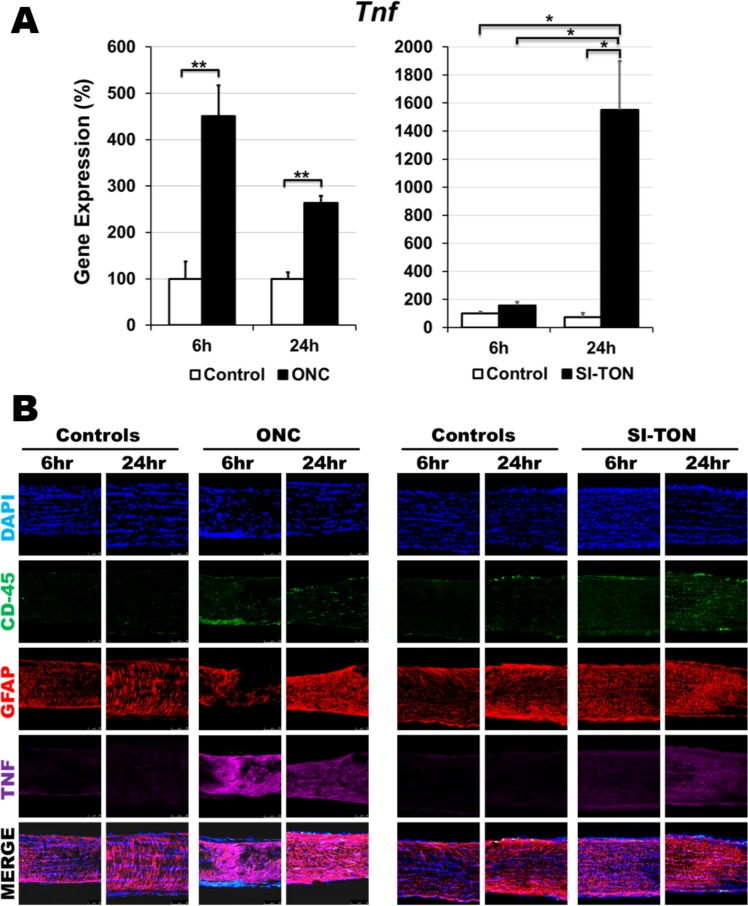
The use of two injury models was selected in order to validate the findings as pertaining to optic nerve injury mechanisms rather than model-specific variables. Immunohistochemical analysis and quantitative RT-PCR showed upregulation of TNF at the gene and protein level within hours after injury (Figs. 1A, 1B). In ONC, Tnf gene expression levels reached 450% ± 66% over controls at 6 hours post crush (P < 0.01) and remained significantly upregulated at 24 hours post injury with an expression level 263% ± 15% higher than that of control nerves (P < 0.01; Fig. 1A, left). In SI-TON, Tnf gene expression was not statistically significantly increased at 6 hours after injury (142% ± 25% of control, P = 0.11). At 24 hours, however, Tnf gene expression levels increased to 1417% ± 317% when compared to control nerves (P < 0.03). This group (SI-TON, 24 hour post injury) experienced the highest increase in Tnf gene expression, which was significantly higher than all the other groups in this experimental injury model (Fig. 1A, right). At the protein level, we observed an increase in TNF immunoreactivity in both models at the 6- and 24-hour postinjury time points (Fig. 1B). Interestingly, this finding coincides with increased immunoreactivity for CD45+ infiltrating leukocytes into the injured nerves and increased expression of GFAP, indicating initiation of an inflammatory process in both injury models (Fig. 1B). Our immunohistochemical evaluation, however, did not allow us to discern specific contributions of soluble TNF from glial cells or leukocytes.
Figure 1.
Increased TNF-α expression in early ONC and SI-TON models. (A) Quantitative PCR Tnf gene expression profiling at 6- and 24-hour post injury in both models showing significant upregulation of Tnf expression following trauma (n = 5 mice per group, *P < 0.05, **P < 0.01). (B) Immunohistochemical assay of injured optic nerves in ONC and SI-TON models showing increased TNF-α, CD-45, and GFAP immunoreactivity at 6- and 24-hours post injury in both animal models.
TNF-α Inhibition Results in Significant RGC Neuroprotection Following Optic Nerve Trauma
To test the neuroprotective effect of reduced TNF levels in TON, we used the TNF inhibitor etanercept, administered daily for 7 days after trauma in ONC and SI-TON injury models. Control animals received vehicle (PBS) injections.
Crush Injury
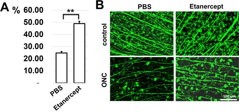
Two weeks after injury (1 week after treatment cessation), retinas were collected and stained for the RGC marker tubulin-βIII (TUJ1). In the control group, the average RGC count in injured eyes was quantified to be 25% ± 1% (range, 23%–29%) of the uninjured contralateral eyes (Fig. 2A), whereas in the etanercept-treated group, the average RGC count in the injured eyes was 49% ± 2% (range, 45%–55%) of the uninjured eye counts (Fig. 2A). This difference between vehicle control and etanercept-treated cohorts was statistically significant (P < 0.01). Immunohistochemically (Fig. 2B), tubulin-βIII staining showed a significant dropout of immunoreactive RGCs in the PBS vehicle control injured eyes when compared to uninjured contralateral eyes 2 weeks after injury. This was accompanied by disruption of tissue ultrastructure. These results are consistent with previously reported effects on retinal morphologic and RGC numbers in ONC animal models.33 In the etanercept-treated animals, injured eyes also presented with loss of RGC immunoreactivity (as confirmed by RGC counting data), although to a lesser extent than in PBS-treated injured eyes. Evidence of tissue ultrastructure damage was still present even after TNF inhibition (Fig. 2B).
Figure 2.
Tnf-α inhibition promotes RGC survival after ONC. (A) RGC survival in retinas of injured animals treated with either etanercept or PBS (control) was calculated as a percentage of the mean cell density in the retinas of fellow control (no surgery) eyes of the same animals (n = 5 mice per group). To identify RGCs, flat-mounted retinas were immunostained for the RGC marker tubulin-βIII (TUJ1) (green). (B) Representative confocal images of tubulin-βIII (TUJ1)–labeled GCLs (green) in flat-mounted retinas were imaged 14 days after injury.
Ultrasound Injury
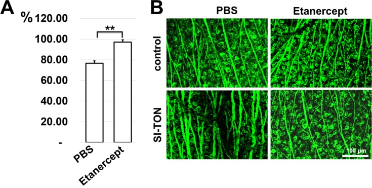
Retinas were collected and stained for tubulin-βIII 4 weeks after injury. As with the ONC model, we found that the use of etanercept immediately after SI-TON exerted prosurvival effects on RGCs. However, RGC dropout and tissue ultrastructure damage were less severe in the SI-TON model than in ONC. Nevertheless, in the PBS vehicle control group, the average RGC count in injured eyes was 77% ± 2% (range, 71%–83%) that of the uninjured contralateral eye counts (Fig. 3A). In contrast, in the etanercept-treated group the average RGC counts in injured eyes was 97% ± 2% (range, 90%–102%) of the uninjured contralateral eyes (Fig. 3A). This difference between vehicle and etanercept-treated cohorts was also statistically significant for this injury model (P < 0.01). Immunohistochemically (Fig. 3B), TNF inhibition in the milder, indirect SI-TON showed significant maintenance of RGC immunoreactivity and tissue morphology when compared to the more severe ONC-injured animals. In the SI-TON model, PBS vehicle treatment leads to reduced RGC immunoreactivity and mild tissue morphologic disruptions consistent with what has previously been reported.17 On the other hand, in the SI-TON injured, etanercept-treated animals, RGC counts and retinal morphology are preserved and indistinguishable from contralateral uninjured retinas 4 weeks after injury (Fig. 3B).
Figure 3.
Treatment with Tnf-α inhibitor results in neuroprotective effects in the GCL after SI-TON. (A) Percentage of RGC lost at 4 weeks following SI-TON in mice treated with etanercept and PBS (control) (**P < 0.01, *P < 0.05, n = 5). (B) Representative confocal images of tubulin-βIII (TUJ1)–labeled (green) RGCs in flat-mounted retinas in sham-operated controls and SI-TON eyes.
TNF Inhibition Leads to Improved Visual Function Following Optic Nerve Trauma
To study whether TNF inhibition improves RGC function, PERG recordings were obtained.
Crush Injury
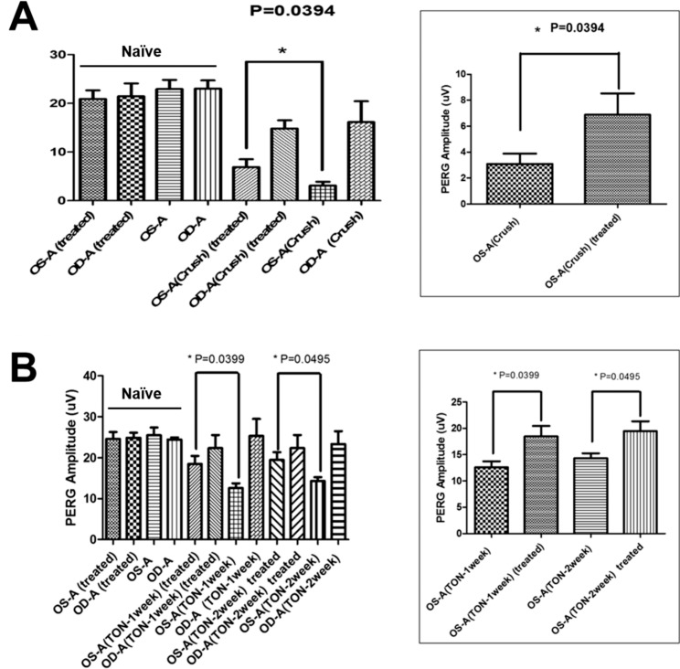
PERG recordings in ONC showed that visual response was significantly diminished in both the injured and uninjured retinas 2 weeks after injury when compared to naïve control recordings (Fig. 4A). This is consistent with previous reports of visual function decline using this injury model.32 Treatment with etanercept had no effect on visual function in uninjured naïve animals as was expected given the drug's safety profile. However, PERG data show that at 2 weeks after injury, the average a-wave amplitude in the injured eyes was 3.01 ± 0.79 μV in the vehicle (PBS)–treated mice, while in injured eyes treated with etanercept, the average a-wave amplitude was 6.88 ± 1.64 μV. The difference between the PBS- and etanercept-treated groups was statistically significant at this time point (P < 0.05; Fig. 4A).
Figure 4.
Tnf-α inhibition in TON improves RGC function measured by PERG. (A) Visual function (as assessed by PERG) was reduced significantly after 14 days in PBS-treated animals (controls) suffering from ONC, but showed no significant reductions in animals treated with etanercept. (B) RGC function decreased after SI-TON was improved in etanercept-treated animals. PERGs were recorded at baseline and 1 and 2 weeks after injury.
Ultrasound Injury
Similarly to the ONC model, PERG results at 1 and 2 weeks after injury showed a significant and maintained improvement in visual function in the animals injured and treated with etanercept (Fig. 4B). At the 1-week time point, the average a-wave amplitude in injured eyes treated with PBS was 12.53 ± 0.99 μV, while the a-wave amplitude in etanercept-treated injured eyes was 17.84 ± 2.41 μV, a statistically significant difference (P < 0.05). Interestingly, this improvement in visual function was maintained through the 2-week time point, 1 week following the cessation of drug administration. At 2 weeks after injury, the a-wave amplitude in the injured eyes of mice treated with PBS was 14.75 ± 0.79 μV, while the a-wave amplitude in etanercept-treated injured eyes was 18.51 ± 2.35 μV, which was statistically significant (P < 0.05) (Fig. 4B).
Discussion
It is clear that following optic nerve injury, a cascading sequence of molecular events ensues leading to eventual RGC death and progressive irreversible visual loss. Yet, elucidation of the specific molecular triggers for this neurodegenerative process has proven challenging. Among the major obstacles has been the lack of adequate animal models to recapitulate the human clinical manifestations of the disease. Recently, we have published a new animal model using ultrasound shockwave energy to indirectly, and focally, injure the optic nerve that leads to progressive TON: SI-TON.19 Being a much milder animal model of TON than ONC, this model allows for a more nuanced characterization of molecular events without the confounding effects of massive tissue damage, vascular disruption, and crush-associated axotomies seen in the ONC model. In our published study, we note that early proinflammatory molecules IL-1B and TNF, as well as inflammatory chemokines Ccl2 and Cxcl10, are significantly upregulated within hours of injury and lead to a sterile inflammatory process within the nerve.19 Given the availability of FDA-approved pharmaceutical agents to inhibit TNF signaling, we decided to explore the effect of TNF inhibition in ameliorating the progressive effects of TON on visual function, using both the ONC and SI-TON animal models.
We chose etanercept, a TNF-binding protein with a known pharmacodynamic profile, which has previously been shown to attenuate traumatic brain injury in rat models.34–36 In a normal state, etanercept usually has difficulty in traversing the blood brain barrier (BBB) because of its high molecular weight.37 Cheong and colleagues34 believe that etanercept works by blocking peripheral TNF but have found increased levels of the drug in the frontal cortex and hippocampus; they hypothesize that breakdown of the BBB allows for CNS penetration. Similarly, in our model, we believe etanercept produces a peripheral blockade of TNF, leading to a reduction in posttraumatic systemic inflammation. However, we cannot discount the possibility that there is some CNS penetration of the drug as a result of BBB breakdown.
Overall, TNF inhibitors have a good safety profile, with the most common side effect being injection site reactions with localized rash and itching.38 Patients are also at increased susceptibility to infections, including tuberculosis and fungal infections while receiving therapy. Recently, a rare but more serious adverse effect of prolonged etanercept use has been described in the form of anti-TNF medication–induced peripheral neuropathies, with a median time to onset of 16.8 months of anti-TNF medication use.39
There are two biologically active forms of TNF: transmembrane (tmTNF) and soluble (solTNF) forms.40,41 TmTNF and solTNF bind and activate two distinct receptors, TNF receptor 1 (TNFR1) and TNF receptor 2 (TNFR2), with different binding affinities.42 SolTNF preferentially signals through TNFR1, while tmTNF binds with and primarily activates TNFR2. These two receptors activate opposite cellular processes: TNFR1 mediates cell death, apoptosis, and chronic inflammation; while TNFR2 mediates cell survival, resolution of inflammation, immunity, and myelination.43,44 Etanercept acts as a pan-TNF inhibitor that blocks both solTNF and tmTNF. We acknowledge that this may not represent the most beneficial approach to neuroprotection in TON given the opposing roles described for soluble and membrane-bound TNF-α activation of their cognate receptors TNFR1 and TNFR2, respectively.21,28,29 Perhaps an approach exclusively addressing the soluble form of TNF and TNFR1 activation may yield a more efficacious neuroprotection for RGCs in TON.
Regardless, our results showed that etanercept use immediately following optic nerve injury, both direct and indirect, confers a neuroprotective effect on RGCs. In both injury arms, this effect was statistically significant and maintained. As was expected given the nature of the injury, a more severe RGC loss was observed in the ONC animals. However, the percentage-based increase in RGC counts for eyes treated with etanercept was comparable in both injury models (24% in ONC versus 20% in SI-TON). The percentage of RGC salvaged was higher in the etanercept-treated eyes in the SI-TON model than in ONC. The use of etanercept to address acute neuroinflammatory conditions has recently been reported with encouraging results.45 Similarly, Bae et al.46 have reported on the use of etanercept in a rat model of retinal ischemia that results in axonal preservation and reduces microglia activation in all dosing cohorts. Topdag et al.30 have explored the use of etanercept following crush injury to the facial nerve of rats, demonstrating that etanercept administration not only results in reduced immunoreactivity for inflammatory markers, but also significantly improves recovery of neuronal function when compared to controls or steroid-treated animal cohorts.
Functionality is the ultimate outcome measure for evaluating a treatment's effectiveness. We used PERG, an electrophysiological measure of optic nerve function dominated by the RGC response,32 as a measure of visual function. Our findings indicate that eyes treated with etanercept had significantly higher a-wave amplitudes than untreated injured controls. The improved PERG function coincides with increased quantitative RGC salvage following etanercept treatment. It is worth noting that despite the recovery in PERG a-wave amplitude in etanercept-treated animals, the a-wave amplitude recordings in these cohorts were still lower than those recorded for naïve uninjured animals, indicating that further improvements can be made to the therapeutic approach. One possible area for improvement in the targeted approach is addressing synergistic intracellular cascades contributing or acting downstream to TNF's deleterious effect. To this end, understanding the mechanisms and pathways altered by TNF is crucial. Venters et al.20 report that activation of TNF receptors leads to neuronal death through silencing of survival signals, such as the phosphatidylinositol 3′ kinase (PI3K), an effect that could be addressed by concomitant administration of PI3K potentiating drugs. Similarly, Kitaoka et al.23 suggest that TNF-mediated neurodegeneration is transduced via activation of Rho kinases, which can be inhibited by a number of pharmacologic agents. In contrast, Liefner et al.22 suggest that TNF acts as a macrophage chemotactic agent, leading to subsequent myelin phagocytosis by infiltrating macrophages, subsequently leading to Wallerian degeneration of the sciatic nerve. Dvoriantchikova and Ivanov26 propose that the lack of NF-κB activity in RGCs in the presence of TNF, as well as sustained c-jun N-terminal kinase (JNK) activation, both contribute to RGC death, mostly by necrosis (necroptosis). Finally, a recent study by Russell et al.24 demonstrates that TNF-α causes a rapid decline in mitochondrial function, leading to neurotoxicity. This study is of particular interest in TON as in our SI-TON model, a rapid onset of mitochondrial reactive oxygen species was observed in RGC somas within minutes of optic nerve injury.19
A noted limitation with the translation of the results presented here was the immediacy with which etanercept was administered to the injured animals. While these results represent a proof-of-principle study showing that early inhibition of TNF leads to improved visual outcomes in TON, in the clinical setting, it is unreasonable to expect initiation of therapy immediately after injury. However, there is still a timeframe in which initiation of therapy can confer sufficient neuroprotection to yield acceptable visual outcomes. We have begun “window of opportunity” studies to better define this timeframe. We believe the findings presented here support the validity of including TNF inhibition in the acute phase of TON as a targeted approach to deal with the molecular events leading to its pathogenesis. We expect this study to set the stage for future refinement of the clinical management of TON.
Acknowledgments
The authors thank Tsung-Han Chou, PhD, and Vittorio Porciatti, PhD, for their assistance with the PERG recording procedures.
Supported in part by the National Institutes of Health Center Core Grant P30EY014801, Research to Prevent Blindness Unrestricted Grant, Inc. (New York, NY, USA), National Institutes of Health/National Eye Institutes Grant R01 EY027311 (DI), National Institutes of Health/National Institute on Aging Grant R56 AG053369 (DI), and the Dr. Nasser Ibrahim Al-Rashid Orbital Vision Research Fund. The authors alone are responsible for the content and writing of this paper.
Disclosure: B.C. Tse, None; G. Dvoriantchikova, None; W. Tao, None; R.A. Gallo, None; J.Y. Lee, None; S. Pappas, None; R. Brambilla, None; D. Ivanov, None; D.T. Tse, None; D. Pelaez, None
References
- 1. Guy WM, Soparkar CN, Alford EL, Patrinely JR, Sami MS, Parke RB. . Traumatic optic neuropathy and second optic nerve injuries. JAMA Ophthalmol. 2014; 132: 567– 571. [DOI] [PubMed] [Google Scholar]
- 2. Kumaran AM, Sundar G, Chye LT. . Traumatic optic neuropathy: a review. Craniomaxillofac Trauma Reconstr. 2015; 8: 31– 41. [DOI] [PMC free article] [PubMed] [Google Scholar]
- 3. Carta A, Ferrigno L, Leaci R, Kosmarikou A, Zola E, Gomarasca S. . Long-term outcome after conservative treatment of indirect traumatic optic neuropathy. Eur J Ophthalmol. 2006; 16: 847– 850. [DOI] [PubMed] [Google Scholar]
- 4. Steinsapir KD. . Traumatic optic neuropathy. Curr Opin Ophthalmol. 1999; 10: 340– 342. [DOI] [PubMed] [Google Scholar]
- 5. Singman EL, Daphalapurkar N, White H,et al. Indirect traumatic optic neuropathy. Mil Med Res. 2016; 3: 2. [DOI] [PMC free article] [PubMed] [Google Scholar]
- 6. Giza CC, Hovda DA. . The neurometabolic cascade of concussion. J Athl Train. 2001; 36: 228– 235. [PMC free article] [PubMed] [Google Scholar]
- 7. Medeiros FA, Moura FC, Vessani RM, Susanna R Jr.. Axonal loss after traumatic optic neuropathy documented by optical coherence tomography. Am J Ophthalmol. 2003; 135: 406– 408. [DOI] [PubMed] [Google Scholar]
- 8. Chaon BC, Lee MS. . Is there treatment for traumatic optic neuropathy? Curr Opin Ophthalmol. 2015; 26: 445– 449. [DOI] [PubMed] [Google Scholar]
- 9. Yan W, Chen Y, Qian Z,et al. Incidence of optic canal fracture in the traumatic optic neuropathy and its effect on the visual outcome. Br J Ophthalmol. 2017; 101: 261– 267. [DOI] [PubMed] [Google Scholar]
- 10. Goldberg RA, Steinsapir KD. . Extracranial optic canal decompression: indications and technique. Ophthal Plast Reconstr Surg. 1996; 12: 163– 170. [DOI] [PubMed] [Google Scholar]
- 11. Yu B, Ma Y, Tu Y, Wu W. . The outcome of endoscopic transethmosphenoid optic canal decompression for indirect traumatic optic neuropathy with no-light-perception. J Ophthalmol. 2016; 2016: 6492858. [DOI] [PMC free article] [PubMed] [Google Scholar]
- 12. Kashkouli MB, Yousefi S, Nojomi M,et al. Traumatic optic neuropathy treatment trial (TONTT): open label, phase 3, multicenter, semi-experimental trial. Graefes Arch Clin Exp Ophthalmol. 2017; 256: 209– 218. [DOI] [PubMed] [Google Scholar]
- 13. Yu-Wai-Man P, Griffiths PG. . Surgery for traumatic optic neuropathy. Cochrane Database Syst Rev. 2013; 6: CD005024. [DOI] [PMC free article] [PubMed] [Google Scholar]
- 14. Levin LA, Beck RW, Joseph MP, Seiff S, Kraker R. . The treatment of traumatic optic neuropathy: the International Optic Nerve Trauma Study. Ophthalmology. 1999; 106: 1268– 1277. [DOI] [PubMed] [Google Scholar]
- 15. Carta A, Ferrigno L, Salvo M, Bianchi-Marzoli S, Boschi A, Carta F. . Visual prognosis after indirect traumatic optic neuropathy. J Neurol Neurosurg Psychiatry. 2003; 74: 246– 248. [DOI] [PMC free article] [PubMed] [Google Scholar]
- 16. Chierzi S, Strettoi E, Cenni MC, Maffei L. . Optic nerve crush: axonal responses in wild-type and bcl-2 transgenic mice. J Neurosci. 1999; 19: 8367– 8376. [DOI] [PMC free article] [PubMed] [Google Scholar]
- 17. Levkovitch-Verbin H. . Animal models of optic nerve diseases. Eye (Lond). 2004; 18: 1066– 1074. [DOI] [PubMed] [Google Scholar]
- 18. Xue F, Wu K, Wang T, Cheng Y, Jiang M, Ji J. . Morphological and functional changes of the optic nerve following traumatic optic nerve injuries in rabbits. Biomed Rep. 2016; 4: 188– 192. [DOI] [PMC free article] [PubMed] [Google Scholar]
- 19. Tao W, Dvoriantchikova G, Tse BC,et al. A novel mouse model of traumatic optic neuropathy using external ultrasound energy to achieve focal, indirect optic nerve injury. Sci Rep. 2017; 7: 11779. [DOI] [PMC free article] [PubMed] [Google Scholar]
- 20. Venters HD, Dantzer R, Kelley KW. . A new concept in neurodegeneration: TNFalpha is a silencer of survival signals. Trends Neurosci. 2000; 23: 175– 180. [DOI] [PubMed] [Google Scholar]
- 21. Yuan L, Neufeld AH. . Tumor necrosis factor-alpha: a potentially neurodestructive cytokine produced by glia in the human glaucomatous optic nerve head. Glia. 2000; 32: 42– 50. [PubMed] [Google Scholar]
- 22. Liefner M, Siebert H, Sachse T, Michel U, Kollias G, Bruck W. . The role of TNF-alpha during Wallerian degeneration. J Neuroimmunol. 2000; 108: 147– 152. [DOI] [PubMed] [Google Scholar]
- 23. Kitaoka Y, Sase K, Tsukahara C,et al. Axonal protection by ripasudil, a rho kinase inhibitor, via modulating autophagy in TNF-induced optic nerve degeneration. Invest Ophthalmol Vis Sci. 2017; 58: 5056– 5064. [DOI] [PubMed] [Google Scholar]
- 24. Russell AE, Doll DN, Sarkar SN, Simpkins JW. . TNF-alpha and beyond: rapid mitochondrial dysfunction mediates TNF-alpha-induced neurotoxicity. J Clin Cell Immunol. 2016; 7: 467– 470. [DOI] [PMC free article] [PubMed] [Google Scholar]
- 25. Viviani B, Corsini E, Galli CL, Marinovich M. . Glia increase degeneration of hippocampal neurons through release of tumor necrosis factor-alpha. Toxicol Appl Pharmacol. 1998; 150: 271– 276. [DOI] [PubMed] [Google Scholar]
- 26. Dvoriantchikova G, Ivanov D. . Tumor necrosis factor-alpha mediates activation of NF-kappaB and JNK signaling cascades in retinal ganglion cells and astrocytes in opposite ways. Eur J Neurosci. 2014; 40: 3171– 3178. [DOI] [PMC free article] [PubMed] [Google Scholar]
- 27. Dvoriantchikova G, Santos AR, Saeed AM, Dvoriantchikova X, Ivanov D. . Putative role of protein kinase C in neurotoxic inflammation mediated by extracellular heat shock protein 70 after ischemia-reperfusion. J Neuroinflamm. 2014; 11: 81. [DOI] [PMC free article] [PubMed] [Google Scholar]
- 28. Tezel G, Yang X, Yang J, Wax MB. . Role of tumor necrosis factor receptor-1 in the death of retinal ganglion cells following optic nerve crush injury in mice. Brain Res. 2004; 996: 202– 212. [DOI] [PubMed] [Google Scholar]
- 29. Fontaine V, Mohand-Said S, Hanoteau N, Fuchs C, Pfizenmaier K, Eisel U. . Neurodegenerative and neuroprotective effects of tumor necrosis factor (TNF) in retinal ischemia: opposite roles of TNF receptor 1 and TNF receptor 2. J Neurosci. 2002; 22: RC216. [DOI] [PMC free article] [PubMed] [Google Scholar]
- 30. Topdag M, Iseri M, Topdag DO, Kokturk S, Ozturk M, Iseri P. . The effect of etanercept and methylprednisolone on functional recovery of the facial nerve after crush injury. Otol Neurotol. 2014; 35: 1277– 1283. [DOI] [PubMed] [Google Scholar]
- 31. Dvoriantchikova G, Pappas S, Luo X,et al. Virally delivered, constitutively active NFkappaB improves survival of injured retinal ganglion cells. Eur J Neurosci. 2016; 44: 2935– 2943. [DOI] [PMC free article] [PubMed] [Google Scholar]
- 32. Chou TH, Bohorquez J, Toft-Nielsen J, Ozdamar O, Porciatti V. . Robust mouse pattern electroretinograms derived simultaneously from each eye using a common snout electrode. Invest Ophthalmol Vis Sci. 2014; 55: 2469– 2475. [DOI] [PMC free article] [PubMed] [Google Scholar]
- 33. Kalesnykas G, Oglesby EN, Zack DJ,et al. Retinal ganglion cell morphology after optic nerve crush and experimental glaucoma. Invest Ophthalmol Vis Sci. 2012; 53: 3847– 3857. [DOI] [PMC free article] [PubMed] [Google Scholar]
- 34. Cheong CU, Chang CP, Chao CM, Cheng BC, Yang CZ, Chio CC. . Etanercept attenuates traumatic brain injury in rats by reducing brain TNF-alpha contents and by stimulating newly formed neurogenesis. Mediators Inflamm. 2013; 2013: 620837. [DOI] [PMC free article] [PubMed] [Google Scholar]
- 35. Chio CC, Lin JW, Chang MW,et al. Therapeutic evaluation of etanercept in a model of traumatic brain injury. J Neurochem. 2010; 115: 921– 929. [DOI] [PubMed] [Google Scholar]
- 36. Korth-Bradley JM, Rubin AS, Hanna RK, Simcoe DK, Lebsack ME. . The pharmacokinetics of etanercept in healthy volunteers. Ann Pharmacother. 2000; 34: 161– 164. [DOI] [PubMed] [Google Scholar]
- 37. Ignatowski TA, Spengler RN, Dhandapani KM, Folkersma H, Butterworth RF, Tobinick E. . Perispinal etanercept for post-stroke neurological and cognitive dysfunction: scientific rationale and current evidence. CNS Drugs. 2014; 28: 679– 697. [DOI] [PMC free article] [PubMed] [Google Scholar]
- 38. Murdaca G, Spano F, Puppo F. . Selective TNF-alpha inhibitor-induced injection site reactions. Expert Opin Drug Saf. 2013; 12: 187– 193. [DOI] [PubMed] [Google Scholar]
- 39. Tsouni P, Bill O, Truffert A,et al. Anti-TNF alpha medications and neuropathy. J Peripher Nerv Syst. 2015; 20: 397– 402. [DOI] [PubMed] [Google Scholar]
- 40. Black RA, Rauch CT, Kozlosky CJ,et al. A metalloproteinase disintegrin that releases tumour-necrosis factor-alpha from cells. Nature. 1997; 385: 729– 733. [DOI] [PubMed] [Google Scholar]
- 41. Kriegler M, Perez C, DeFay K, Albert I, Lu SD. . A novel form of TNF/cachectin is a cell surface cytotoxic transmembrane protein: ramifications for the complex physiology of TNF. Cell. 1988; 53: 45– 53. [DOI] [PubMed] [Google Scholar]
- 42. Richter C, Messerschmidt S, Holeiter G,et al. The tumor necrosis factor receptor stalk regions define responsiveness to soluble versus membrane-bound ligand. Mol Cell Biol. 2012; 32: 2515– 2529. [DOI] [PMC free article] [PubMed] [Google Scholar]
- 43. Brambilla R, Ashbaugh JJ, Magliozzi R,et al. Inhibition of soluble tumour necrosis factor is therapeutic in experimental autoimmune encephalomyelitis and promotes axon preservation and remyelination. J Neurol. 2011; 134: 2736– 2754. [DOI] [PMC free article] [PubMed] [Google Scholar]
- 44. Madsen PM, Clausen BH, Degn M,et al. Genetic ablation of soluble tumor necrosis factor with preservation of membrane tumor necrosis factor is associated with neuroprotection after focal cerebral ischemia. J Cereb Blood Flow Metab. 2016; 36: 1553– 1569. [DOI] [PMC free article] [PubMed] [Google Scholar]
- 45. Ye J, Jiang R, Cui M,et al. Etanercept reduces neuroinflammation and lethality in mouse model of Japanese encephalitis. J Infect Dis. 2014; 210: 875– 889. [DOI] [PubMed] [Google Scholar]
- 46. Bae HW, Lee N, Seong GJ, Rho S, Hong S, Kim CY. . Protective effect of etanercept, an inhibitor of tumor necrosis factor-alpha, in a rat model of retinal ischemia. BMC Ophthalmol. 2016; 16: 75. [DOI] [PMC free article] [PubMed] [Google Scholar]


