Abstract
Purpose
Little is known about the isometry of anatomic single-bundle anterior cruciate ligament (ACL) tunnel positions in vivo although it is closely related to graft tension throughout the range of motion. The purpose of this study was to evaluate intraoperative graft isometry in anatomic single-bundle ACL reconstruction in vivo.
Materials and Methods
Graft length changes were assessed before bio-screw fixation in the tibial tunnel by pulling the graft with tensions of 20 lbs and 30 lbs in full extension at flexion angles of 30°, 60°, 90°, and 120°.
Results
At the flexion angle of 30°, 20 lbs and 30 lbs of tension showed −0.4 mm and −0.6 mm length changes, respectively. The greater the flexion angle of the knee, the shorter the graft length in the joint. At the flexion angles of 90° and 120°, there was significant difference in the graft length change between 20 lbs and 30 lbs of tension.
Conclusions
Anatomic single-bundle ACL reconstruction was non-isometric. The graft length was the longest in full extension. The tension of graft became loose in flexion. At the flexion angles of 90° and 120°, there was significant difference in the graft length change between 20 lbs and 30 lbs of tension.
Keywords: Knee, Anterior cruciate ligament, Reconstruction, Single bundle, Isometry
Introduction
The transtibial technique used to be the most popular method for creating the femoral tunnel in anterior cruciate ligament (ACL) reconstruction1). Although the femoral tunnel was originally placed at the so-called ACL isometric point with this technique, findings of detailed studies did not support the concept of ACL isometry2). It is known to have significant drawbacks such as knee pain and rotatory instability due to separation from the real ACL femoral footprint caused by constraint in the direction of the tibial tunnel3). As a result, changes in the surgical technique toward anatomic ACL reconstruction have been made over the past 10 years. Anatomic ACL reconstruction can be defined as functional restoration of the ACL to its native dimensions, collagen orientation, and insertion sites4). In 2013, most (68%) surgeons were using tibia-independent drilling techniques for making anatomic femoral tunnels in America5). Many studies have reported that anatomic ACL reconstruction can restore knee function significantly and more closely to normal knee compared to non-anatomic procedures6,7). However, little has been described about the isometry of anatomic single-bundle ACL tunnel positions in vivo, although it is closely related to graft tension throughout the range of motion (ROM)8). It is crucial for orthopedic surgeons to understand the effect of graft isometry in order to avoid graft failure which may occur when tensioning and fixation are done at an incorrect knee flexion angle. Therefore, the purpose of this study was to evaluate intraoperative graft isometry of anatomic single bundle ACL reconstruction with the outside-in technique and determine differences in isometry according to the tension during ACL graft fixation. Isometry was defined as less than 2 mm of length change during ROM. Our hypotheses of this study were as follows: (1) in anatomic single-bundle ACL reconstruction, length changes of graft during ROM would be isometric, and (2) there would be no difference in isometry between 20 lbs and 30 lbs of tension during ACL graft fixation.
Materials and Methods
1. Subjects
From October 2014 to June 2016, 60 patients with unilateral ACL deficiency who underwent anatomic single-bundle ACL reconstruction with autogenous hamstring graft were included in this study. ACL reconstruction was performed using the tibial tunnel-independent outside-in technique by a single surgeon (JDY). Consecutive patients for whom postoperative computed tomography (CT) scan was possible were enrolled in this study without randomization.
2. Surgical Technique
A routine arthroscopic examination of the knee joint was performed. After careful diagnostic arthroscopy, the tibial tunnel was prepared with the traditional transtibial technique. A femoral socket was placed as close as possible to the center point of the ACL footprint referring to bony landmarks for direct insertion of the ACL graft. The femoral socket was made with the outside-in technique using retractable retrograde cutting bits (Flipcutter; Arthrex, Naples, FL, USA) which only required a portal-sized stab wound. After positioning the graft, TightRope (Arthrex) was used for femoral fixation of the graft. The graft on the tibial side was fixed at 10°–20° of flexion, using bioabsorbable interference screws. Additional fixation on the tibial side was performed using and a screw and a spiked washer9).
3. Three-Dimensional Reconstruction of CT
Three days after the surgery, three-dimensional (3D) CT was performed with the knee extended. 3D CT images can depict bone tunnel apertures in three dimensions from all viewpoints10). 3D CT images were reconstructed to obtain sagittal, coronal, and axial plane views. For geometric measurement, the file was converted to DICOM format and transmitted to picture archiving and communication system (PiViewSTAR 5.0; INFINITT, Seoul, Korea).
4. Tunnel Position on 3D CT
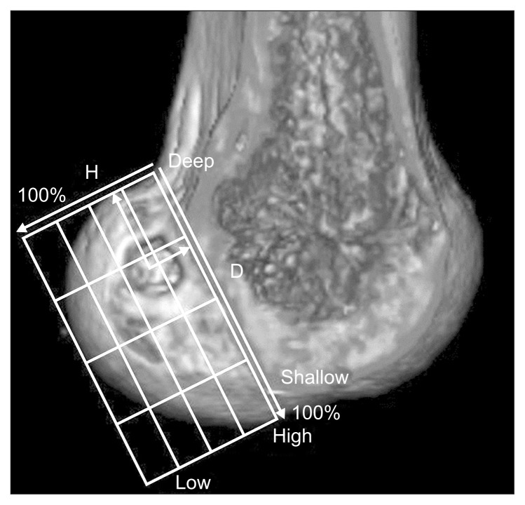
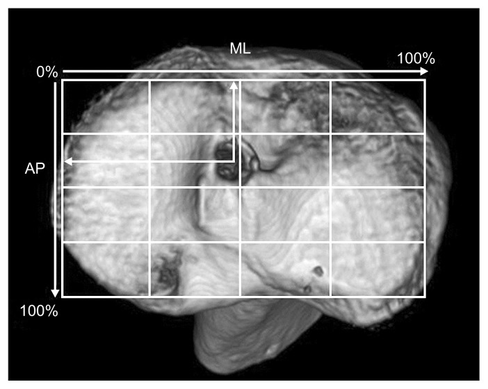
The femoral tunnel position was assessed in postoperative 3D CT (Fig. 1). The quadrant method (Bernard quadrant method) was used to locate the femoral tunnel11). This method is commonly used with simple radiography and 3D CT12). On 3D CT scans, a line connecting the most anterior and posterior edges of the intercondylar roof was the reference for Blumensaat’s line. The inferior border of the rectangle was a line tangent to the most distal point in the lateral condyle. Anterior and posterior edges of the lateral femoral condyle served as the other two borders to make a grid. ACL positioning was defined as a percentage of total sagittal diameter of lateral condyle and intercondylar notch height13). To evaluate tibial tunnel position, a true proximal-to-distal view on tibial plateau was used as described by Tsuda et al.14) (Fig. 2). Femoral and tibial tunnel positions were assessed twice by two clinical knee fellows. Interobserver reliability was then determined.
Fig. 1.
Location of the center of the femoral tunnel defined as the percentage of the distance from the most posterior contour in reference to the total length of the lateral condyle parallel to the Blumensaat’s line and the percentage of the distance from the intercondylar roof with respect to the total depth of the intercondylar notch perpendicular to the Blumensaat’s line. H: perpendicular to the Blumensaat’s line, D: parallel to the Blumensaat’s line.
Fig. 2.
Location of the center of the tibial tunnel defined as the percentage of the distance from the most medial contour with respect to the mediolateral (ML) width of the tibial plateau and the percentage of the distance from the most anterior contour with respect to the anteroposterior (AP) length of the tibial plateau.
5. Intraoperative Graft Isometry Assessment
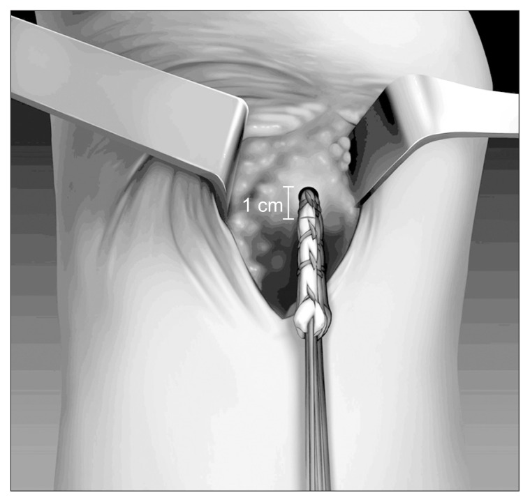
The graft length change was assessed before bio-screw fixation in the tibial tunnel by pulling the graft with tensions of 20 lbs and 30 lbs in full extension at flexion angles of 30°, 60°, 90°, and 120°. The graft position was marked one centimeter apart from the outer orifice of tibial tunnel in full extension which was used as a reference length (Fig. 3). During flexion of the knee, the marked point of graft was identified. When the graft was moved outward, the graft length in the joint was shortened, resulting in a negative value of the graft length change. When the graft was moved inward, the graft length in the joint was lengthened, resulting in a positive value of the graft length change (Fig. 4). Graft length change was assessed twice by two clinical knee fellows. Interobserver reliability was then determined.
Fig. 3.
Marking of graft position at one centimeter apart from the outer orifice of the tibial tunnel in full extension as a reference length.
Fig. 4.
Checking graft position change at each knee flexion angle. When the graft moves outward, the graft length in the joint is shortened, resulting in a negative value of graft length change. When the graft moves inward, the graft length in the joint is lengthened, resulting in a positive value of graft length change.
6. Statistical Analysis
Interobserver reliability was determined using intraclass correlation coefficient (ICC) test. Interpretation of ICC was based on the criteria of Fleiss as follows: excellent, ICC≥0.75; fair-to-good, 0.40≤ICC<0.75; and poor, ICC<0.40. Graft length changes at each flexion angle between tensions of 20 lbs and 30 lbs were compared by two-way repeated-measures of analysis of variance. All statistical analyses were performed using SPSS ver. 18.0 (SPSS Inc., Chicago, IL, USA).
Results
1. Subjects
A total of 60 patients were enrolled in the study, including 52 males and 8 females. Their mean age was 32.4 years (range, 15 to 56 years). Their mean height and body weight were 168±7 cm and 66±5 kg, respectively.
2. Tunnel Position
The center of the femoral tunnel was located at 31.58±5.48% in the shallow/deep direction along Blumensaat’s line and at 34.82±7.78% in the high/low direction from the intercondylar roof. The center of the tibial tunnel was located at 38.58±7.93% of the distance from the anterior edge of the tibia in anteroposterior direction and at 48.25±2.78% in mediolateral direction. This value is the average of measurements performed by the two clinical knee fellows. The ICC of interobserver reliability was 0.91, indicating excellent reliability.
3. Intraoperative Graft Isometry
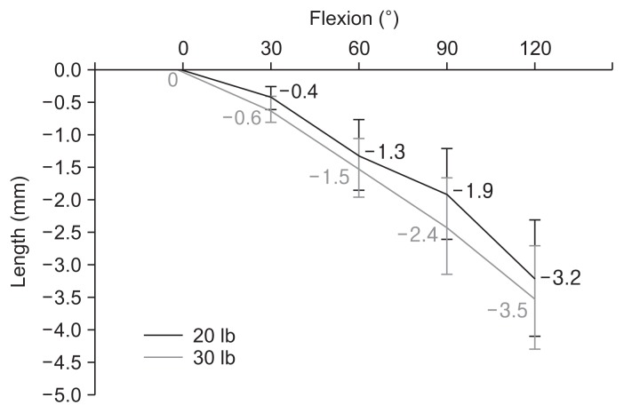
At 30° of flexion angle, 20 lbs and 30 lbs of tension resulted in length changes of −0.4 mm and −0.6 mm, respectively. The more the flexion angle of the knee, the shorter the graft length in the joint. At the flexion angles of 90° and 120°, there was significant (p<0.05) difference in the graft length change between 20 lbs and 30 lbs of tension. The ICC of interobserver reliability was 0.721, indicating fair-to-good reliability (Table 1, Fig. 5).
Table 1.
Length Change (mm) through Range of Motion with Tensions of 20 lbs and 30 lbs
| Variable | Flexion (°) | Maximum excursion | ||||
|---|---|---|---|---|---|---|
|
| ||||||
| 0 | 30 | 60 | 90 | 120 | ||
| 20 lb | 0 | −0.4±0.4 | −1.3±1.1 | −1.9±1.4 | −3.2±1.8 | 3.2 |
| 30 lb | 0 | −0.6±0.5 | −1.5±0.9 | −2.4±1.5 | −3.5±1.6 | 3.5 |
| p-value | 0.12 | 0.09 | <0.05 | <0.05 | <0.05 | |
Values are presented as mean±standard deviation.
Fig. 5.
Length change through range of motion with tensions of 20 lbs and 30 lbs.
Discussion
Graft isometry is relevant to graft tension during early postoperative phase, and it is also related to graft loosening. A cadaveric study has evaluated graft isometry in anatomic ACL reconstruction; however, a cadaveric knee model cannot reproduce muscle tension or tissue. Therefore, we evaluated isometry in anatomic ACL reconstruction in vivo. To the best of our knowledge, this is the first in vivo study that determines intraoperative graft isometry in anatomic ACL reconstruction. In this study, we defined a graft length change of less than 2 mm as isometric. According to this definition, the graft was isometric when the flexion angle of the knee was below 90°; it was non-isometric when the flexion angle was 90° or over. However, some authors defined graft isometry as a length change of less than 1 mm. According to such standard, the graft was isometric when flexion of the knee was less than 30° while it was non-isometric when it was 30° or above. Overall, anatomic single-bundle ACL reconstruction was non-isometric. The difference in graft length change between 20 lbs and 30 lbs of tension was not statistically significant at knee flexion below 90°. However, there was significant difference in the graft length change between the two tension groups at knee flexion angles of 90° or over. This indicates that the graft should be fixed using 20 lbs of tension rather than 30 lbs of tension to reduce the degree of non-isometry. In this study, 5 cases had a graft length change of more than 2 mm at the knee flexion angle of 30°. The mean graft length change was 2.3 mm. The femoral tunnel was located at 39.2% in depth and 40.5% in height. This indicates that the femoral tunnel in the 5 cases was formed at shallower and lower positions than that in the other cases. A long-term follow-up is needed for these cases.
Anatomical placement of a femoral tunnel is very important in ACL reconstruction. Kim et al.15) applied the Bernard quadrant method on 3D CT and found that it showed a high correlation with the conventional method using lateral radiographs. Therefore, the Bernard quadrant method can be used reliably for localizing a reconstructed ACL. This method was also used in this study to determine the femoral tunnel position. Colombet et al.16) reported that the anteromedial bundle was located at 26.4% in the shallow/deep direction and 25.3% in the high/low direction while the posterolateral bundle was located at 32.3% of the length and 47.6% of the height in their cadaveric study. Zantop et al.17) and Forsythe et al.18) also reported similar results in their studies. In the present study, the femoral tunnel was located at 31.58% of the length and 34.82% of the height. These results were within the ranges of those reported in previous studies.
In the cadaveric study of Lee et al.8), the anatomic femoral tunnel was non-isometric and graft length was the longest in full extension. When a femoral anatomic tunnel was chosen for ACL reconstruction, the anterior tibial tunnel offered greater isometric benefits than the conventional tibial tunnel. Our results were in line with results of this cadaveric study. In another cadaveric study by Smith et al.19), anatomic ACL positioning resulted in comparable anisometry to the native ACL. According to their results, a combination of a high anteromedial bundle femoral tunnel with an anteromedial bundle tibial tunnel resulted in the most isometric benefit among several options. Another important finding of this study was that ACL graft fixation in full extension would result in knee laxity. According to Lubowitz20), ACL laxity during flexion can be better tolerated by patients than irreversible graft stretch and graft rupture or extension loss after ACL reconstructive surgery.
The strength of this study was that it was the first in vivo study that evaluated intraoperative graft isometry in anatomic single-bundle ACL reconstruction. However, this study has some limitations. First, there was a lack of data on correlation between intraoperative graft isometry and follow-up clinical study, which should be investigated in a further study. Second, there might be measurement errors due to small length changes.
Conclusions
Anatomic single-bundle ACL reconstruction is non-isometric: the graft length is the longest in full extension, and tension of graft becomes loose in flexion. At a knee flexion angle of 90° or over, there was significant difference in graft length change between 20 lbs of tension and 30 lbs of tension. It would be appropriate to maintain a tension of 20 lbs for graft fixation.
Footnotes
Conflict of Interest
No potential conflict of interest relevant to this article was reported.
References
- 1.Lee DH, Kim HJ, Ahn HS, Bin SI. Comparison of femur tunnel aperture location in patients undergoing transtibial and anatomical single-bundle anterior cruciate ligament reconstruction. Knee Surg Sports Traumatol Arthrosc. 2016;24:3713–21. doi: 10.1007/s00167-015-3657-x. [DOI] [PubMed] [Google Scholar]
- 2.Chung JY, Ha CW, Lee DH, Park YG, Park YB, Awe SI. Anatomic placement of the femoral tunnel by a modified transtibial technique using a large-offset femoral tunnel guide: a cadaveric study. Knee. 2016;23:659–65. doi: 10.1016/j.knee.2015.10.001. [DOI] [PubMed] [Google Scholar]
- 3.Lee MC, Seong SC, Lee S, Chang CB, Park YK, Jo H, Kim CH. Vertical femoral tunnel placement results in rotational knee laxity after anterior cruciate ligament reconstruction. Arthroscopy. 2007;23:771–8. doi: 10.1016/j.arthro.2007.04.016. [DOI] [PubMed] [Google Scholar]
- 4.van Eck CF, Lesniak BP, Schreiber VM, Fu FH. Anatomic single- and double-bundle anterior cruciate ligament reconstruction flowchart. Arthroscopy. 2010;26:258–68. doi: 10.1016/j.arthro.2009.07.027. [DOI] [PubMed] [Google Scholar]
- 5.Chechik O, Amar E, Khashan M, Lador R, Eyal G, Gold A. An international survey on anterior cruciate ligament reconstruction practices. Int Orthop. 2013;37:201–6. doi: 10.1007/s00264-012-1611-9. [DOI] [PMC free article] [PubMed] [Google Scholar]
- 6.Yasuda K, van Eck CF, Hoshino Y, Fu FH, Tashman S. Anatomic single- and double-bundle anterior cruciate ligament reconstruction, part 1: basic science. Am J Sports Med. 2011;39:1789–99. doi: 10.1177/0363546511402659. [DOI] [PubMed] [Google Scholar]
- 7.Abebe ES, Utturkar GM, Taylor DC, Spritzer CE, Kim JP, Moorman CT, 3rd, Garrett WE, DeFrate LE. The effects of femoral graft placement on in vivo knee kinematics after anterior cruciate ligament reconstruction. J Biomech. 2011;44:924–9. doi: 10.1016/j.jbiomech.2010.11.028. [DOI] [PMC free article] [PubMed] [Google Scholar]
- 8.Lee JS, Kim TH, Kang SY, Lee SH, Jung YB, Koo S, Chang SH, Lee WB, Jung HJ. How isometric are the anatomic femoral tunnel and the anterior tibial tunnel for anterior cruciate ligament reconstruction? Arthroscopy. 2012;28:1504–12. doi: 10.1016/j.arthro.2012.03.010. [DOI] [PubMed] [Google Scholar]
- 9.Ko YW, Rhee SJ, Kim IW, Yoo JD. The correlation of tunnel position, orientation and tunnel enlargement in outside-in single-bundle anterior cruciate ligament reconstruction. Knee Surg Relat Res. 2015;27:247–54. doi: 10.5792/ksrr.2015.27.4.247. [DOI] [PMC free article] [PubMed] [Google Scholar]
- 10.Ahn JH, Jeong HJ, Ko CS, Ko TS, Kim JH. Three-dimensional reconstruction computed tomography evaluation of tunnel location during single-bundle anterior cruciate ligament reconstruction: a comparison of transtibial and 2-incision tibial tunnel-independent techniques. Clin Orthop Surg. 2013;5:26–35. doi: 10.4055/cios.2013.5.1.26. [DOI] [PMC free article] [PubMed] [Google Scholar]
- 11.Bernard M, Hertel P, Hornung H, Cierpinski T. Femoral insertion of the ACL: radiographic quadrant method. Am J Knee Surg. 1997;10:14–21. [PubMed] [Google Scholar]
- 12.Hoser C, Tecklenburg K, Kuenzel KH, Fink C. Postoperative evaluation of femoral tunnel position in ACL reconstruction: plain radiography versus computed tomography. Knee Surg Sports Traumatol Arthrosc. 2005;13:256–62. doi: 10.1007/s00167-004-0548-y. [DOI] [PubMed] [Google Scholar]
- 13.Fernandes TL, Martins NM, de Watai FA, Albuquerque C, Jr, Pedrinelli A, Hernandez AJ. 3D computer tomography for measurement of femoral position in acl reconstruction. Acta Ortop Bras. 2015;23:11–5. doi: 10.1590/1413-78522015230100993. [DOI] [PMC free article] [PubMed] [Google Scholar]
- 14.Tsuda E, Ishibashi Y, Fukuda A, Yamamoto Y, Tsukada H, Ono S. Tunnel position and relationship to postoperative knee laxity after double-bundle anterior cruciate ligament reconstruction with a transtibial technique. Am J Sports Med. 2010;38:698–706. doi: 10.1177/0363546509351561. [DOI] [PubMed] [Google Scholar]
- 15.Kim DH, Lim WB, Cho SW, Lim CW, Jo S. Reliability of 3-dimensional computed tomography for application of the bernard quadrant method in femoral tunnel position evaluation after anatomic anterior cruciate ligament reconstruction. Arthroscopy. 2016;32:1660–6. doi: 10.1016/j.arthro.2016.01.043. [DOI] [PubMed] [Google Scholar]
- 16.Colombet P, Robinson J, Christel P, Franceschi JP, Djian P, Bellier G, Sbihi A. Morphology of anterior cruciate ligament attachments for anatomic reconstruction: a cadaveric dissection and radiographic study. Arthroscopy. 2006;22:984–92. doi: 10.1016/j.arthro.2006.04.102. [DOI] [PubMed] [Google Scholar]
- 17.Zantop T, Wellmann M, Fu FH, Petersen W. Tunnel positioning of anteromedial and posterolateral bundles in anatomic anterior cruciate ligament reconstruction: anatomic and radiographic findings. Am J Sports Med. 2008;36:65–72. doi: 10.1177/0363546507308361. [DOI] [PubMed] [Google Scholar]
- 18.Forsythe B, Kopf S, Wong AK, Martins CA, Anderst W, Tashman S, Fu FH. The location of femoral and tibial tunnels in anatomic double-bundle anterior cruciate ligament reconstruction analyzed by three-dimensional computed tomography models. J Bone Joint Surg Am. 2010;92:1418–26. doi: 10.2106/JBJS.I.00654. [DOI] [PubMed] [Google Scholar]
- 19.Smith JO, Yasen S, Risebury MJ, Wilson AJ. Femoral and tibial tunnel positioning on graft isometry in anterior cruciate ligament reconstruction: a cadaveric study. J Orthop Surg (Hong Kong) 2014;22:318–24. doi: 10.1177/230949901402200310. [DOI] [PubMed] [Google Scholar]
- 20.Lubowitz JH. Anatomic ACL reconstruction produces greater graft length change during knee range-of-motion than transtibial technique. Knee Surg Sports Traumatol Arthrosc. 2014;22:1190–5. doi: 10.1007/s00167-013-2694-6. [DOI] [PubMed] [Google Scholar]


