Resumo
A persistência da artéria isquiática é uma anomalia vascular congênita rara cuja principal complicação é a dilatação aneurismática. O quadro clínico pode incluir sintomas decorrentes da dilatação arterial e da isquemia, causada por trombose ou embolização distal. O tratamento dessa afecção rara conta com opções diversas que abrangem desde a ligadura do aneurisma até a correção endovascular. O presente relato descreve o caso de uma paciente do sexo feminino com queixa de tumoração pulsátil na região glútea. Foi encaminhada ao serviço de referência e realizou angiotomografia, que evidenciou persistência completa da artéria isquiática bilateralmente, com dilatação aneurismática à esquerda. A paciente foi submetida a tratamento endovascular do aneurisma, através de punção contralateral, com implante de dois stents revestidos com manutenção da perviedade distal da artéria. A manutenção da perviedade é particularmente importante nos casos da forma completa dessa variação anatômica. A paciente cursou com boa evolução.
Palavras-chave: aneurisma, procedimentos endovasculares, persistência da artéria isquiática, extremidade inferior
INTRODUÇÃO
A persistência da artéria isquiática é uma anomalia vascular rara, com incidência estimada em 0,01 a 0,06%1 , 2. Consiste em uma anomalia congênita na qual a artéria isquiática persistente (AIP) se situa em continuidade com a artéria ilíaca interna após a emergência das artérias glútea superior e pudenda interna3. A persistência da artéria isquiática pode ser classificada como forma completa ou incompleta. Na forma completa, ela continua até a artéria poplítea, sendo a artéria dominante da extremidade1. Na forma incompleta, a artéria isquiática persistente é hipoplásica, sendo o sistema femoral dominante, e pode haver comunicação entre a AIP e a artéria poplítea através de ramos colaterais1.
A dilatação aneurismática é uma das alterações que podem ocorrer na AIP, afetando cerca de 44 a 48% dos casos1 , 2. A etiologia da dilatação aneurismática não está clara; pode estar relacionada a repetidos traumas, predisposição a aterosclerose e hipoplasia do tecido conectivo1. Sintomas clínicos variáveis, com casos de isquemia e dilatação aneurismática, são relatados na literatura1 - 6.
Este trabalho relata um caso de persistência bilateral da artéria isquiática com dilatação aneurismática à esquerda. Trata-se de um caso diagnosticado e tratado através da técnica endovascular pelo Serviço de Cirurgia Vascular no Complexo Hospitalar Universitário Professor Edgard Santos/Hospital Ana Nery na cidade de Salvador (BA).
DESCRIÇÃO DO CASO
Paciente do sexo feminino, 76 anos, com história de sensação de pulsação anormal, indolor, na região glútea esquerda por aproximadamente cinco anos. Nos cinco meses anteriores, percebeu pulsação na genitália e dor na região glútea esquerda associada aos movimentos, como ao se sentar. Negava história de trauma local, claudicação intermitente ou outras queixas nos membros inferiores. Não relatava outras comorbidades, apenas passado de tabagismo havia mais de 20 anos. Procurou atendimento em sua cidade de origem, onde foi inicialmente avaliada com ultrassom de partes moles, que identificou dilatação vascular medindo 2,7 cm no tecido celular subcutâneo da região glútea esquerda. Após a realização do exame ultrassonográfico, a paciente foi encaminhada para avaliação no Serviço de Cirurgia Vascular do Complexo Hospitalar Universitário Professor Edgard Santos/Hospital Ana Nery.
A paciente foi admitida no ambulatório do serviço e, ao exame físico, apresentava massa pulsátil com cerca de 5 x 7 cm de diâmetro no quadrante inferolateral da região glútea esquerda, indolor, com frêmito à palpação. Ao exame vascular, todos os pulsos estavam presentes e simétricos.
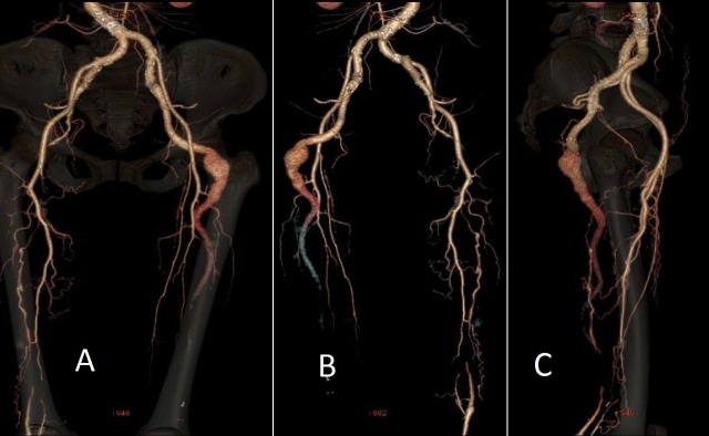
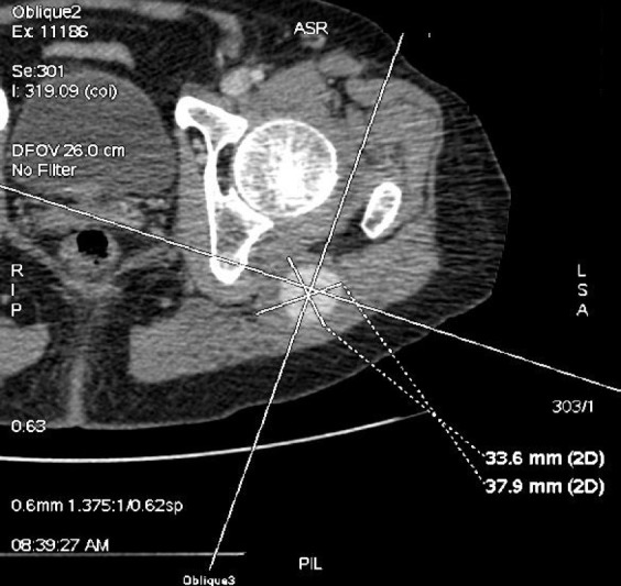
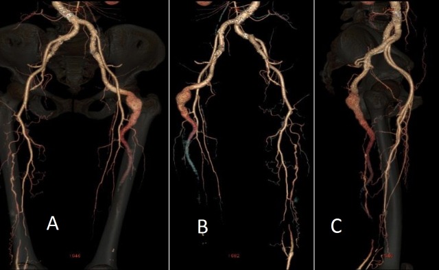
Diante do quadro clínico e dos exames físico e ultrassonográfico, formulou-se a hipótese diagnóstica de aneurisma da AIP. Foi solicitada uma angiotomografia de aorta abdominal e de membros inferiores, que demonstrou aorta abdominal de calibre normal e trajeto levemente tortuoso em segmento infrarrenal, com ateromatose parietal difusa e sem estenoses. Evidenciou-se também perviedade das artérias ilíacas comuns, internas e externas, destacando-se hipoplasia das ilíacas externas e predominância das ilíacas internas, que continuavam bilateralmente com a artéria isquiática na face posterior de ambas as coxas. Observava-se, ainda, ectasia da AIP à esquerda, com aneurisma fusiforme e trombo mural, com maior diâmetro de 3,7 x 3,3 cm e extensão de 6,4 cm (Figuras 1 e 2).
Figura 1. Angiotomografia em corte axial evidenciando o diâmetro (37,9 x 33,6 mm) do aneurisma da artéria isquiática persistente.
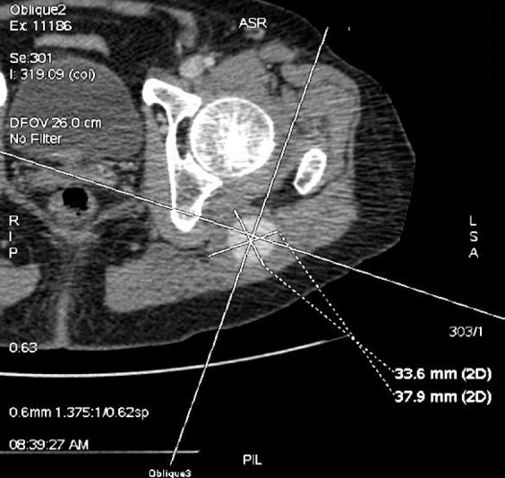
Figura 2. Angiotomografia evidenciando persistência bilateral da artéria isquiática com formação aneurismática à esquerda: imagens de reconstrução na visão anterior (A), posterior (B) e lateral (C).
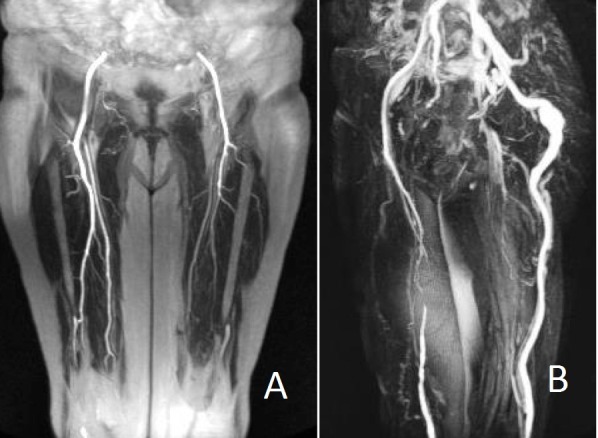
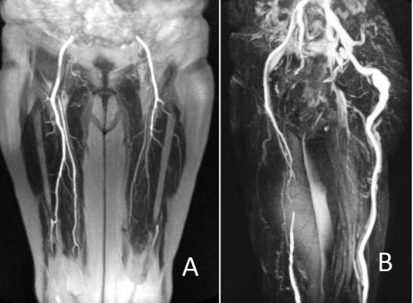
Foi realizada uma angiorressonância para detalhar as artérias dos membros inferiores. Havia hipoplasia de ambas as artérias femorais superficiais e as artérias femorais comuns e profundas estavam com calibres preservados. A artéria isquiática continuava, bilateralmente, com a artéria poplítea, caracterizando a forma completa dessa anomalia vascular. As artérias infrageniculares encontravam-se pérvias e com calibres normais (Figura 3).
Figura 3. Angiorressonância: hipoplasia de artérias femorais superficiais e profundas (A) e artérias isquiáticas dando origem às artérias poplíteas, o que caracteriza a forma completa de persistência da artéria isquiática bilateralmente. Dilatação aneurismática fusiforme da artéria isquiática persistente à esquerda (B).
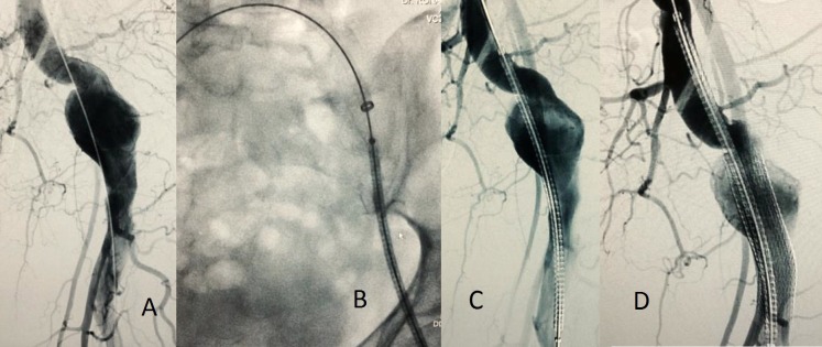
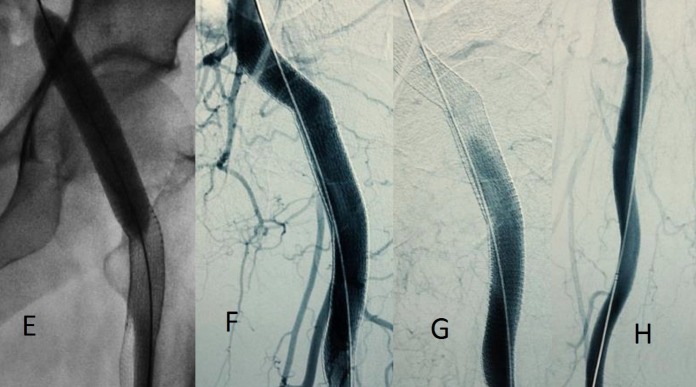
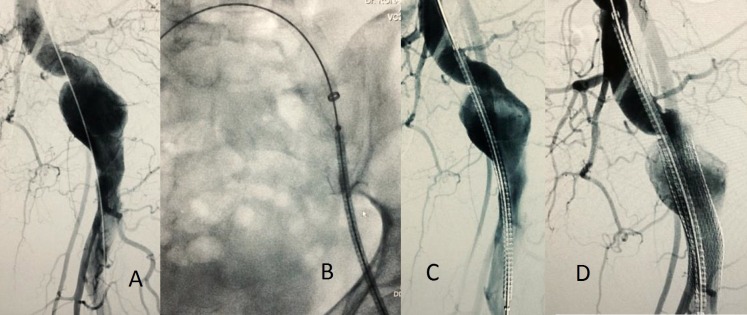
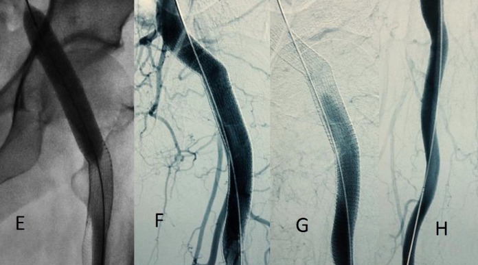
A paciente foi então encaminhada para tratamento endovascular do aneurisma sintomático da AIP esquerda, visto que mantinha quadro de dor local progressiva e constante. O tratamento proposto foi a correção endovascular do aneurisma de AIP com dois stents revestidos (Viabahn® - Gore®), para exclusão completa do saco aneurismático e manutenção da patência da artéria isquiática. Por se tratar de um caso de persistência completa, a AIP fornece o principal aporte sanguíneo para o membro inferior esquerdo, tendo continuidade com a artéria poplítea. O tratamento endovascular do aneurisma de AIP à esquerda foi iniciado através de acesso contralateral pela artéria femoral comum direita, sendo realizado o implante do stent revestido distal (13 x 10 mm), seguido do implante do segundo stent revestido proximal (11 x 10 mm), com bom aspecto (Figuras 4 e 5).
Figura 4. Tratamento endovascular do aneurisma da artéria isquiática persistente à esquerda: (A) angiografia através de acesso contralateral pela artéria femoral comum direita; (B) e (C) posicionamento do primeiro stent revestido (distal) 13 x 10 mm. (D) posicionamento do segundo stent revestido (proximal) 11 x 10 mm.
Figura 5. Tratamento endovascular do aneurisma da artéria isquiática persistente: (E) balonamento proximal, distal e em conexão dos stents revestidos com balão 12 x 60 mm; (F) e (G) angiografia de controle evidenciando exclusão do aneurisma e ausência de vazamento; (H) angiografia de artéria isquiática distalmente ao tratamento do aneurisma mostrando perviedade.
Não houve intercorrência durante a correção endovascular do aneurisma de AIP, e a paciente se manteve estável durante todo o procedimento, recebendo alta hospitalar dois dias após o procedimento, com melhora da dor e ausência de massa pulsátil na região glútea esquerda. A paciente concordou em participar do relato de caso através do termo de consentimento livre e esclarecido. Após um ano do tratamento endovascular, a paciente mantinha-se assintomática em acompanhamento ambulatorial no serviço, com pulsos distais presentes. Nesse período, foi realizada uma angiotomografia de controle, que evidenciou stents revestidos pérvios e sem vazamentos, com regressão completa da dilatação aneurismática na AIP esquerda (Figura 6).
Figura 6. (A) Angiotomografia realizada após o tratamento endovascular, com reconstrução tridimensional em anteroposterior, mostrando stents revestidos na artéria isquiática persistente pérvios e sem vazamentos; (B) corte axial da angiotomografia evidenciando regressão completa da dilatação aneurismática na artéria isquiática persistente esquerda.

DISCUSSÃO
A artéria isquiática é uma continuação da artéria ilíaca interna, sendo a principal responsável pelo suprimento vascular para o membro inferior durante o período embrionário3. No terceiro mês de desenvolvimento embrionário, a artéria isquiática regride e o sistema femoral torna-se o principal responsável pelo fluxo sanguíneo do membro inferior3 , 7. Apesar de a AIP ser uma ocorrência rara na população, a possibilidade de isquemia e dilatação aneurismática torna o diagnóstico diferencial importante na prática clínica7.
O quadro clínico pode variar desde o paciente assintomático, com queixa de claudicação intermitente ou presença de massa pulsátil, até quadros de emergência como isquemia aguda de membro inferior ou aneurisma roto2 , 8 , 9. Uma revisão da literatura realizada por Van Hooft et al. mostrou que 80% dos doentes eram sintomáticos à época do diagnóstico2. Ao exame físico, o sinal de Cowie, que consiste na ausência do pulso femoral com pulso poplíteo palpável, foi descrito em poucos casos2 , 10.
Em alguns trabalhos, a arteriografia vem sendo descrita como principal método diagnóstico nos casos de AIP1 - 3. No entanto, um estudo realizado com uso de angiotomografia mostrou que esse método também pode ser utilizado na avaliação da AIP11. Jung et al. avaliaram 307 angiotomografias e encontraram AIP em seis pacientes (1,63% dos exames), com dilatação aneurismática em dois casos1.
O tratamento da AIP varia de acordo com a apresentação clínica do doente. Os pacientes assintomáticos, sem dilatação, provavelmente podem ser acompanhados regularmente através dos exames físico e de imagem1. Nos quadros de isquemia de membro inferior, os casos relatados mostraram a realização de pontes femoropoplíteas ou femorodistais com a utilização da veia safena magna como substituto arterial4 , 6 , 9 , 12 - 15. A utilização de prótese de dácron e a angioplastia de AIP também são descritas na literatura16 , 17. Nos casos de claudicação, o acompanhamento sem tratamento cirúrgico foi indicado em alguns casos3 , 13 , 18. Nas persistências incompletas, a ligadura da AIP, sem revascularização, também já foi descrita3 , 19. Uma revisão da literatura com 146 casos de AIP encontrou cinco casos de amputações maiores2. Nas dilatações aneurismáticas, os tratamentos descritos mostram as possibilidades de ligadura do aneurisma através da cirurgia convencional ou endovascular pela embolização, com ou sem necessidade de revascularização, devendo ser avaliado se a persistência é completa ou incompleta2 , 6 , 13 , 19.
Na nossa paciente, optamos pelo tratamento endovascular com uso de dois stents revestidos, por se tratar de persistência completa, com bom resultado. Há relato na literatura de um paciente de 53 anos com aneurisma da AIP de 7 cm associado à isquemia distal, em que os autores descrevem o tratamento endovascular através da utilização de dois stents revestidos (Hemobahn®)5. Apesar da raridade do diagnóstico, o tratamento endovascular do aneurisma da AIP pode evitar potenciais complicações relacionadas ao acesso cirúrgico, devido à proximidade do nervo ciático e veia7. Quanto à possibilidade de fratura do stent revestido, um estudo realizado com aneurismas da artéria poplítea tratados através dessa técnica revelou um índice de fratura de 16,7%, sem influência significativa na patência do stent 20.
O aneurisma da AIP é uma doença rara, que requer um alto índice de suspeição para seu diagnóstico. O tratamento passa por uma ampla gama de possibilidades, na dependência do quadro clínico das características anatômicas de cada caso. O estudo completo da circulação pélvica e dos membros inferiores é recomendável para o planejamento do tratamento. Os avanços da cirurgia endovascular podem trazer contribuições para o tratamento da AIP, com sucesso terapêutico, como no caso aqui relatado.
Footnotes
Fonte de financiamento: Nenhuma.
O estudo foi realizado no Complexo Hospitalar Universitário Professor Edgard Santos/Hospital Ana Nery, Universidade Federal da Bahia (UFBA), Salvador, BA, Brasil.
REFERÊNCIAS
- 1.Brantley SK, Rigdon EE, Raju S. Persistent sciatic artery: embryology, pathology and treatment. J Vasc Surg. 1993;18(2):242–248. http://dx.doi.org/10.1016/0741-5214(93)90604-K [PubMed] [Google Scholar]
- 2.van Hooft IM, Zeebregts CJ, van Sterkenburg SMM, de Vries WR, Reijnen MMPJ. The persistent sciatic artery. Eur J Vasc Endovasc Surg. 2009;37(5):585–591. doi: 10.1016/j.ejvs.2009.01.014. http://dx.doi.org/10.1016/j.ejvs.2009.01.014 [DOI] [PubMed] [Google Scholar]
- 3.Mandell VS, Jaques PF, Delany DJ, Oberheu V. Persistent sciatic artery: clinical, embryologic, and angiographic features. AJR. 1985;144(2):245–249. doi: 10.2214/ajr.144.2.245. http://dx.doi.org/10.2214/ajr.144.2.245 [DOI] [PubMed] [Google Scholar]
- 4.Patel MV, Patel NH, Schneider JR, Kim S, Verta MJ. Persistent sciatic artery presenting with limb Ischemia. J Vasc Surg. 2013;57(1):225–229. doi: 10.1016/j.jvs.2012.06.108. http://dx.doi.org/10.1016/j.jvs.2012.06.108 [DOI] [PubMed] [Google Scholar]
- 5.Nuño-Escobar C, Pérez-Durán MA, Ramos-López R, et al. Persistent sciatic artery aneurysm. Ann Vasc Surg. 2013;27(8):1182.e13–6. doi: 10.1016/j.avsg.2013.04.003. http://dx.doi.org/10.1016/j.avsg.2013.04.003 [DOI] [PubMed] [Google Scholar]
- 6.Knight BC, Tait WF. Massive aneurysmen a peersistent sciatic artery. Ann Vasc Surg. 2010;24(8):1135.e13–8. doi: 10.1016/j.avsg.2010.05.017. http://dx.doi.org/10.1016/j.avsg.2010.05.017 [DOI] [PubMed] [Google Scholar]
- 7.Savov JD, Wassilev WA. Bilateral persistent complete sciatic artery. Clin Anat. 2000;13(6):456–460. doi: 10.1002/1098-2353(2000)13:6<456::AID-CA12>3.0.CO;2-T. http://dx.doi.org/10.1002/1098-2353(2000)13:6<456::AID-CA12>3.0.CO;2-T [DOI] [PubMed] [Google Scholar]
- 8.Rezayat C, Sambol E, Goldstein L, et al. Ruptured persistent sciatic artery aneurysm: managed by endovascular embolization. Ann Vasc Surg. 2010;24(1):115.e5–9. doi: 10.1016/j.avsg.2009.07.003. http://dx.doi.org/10.1016/j.avsg.2009.07.003 [DOI] [PubMed] [Google Scholar]
- 9.Yang S, Ranum K, Malone M, Nazzal M. Bilateral Persistent Sciatic Artery with Aneurysm Formation and Review of the Literature. Ann Vasc Surg. 2014;28(1):264.e1–7. doi: 10.1016/j.avsg.2013.03.015. http://dx.doi.org/10.1016/j.avsg.2013.03.015 [DOI] [PubMed] [Google Scholar]
- 10.Brancaccio G, Falco E, Pera M, Celoria G, Stefanini T, Puccianti F. Symptomatic persistent sciatic artery. J Am Coll Surg. 2004;198(1):158. doi: 10.1016/j.jamcollsurg.2003.06.008. http://dx.doi.org/10.1016/j.jamcollsurg.2003.06.008 [DOI] [PubMed] [Google Scholar]
- 11.Jung AY, Lee W, Chung JW, et al. Role of computed tomographic angiography in the detection and comprehensive evaluation of persistent sciatic artery. J Vasc Surg. 2005;42(4):678–683. doi: 10.1016/j.jvs.2005.06.001. http://dx.doi.org/10.1016/j.jvs.2005.06.001 [DOI] [PubMed] [Google Scholar]
- 12.Vaz C, Machado R, Rego D, Matos A, Almeida R. Hybrid approach in a case of persistent sciatic artery aneurysm. Ann Vasc Surg. 2014;28(5):1313.e5–7. doi: 10.1016/j.avsg.2013.08.028. http://dx.doi.org/10.1016/j.avsg.2013.08.028 [DOI] [PubMed] [Google Scholar]
- 13.Mayschak DT, Flye MW. Treatment of the persistent sciatic artery. Ann Surg. 1984;199(1):69–74. doi: 10.1097/00000658-198401000-00012. http://dx.doi.org/10.1097/00000658-198401000-00012 [DOI] [PMC free article] [PubMed] [Google Scholar]
- 14.Tiago J, Ministro A, Evangelista A, Damião A, Leitão J, Dinis da Gama A. Lower limb ischemia due to occlusion of a persistent sciatic artery aneurysm- a case report. Angiol Cir Vasc. 2012;8(2):70–72. [Google Scholar]
- 15.Bez LG, Costa-Val R, Bastianetto P, et al. Persistência da artéria isquiática: relato de caso. J Vasc Bras. 2006;5(3):233–236. http://dx.doi.org/10.1590/S1677-54492006000300014 [Google Scholar]
- 16.Nunes MA, Ribeiro RM, Aragão JA, Reis FP, Feitosa VL. Diagnóstico e tratamento de aneurisma da artéria isquiática persistente: relato de caso e revisão da literatura. J Vasc Bras. 2008;7(1):66–71. http://dx.doi.org/10.1590/S1677-54492008000100012 [Google Scholar]
- 17.Szejnfeld D, Belczak SQ, Sincos IR, Aun R. Angioplasty of a persistent sciatic artery: case report. J Vasc Bras. 2011;10(2):168–172. http://dx.doi.org/10.1590/S1677-54492011000200013 [Google Scholar]
- 18.Wang B, Liu Z, Shen L. Bilateral persistent sciatic arteries complicated with chronic lower limb ischemia. Int J Surg Case Rep. 2011;2(8):309–312. doi: 10.1016/j.ijscr.2011.07.010. http://dx.doi.org/10.1016/j.ijscr.2011.07.010 [DOI] [PMC free article] [PubMed] [Google Scholar]
- 19.Handa GI, Coral FE, Buzingnani VZ, et al. Tratamento cirúrgico de pseudoaneurisma de artéria isquiática – relato de caso e revisão da literatura. J Vasc Bras. 2011;10(3):256–260. http://dx.doi.org/10.1590/S1677-54492011000300013 [Google Scholar]
- 20.Tielliu IFJ, Zeebregts CJ, Vourliotakis G, et al. Stent fractures in the Hemobahn/Viabahn stent graft after endovascular popliteal aneurysm repair. J Vasc Surg. 2010;51(6):1413–1418. doi: 10.1016/j.jvs.2009.12.071. http://dx.doi.org/10.1016/j.jvs.2009.12.071 [DOI] [PubMed] [Google Scholar]


