Abstract
A 60-year-old lady presented with symptoms of chronic iron deficiency anaemia. On evaluation with routine chest X-ray, it surprisingly revealed an irregular mass-like opacity in proximal part of stomach suggesting gastric cancer. The diagnosis was later confirmed with upper GI endoscopy and CT scan abdomen. Hence, in modern era of advanced diagnostics, chest X-ray findings should not be overlooked.
Keywords: Gastric cancer, Chest X-ray, Diagnosis, Anaemia
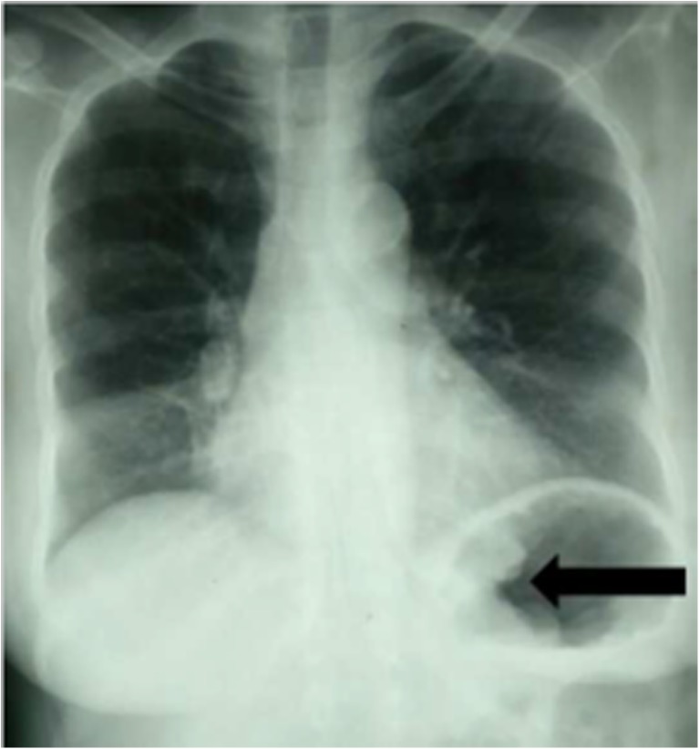
A 60-year-old lady with a known case of diabetes mellitus presented with complaints of episodic black coloured stool, malaise and dizziness for 3 months duration. She has undergone 3 pints of blood transfusion for chronic iron deficiency anaemia. She denies any history of pain abdomen, early satiety or loss of weight. On physical and abdominal examination, there were no significant findings other than pallor. Blood investigations showed haemoglobin of 6.0 g/dl. Liver function and renal function tests were within normal limits. A chest X-ray (CXR) was done which surprisingly revealed an irregular, funnel shaped, soft tissue opacity in cardia and fundus of stomach suggesting a mass lesion in stomach (Fig. 1). Upper GI endoscopy and biopsy confirmed the finding of advanced proximal gastric adenocarcinoma. As part of staging, high-resolution CT scan of abdomen was done which showed the similar lesion in stomach corresponding to CXR findings (Fig. 2). The patient underwent total gastrectomy followed by adjuvant therapy. At 1 year of follow-up, the patient is doing well.
Fig. 1.
Chest X-ray showing irregular soft tissue opacity in proximal part of stomach
Fig. 2.
CT scan abdomen showing irregular thickened cardia and fundus of stomach
Diagnosis of gastric cancer is generally made by upper GI endoscopy and CT scan [1]. Chest X-ray radiograph has minimal role as part of routine investigation. As technology has progressed, new diagnostic and staging modalities like multidetector CT scan, endoscopic ultrasound (EUS), magnetic resonance imaging (MRI) and recently PET/CT scan has emerged as initial investigation of choice [2, 3]. Chest X-ray as a diagnostic modality falls back and is overlooked in modern era of advances in diagnostics. Hence, to the beginners and residents point of view, CXR is an important investigation which can reveal much more information, and if the minds are kept open, sometimes gastric cancer can be diagnosed with CXR as seen in the present case.
Compliance with Ethical Standards
We declare that this manuscript represents valid work and that neither this manuscript nor one with substantially similar content under the present authorship has been published or is being considered for publication elsewhere and the authorship of this article will not be contested by anyone whose name(s) is/are not listed here, and that the order of authorship as placed in the manuscript is final and accepted by the co-authors.
There is no prepublished information/material.
No support in financial or other manner is received from any agencies.
Conflict of Interest
The authors declare that they have no conflict of interest.
Contributor Information
Narendra Pandit, Email: narendrapandit111@gmail.com.
Harjeet Singh, Email: harjeetsingh1982@gmail.com.
Lokesh Shekher Jaiswal, Email: lokesh.jaiswal@bpkihs.edu.
References
- 1.Schwarz RE. Current status of management of malignant disease: current management of gastric cancer. J Gastrointest Surg. 2015;19(4):782–788. doi: 10.1007/s11605-014-2707-x. [DOI] [PubMed] [Google Scholar]
- 2.Russell MC, Mansfield PF. Surgical approaches to gastric cancer. J Surg Oncol. 2013;107(3):250–258. doi: 10.1002/jso.23180. [DOI] [PubMed] [Google Scholar]
- 3.Waddell T, Verheij M, Allum W, et al. Gastric cancer: ESMO-ESSO-ESTRO clinical practice guidelines for diagnosis, treatment and follow-up. Eur J Surg Oncol. 2014;40:584–591. doi: 10.1016/j.ejso.2013.09.020. [DOI] [PubMed] [Google Scholar]


