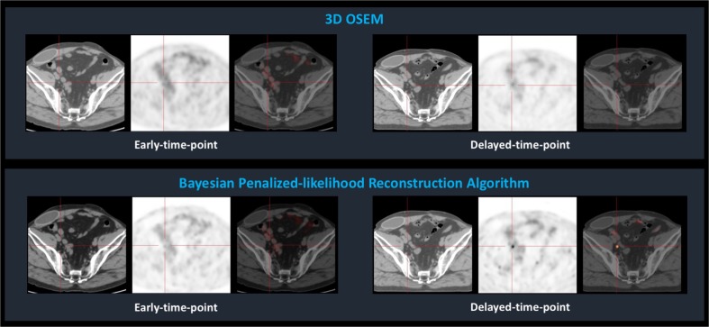
The benefit of using Ga-68-PSMA PET for patients with biochemical recurrence after radical prostatectomy is high [1, 2], and a delayed-time-point (DTP) acquisition protocol has proved to improve the capacity of Ga-68-PSMA PET to detect prostate cancer metastases [3]. New hardware and reconstruction methods are challenging the resolution limits of PET imaging, and Bayesian penalized-likelihood reconstruction algorithms (BPLA) are paradigmatic of this ongoing revolution. BPLA allows an effective convergence, in contrast to Ordered Subset Expectation Maximization (OSEM), which must be stopped before contrast convergence to prevent excessive image noise [4]. We present the Ga-68-PSMA PET images of a man with biochemical recurrence of prostate cancer (PSA = 0.51 ng/ml). A dual-time-point acquisition protocol (1 h and 2 h after radiopharmaceutical administration) was conducted in a Discovery IQ4R, and images were reconstructed using OSEM and BPLA. Early-time-point PET did not reveal foci of abnormally increased uptake, either using OSEM or BPLA. DTP images revealed a focus of increased uptake in a 3 mm right external iliac lymph node when using BPLA, not visible with OSEM. After robotic radiosurgery, PSA values decreased to undetectable, confirming the focus as a metastasis. Ga-68-PSMA PET is advantageous over other imaging techniques to detect prostate cancer recurrence, especially in patients with low PSA levels, but the reported detection rate of metastatic sites in patients with biochemical recurrence after radical prostatectomy in a PSA-range of 0.5–1.0 ng/ml does not surpass 73% [5]. Applying a BPLA to a DTP protocol may further improve the detection rate of Ga-68-PSMA PET for recurrent prostate cancer.
Compliance with ethical standards
Conflicts of interest
None.
References
- 1.Rauscher I, Maurer T, Fendler WP, Sommer WH, Schwaiger M, Eiber M. 68Ga-PSMA ligand PET/CT in patients with prostate cancer: How we review and report. Cancer Imaging. 2016;16:14. doi: 10.1186/s40644-016-0072-6. [DOI] [PMC free article] [PubMed] [Google Scholar]
- 2.Fendler WP, Eiber M, Beheshti M, Bomanji J, Ceci F, Cho S, et al. 68Ga-PSMA PET/CT: Joint EANM and SNMMI procedure guideline for prostate cancer imaging: version 1.0. Eur J Nucl Med Mol Imaging. 2017;44:1014–1024. doi: 10.1007/s00259-017-3670-z. [DOI] [PubMed] [Google Scholar]
- 3.Afshar-Oromieh A, Malcher A, Eder M, Eisenhut M, Linhart HG, Hadaschik BA, et al. PET imaging with a [68Ga]gallium-labelled PSMA ligand for the diagnosis of prostate cancer: biodistribution in humans and first evaluation of tumour lesions. Eur J Nucl Med Mol Imaging. 2013;40:486–495. doi: 10.1007/s00259-012-2298-2. [DOI] [PubMed] [Google Scholar]
- 4.Asma E, Ahn S, Ross SG, Chen A, Manjeshwar RM. Accurate and consistent lesion quantitation with clinically acceptable penalized likelihood images. 2012 I.E. Nuclear Science Symposium and Medical Imaging Conference Record (NSS/MIC); 2012. p. 4062–6.
- 5.Eiber M, Maurer T, Souvatzoglou M, Beer AJ, Ruffani A, Haller B, et al. Evaluation of Hybrid 68Ga-PSMA Ligand PET/CT in 248 Patients with Biochemical Recurrence After Radical Prostatectomy. J Nucl Med. 2015;56:668–674. doi: 10.2967/jnumed.115.154153. [DOI] [PubMed] [Google Scholar]


