Abstract
Typha is a cosmopolitan aquatic plant genus that includes species with widespread distributions. It is a relatively ancient genus with an abundant fossil record dating back to the Paleogene. However, the details of its biogeographic history have remained unclear until now. In this study, we present a revised molecular phylogeny using sequences of seven chloroplast DNA markers from nine species sampled from various regions in order to infer the biogeographic history of the genus. Two clades were recovered with robust support. Typha minima and T. elephantina comprised one clade, and the other clade included the remaining seven species, which represented a polytomy of four robustly supported subclades. Two widespread species, T. angustifolia and T. domingensis, were revealed to be paraphyletic, indicating the need for taxonomic revision. Divergence time estimation suggested that Typha had a mid-Eocene crown origin, and its diversification occurred in the Middle and Late Miocene. Ancestral area reconstruction showed that Typha possibly originated from eastern Eurasia. Both dispersal via the Beringian Land Bridge and recent transoceanic dispersal may have influenced the intercontinental distribution of Typha species.
Subject terms: Phylogenetics, Plant evolution
Introduction
Typha L. (Typhaceae), also known as cattail, is a globally distributed aquatic plant genus. It grows in a variety of aquatic habitats on all continents except Antarctica1. Cattail is often dominant in wetlands and it is of concern in some regions due to its economic and ecological impact2–4. Several species are considered serious weeds that reduce biodiversity because they are highly productive by clonal growth, forming very large, persistent, and often monospecific stands2,5,6.
Typha includes 10–13 species, and most species have a widespread distribution1,7. Currently, taxonomic studies of Typha are mainly limited to a specific country or region, such as India8, Europe9, Iran and Pakistan10,11, Australia12, North America13, or China14. Due to the high morphological variability and frequent interspecific hybridization1,15, the taxonomy of Typha has been a longstanding debate. Traditionally, the genus was classified into two sections (Ebracteolatae and Bracteolatae) based on the presence or absence of bracteoles in the pistillate flowers, respectively16,17. In 1987, Smith1 made a taxonomic revision of Typha and recognized 8–13 species in six groups (without sections or subsections) based on the presence or absence of bracteole, in addition to morphological characteristics of the stigma and pollen grains. Fifteen new species were published after 198718. All these species were local species, and none of them were presented with the support of molecular evidence. Some studies showed that the morphological characters of some new species overlapped with those of existing species. For example, Zhu19 measured a large number of specimens and found that the key characters of two new species, T. tzvelevii sp. nova and T. joannis sp. nova20, were remarkably similar to T. laxmannii and T. orientalis, respectively. Zhu therefore questioned the validity of the two new species19. Similarly, the validity of three endemic Chinese species (T. przewalskii, T. davidiana, and T. changbaiensis) was placed in doubt by two morphological studies19,21, which were supported by a molecular study with extensive sampling throughout China22. In a recently published handbook, Mabberley listed Typha with 10–12 species7. Therefore, we described Typha with 10–13 species based on the work of Smith1 and Mabberley7.
Typha is a relatively ancient genus. The earliest Typha fossil records that have been found were seeds assigned to T. ochreaceae Knobloch and Mai and T. protogaea Knobloch and Mai from the Late Cretaceous (Maastrichtian) period in Eisleben, Germany23. The earliest fossil record of Sparganium L. (the other genus of Typhaceae) is from the late Maastrichtian in Alberta, Canada24. In China, the earliest record of pollen grains assigned to Typhaceae was from the uppermost Maastrichtian (Senonian) to Paleocene sediments25. Both Typha and Sparganium have extensive and distinctive fossil records dating back to the Paleogene26–29. These fossil records can provide useful information for calibration in molecular dating to infer the biogeographical history of Typha.
A previous molecular phylogenetic study outlined the phylogenetic relationships among nine Typha species30. However, a recent study with broad sampling identified T. angustifolia as a paraphyletic species with two highly divergent lineages31. Therefore, it is necessary to reevaluate the molecular systematics of Typha. The intercontinental dispersal of several widespread Typha species has been recently investigated31,32, whereas the origin and diversification of the genus have remained unclear until now. It is time to reconstruct the historical biogeography of the Typha genus.
In this study, we used sequences from seven chloroplast DNA regions to reconstruct the phylogenetic tree of Typha. In addition, we estimated the evolutionary timescale of Typha based on fossil records in order to explore the historical biogeography of this cosmopolitan genus.
Results
Phylogenetic analyses
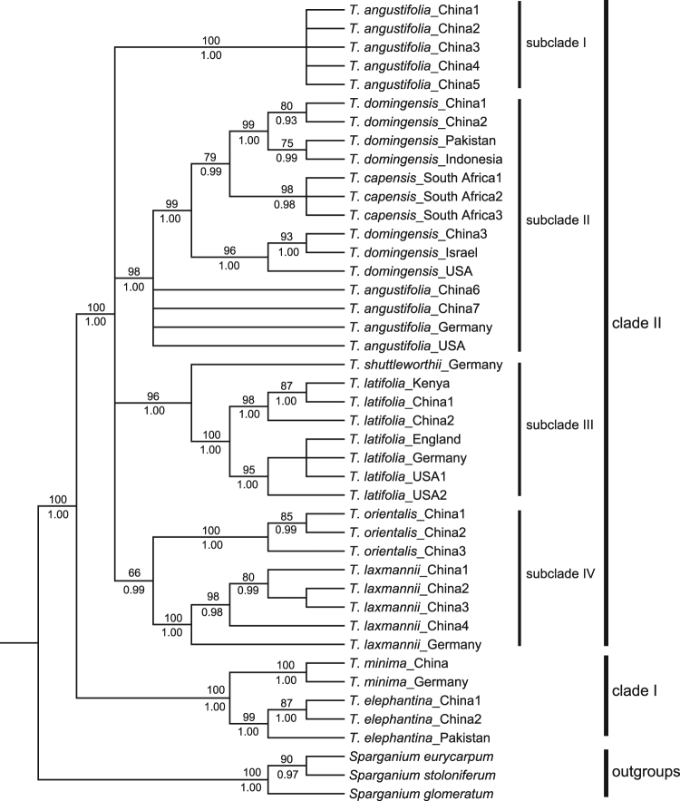
The aligned and concatenated sequences were 6,106 bp long with 988 variable sites. Of these, 426 were parsimony-informative. Phylogenetic relationships were inferred using maximum likelihood (ML) analysis and Bayesian inference (BI). The ML and BI trees were identical in topology (Fig. 1). The monophyly of Typha was strongly supported by both analyses (ML bootstrap support [BS] 100%, BI posterior probability [PP] 1.00). The genus was divided into two clades with strong support. The first clade (clade I) consisted of T. minima and T. elephantina (BS 100%, PP 1.00), and each species was determined to be monophyletic. The second clade (clade II; BS 100%, PP 1.00) included all remaining species and represented a polytomy of four robustly supported subclades. Subclade I (BS 100%, PP 1.00) included T. angustifolia only. Subclade II (BS 98%, PP 1.00) included T. angustifolia, T. domingensis, and T. capensis. Within this subclade, T. domingensis and T. capensis formed a highly supported group (BS 99%, PP 1.00), which was polytomic with three accessions of T. angustifolia, while T. capensis was nested in T. domingensis. Within subclade III (BS 96%, PP 1.00), T. latifolia was sister to T. shuttleworthii and it was further divided into two strongly supported groups. Subclade IV (BS 66%, PP 0.99) consisted of T. orientalis and T. laxmannii, which both formed their own monophyletic groups (Fig. 1).
Figure 1.
Bayesian consensus tree for Typha and three Sparganium species. The phylogenetic tree has been reconstructed based on seven chloroplast DNA regions (atpB-rbcL, psbA-trnH, psbD-trnT, rpl32-trnL, rps16 intron, rps16-trnK, and trnL-trnF). Numbers below the branches are Bayesian posterior probabilities (PP), and numbers above the branches are the ML bootstrap values (BS).
Divergence time estimation
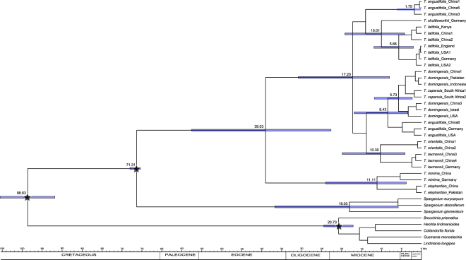
The respective crown ages of Typha and Sparganium were estimated to be 39.03 Mya (95% HPD: 22.64–57.60 Mya) and 18.03 Mya (5.79–36.69 Mya), respectively, based on combined data that included five Bromeliaceae sequences (Fig. 2). The beginning of diversification of the first clade, which included T. minima and T. elephantina, was dated to 11.11 Mya (3.78–24.01 Mya), and the second clade, which included the remaining species, was dated to 17.20 Mya (7.99–30.86 Mya). In the second clade, all three multiple-species subclades were estimated to begin to diversify in the Late Miocene (Fig. 2).
Figure 2.
Chronogram of Typha, three Sparganium species, and five Bromeliaceae species inferred from BEAST. Blue bars represent the 95% highest posterior density intervals for node ages. Stars indicated three calibration points.
Historical biogeography inference
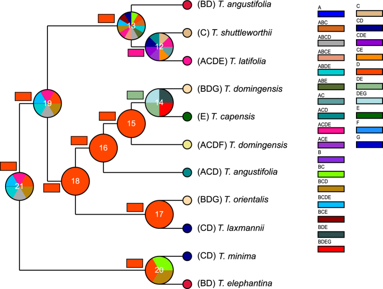
Ancestral area reconstruction based on the dispersal-extinction-cladogenesis (DEC) analyses revealed East Eurasia as the ancestral area for the crown node of Typha genus, clade I, and clade II, while statistical dispersal-vicariance (S-DIVA) analyses determined that East Eurasia or other multiple areas were the ancestral area for the three nodes (Fig. 3, Supplementary Table S1). Both S-DIVA and DEC analyses supported East Eurasia as the ancestral area for the two multiple-species subclades, subclade II and IV, and multiple areas for subclade III, T. latifolia/T. shuttleworthii (Fig. 3). Eighteen dispersal events and only one vicariant event were revealed using the DEC analyses, while 22 dispersal events and two vicariant events were obtained using S-DIVA analyses.
Figure 3.
Reconstruction of the ancestral area of Typha. The pie charts at each node were obtained using S-DIVA analysis, and the rectangle charts beside each node were obtained from DEC analysis. The colors of the charts correspond to the most likely ancestral areas inferred. Letters represent the following biogeographic regions: (A) North America, (B) Indo-Pacific, (C) West Eurasia, (D) East Eurasia, (E) Africa, (F) South America, and (G) Australia.
Discussion
In this study, we revealed that Typha is divided into two strongly supported clades. The first clade consists of two species, T. minima and T. elephantina. The second clade includes seven other species (Fig. 1). Our results are incongruent with a previous study that reported that T. minima is a clade and all other species form the other clade, including T. elephantina30. Typha elephantina and T. minima are morphologically distinct and can be easily distinguished from other species. Typha elephantina is distinct due to its robust habit, deep-set rhizome system, and stiff trigonal leaf blades1,33. Typha minima usually exhibits narrow, needle-like leaves and a stiff, unbranched central flower stalk9. Although their stems and leaves look very different from each other, T. elephantina and T. minima share four reproductive traits, including the presence of pistillate bracteoles, pollen in tetrads, filiform stigmas, and a gap between the male and female inflorescences, which have been used in taxonomic keys to identify Typha species1,14,30. Recently, Witztum and Wayne34,35 examined fiber cables in leaf blades of Typha species and found that the absence of fiber cables in leaf blades only occurs in T. elephantina and T. minima. The fact that T. elephantina and T. minima share the same five morphological characteristics suggests a closer affinity than has been previously considered. This supports our findings that T. elephantina and T. minima are sister species.
We found that T. angustifolia and T. domingensis are paraphyletic species, which suggests incongruence with a previous phylogenetic study of Typha30. Two highly divergent lineages were identified in T. angustifolia. One is subclade I, and the other nests in subclade II (Fig. 1). This was also observed by Ciotir and Freeland31, and it was named a core lineage and a divergent lineage by Freeland et al.36. Freeland et al.36 rejected the hypothesis that the divergent chloroplast DNA (cpDNA) lineage of T. angustifolia represents a cryptic species because it fell in the same genetic cluster as the core lineage based on nuclear genetic data from four microsatellite loci and the LEAFY gene. It was suggested that historical hybridization and introgression are the most likely explanation for this observation. In contrast, high divergence in nuclear ADH gene sequences was found in sympatric populations from both cpDNA lineages from northwest China22. This suggests that further investigation is needed to clarify the relationship between the two cpDNA lineages of T. angustifolia. In T. domingensis, two lineages were identified. One lineage was more closely related to T. capensis than to the other lineage, which formed a monophyletic group (Fig. 1). Similarly, Ciotir and Freeland31 observed paraphyly in T. domingensis and explained that it derived from incomplete lineage sorting following speciation. They also showed that these two lineages were distributed in different geographical ranges31. Further phylogeographic investigation is necessary in order to test whether or not T. domingensis includes cryptic species.
Although the earliest fossil of Typha is from the Late Cretaceous and fossils from the Paleogene are abundant23,25–29, these fossils do not exactly match extant Typha species. Therefore, we cannot rule out the possibility that these fossils are stem relatives, and we therefore treat them as representatives of the Typha stem lineage. The same treatment was used in some molecular dating studies37–39. Molecular dating results show that the crown origin of Typha occurred in the Middle Eocene (Fig. 2). East Eurasia was inferred as the ancestral area for the crown node of Typha in DEC analyses, and East Eurasia or other areas were inferred by S-DIVA analyses (Fig. 3). This suggests that crown-group Typha most likely originated in East Eurasia and then dispersed into other areas. It should be noted that this inference is not so convincing, because the relationship among the four subclades in clade II has not been fully resolved (Fig. 1).
Clade I consists of T. minima and T. elephantina, for which the most likely ancestral area was East Eurasia (Fig. 3), and the crown age was estimated to date to the Late Miocene (Fig. 2). In East Eurasia, the distribution of these two species is well-separated. T. elephantina is restricted to the south of the Qinghai-Tibetan Plateau (QTP), whereas T. minima is distributed north of the QTP. The QTP uplift is likely one of the factors that drove the divergence between these two species, although no consensus has been reached regarding the precise uplift phases of the QTP until now40. Clade II contained three intercontinentally dispersed subclades, including subclade II (i.e., T. angustifolia, T. domingensis, and T. capensis), subclade III (i.e., T. latifolia and T. shuttleworthii), and subclade IV (i.e., T. orientalis and T. laxmannii). Specifically, subclade II and III consisted of species that were intercontinentally dispersed between Eurasia and North America. The North Atlantic Land Bridge (NALB) and Beringian Land Bridge (BLB) served as migration routes for plants between Eurasia and North America41. The NALB facilitated taxa exchange until the Eocene, while the BLB contributed to intercontinental temperate taxa exchange until about 3.5 Mya42–44. The Late Miocene crown age of subclade II and III (Fig. 2) indicates that the BLB was a possible dispersal route for these temperate groups in Typha. In subclade II, the crown age of the T. domingensis lineage (including Eurasian and North American samples) was dated to about 3 Mya (Fig. 2), indicating a relatively recent transoceanic dispersal for T. domingensis. Similarly, transoceanic dispersal was also observed for T. angustifolia in a phylogeographical study based on sampling from Europe and North America32. In subclade III, T. shuttleworthii is restricted in Europe, and intercontinental dispersals occur in T. latifolia. High genetic divergence existed between the Asian and North American lineages30 (Fig. 1). Likewise, a phylogeographical study revealed that two recent T. latifolia colonizations have occurred: one from Asia into eastern Europe and the other from North America into western Europe31. The crown age of 5.66 Mya for T. latifolia (Fig. 2) suggested that the BLB was likely the route for T. latifolia dispersal between Asia and North America. For subclade IV, it was determined that T. orientalis dispersed from Asia to Australia based on having East Eurasia as the ancestral area. This dispersal route was also previously reported for other plants45,46. The time of dispersal into Australia varied widely amongst different taxa, even within single genera, such as Cucumis47. The time for T. orientalis dispersal is undetermined because no sample from Australia was included in this study.
Methods
Taxon sampling
A total of 43 samples were analyzed, including 40 from nine species of Typha and three outgroups from Sparganium species (Supplementary Table S2). Plant material was collected from Asia, North America, Europe, and Africa, and vouchers were deposited at the herbarium of Wuhan University (WH), South China Botanical Garden Herbarium (IBSC), and the United States National Herbarium (US). Detailed information regarding the samples and the associated GenBank accession numbers are listed in Supplementary Table S2.
DNA extraction, amplification, and sequencing
Genomic DNA was extracted from silica-dried leaves using the DNA secure Plant Kit (Tiangen Biotech., Beijing, China) according to the manufacturer’s protocol. Seven cpDNA non-coding regions (atpB-rbcL, psbA-trnH, psbD-trnT, rpl32-trnL, rps16 intron, rps16-trnK, and trnL-trnF) were amplified and sequenced for this study. Sequences of the primers and their sources are listed in Supplementary Table S3. Polymerase chain reaction (PCR) was performed in a volume of 25 μL containing 10–30 ng genomic DNA, 0.1 μM of each primer, 0.2 mM of each dNTP, 2 mM MgCl2, and 0.6 U of ExTaq DNA polymerase (TaKaRa, Dalian, China). PCR reactions were performed in a Veriti 96-Well Thermal Cycler (Applied Biosystems, Foster City, USA) under the following conditions: 4 min at 95 °C, followed by 35 cycles of 45 s at 95 °C, 45 s at 55 °C, and 90 s at 72 °C, and then a final 10-min extension at 72 °C. The PCR products were purified and sequenced in both the 5′ and 3′ directions by the Beijing Genomic Institute in Wuhan, China.
Phylogenetic analyses
All sequences were edited using Sequencher 4.1.4 (Gene Codes, Ann Arbor, MI, USA). Sequences were aligned using MAFFT 6.748 and then manually checked using Se-Al (http://tree.bio.ed.ac.uk/software/seal/). Gaps were treated as missing data. The seven chloroplast DNA regions were concatenated for subsequent analyses. The phylogenetic relationships were inferred using ML analysis implemented in GARLI 2.049. One-thousand bootstrap repetitions were performed to summarize the ML bootstrap support. BI implemented in MrBayes 3.1.250 was also used for phylogenetic reconstruction. Two independent Markov chain Monte Carlo (MCMC) analysis runs were conducted simultaneously, and each run employed four chains. The analysis ran for 3 × 10⁷ generations with sampling at every 1,000 generations. Chain convergence was checked using Tracer 1.551, and posterior probability values were generated from trees after excluding a burn-in of the first 25% of the trees. In the phylogenetic analyses, we assigned model parameters for each cpDNA region identified by Akaike information criterion (AIC) in jModeltest 2.1.752. The K81uf + G model was selected for psbD-trnT, rps16-trnK, and trnL-trnF, while the HKY + I + G, F81 + I, K81uf + I, and TVM were suggested for atpB-rbcL, psbA-trnH, rpl32-trnL, and rps16 intron, respectively.
Divergence time estimation
The divergence time between clades in Typha was estimated based on the concatenated sequence data containing seven cpDNA regions from 31 accessions of Typha, three species of Sparganium, and five species of Bromeliaceae. Sequences of the five Bromeliaceae samples in five cpDNA regions (atpB-rbcL, psbA-trnH, rpl32-trnL, rps16 intron, and trnL-trnF) were obtained from Givnish et al.38, and those in two cpDNA regions were treated as missing data. The divergence time estimate was conducted in BEAST 1.7.453. The substitution model for each respective region was recalculated using jModeltest. The TIM + G model was selected for atpB-rbcL, the F81 + I model was selected for psbA-trnH, the TPM1uf + G model was selected for psbD-trnT and rps16-trnK, the GTR model was selected for rps16 intron, and the TVM + G model was selected for rpl32-trnL and trnL-trnF. Three calibration points were used. One was the stem age of Typha, which was a minimum age of 70 Mya based on fossil evidence. Although the earliest fossil of Typha dated to the Late Cretaceous and fossils from the Paleogene are abundant23,25–29, these fossils do not exactly match extant Typha species. Therefore, we cannot rule out the possibility that these fossils are stem relatives and treat them as representatives of the Typha stem lineage. The stem age of Typha has also been used in previous studies37–39. The detailed setting was: a lognormal prior with an offset of 70, a mean of 1.5, and a standard deviation of 0.5. The other two were the stem age of Typhaceae (100 ± 3.5 Mya) and the crown age of Bromeliaceae (19.1 ± 2.0 Mya), which were obtained from Givnish et al.38. MCMC analyses of 2 × 10⁸ generations were implemented, and every 1,000 generations were sampled. The first 25% of the generations were discarded as burn-in, and the parameters were checked using the program Tracer. Trees were summarized with Tree Annotator53.
Reconstruction of ancestral areas
Ancestral area reconstruction was conducted using S-DIVA implemented in RASP 3.154 and a likelihood model DEC implemented in Lagrange55. The analyses were conducted on a fully resolved topology from the BEAST analysis containing seven species, two lineages of T. angustifolia, and two lineages of T. domingensis. Seven geographical areas were defined based on the worldwide distribution of Typha according to Morse56: (A) North America, (B) Indo-Pacific, (C) West Eurasia, (D) East Eurasia, (E) Africa, (F) South America, and (G) Australia. The distribution of each species was determined based on our collecting localities and data from published papers1,9,12,14,57,58. Although T. domingensis from South America and Australia and T. orientalis from Australia were not included, these two geographical areas were also coded. For the DEC analysis, the dispersal probability between areas were set from 0.1 (for well-separated areas) to 1.0 (for adjacent areas) based on geological history and paleogeography reconstruction59,60 (Supplementary Table S4). The number of maximum areas was set to four because each lineage of T. domingensis and other species did not occur in more than four areas.
Electronic supplementary material
Acknowledgements
This study was supported by grants from the National Natural Science Foundation of China (31270265) to Xinwei Xu and by the Laboratory of Analytical Biology of the Smithsonian Institution. We thank the members of Dan Yu’s group for field assistance, in addition to S. Renner, S. Volis, S.K. Marwat, S. Compton, T. Reader, and M.L. Moody for providing samples.
Author Contributions
T.T., J.W., and X.X. designed the research; B.Z., T.T., and F.K. conducted the laboratory experiments; B.Z. analyzed the data; all authors participated in writing the manuscript.
Competing Interests
The authors declare no competing interests.
Footnotes
Publisher's note: Springer Nature remains neutral with regard to jurisdictional claims in published maps and institutional affiliations.
Beibei Zhou and Tieyao Tu contributed equally to this work.
Change history
6/12/2020
An amendment to this paper has been published and can be accessed via a link at the top of the paper.
Contributor Information
Jun Wen, Email: wenj@si.edu.
Xinwei Xu, Email: xuxw@whu.edu.cn.
Electronic supplementary material
Supplementary information accompanies this paper at 10.1038/s41598-018-27279-3.
References
- 1.Smith SG. Typha: Its taxonomy and the ecological significance of hybrids. Arch. Hydrobiol. 1987;27:129–138. [Google Scholar]
- 2.Morton JFC. Econ. Bot. 1975. (Typha spp.): Weed problem or potential crop? pp. 7–29. [Google Scholar]
- 3.Finlayson C, Roberts J, Chick A, Sale P. The biology of Australian weeds. II. Typha domingensis Pers. and Typha orientalis Presl. J. Aust. Inst. of Agric. Sci. 1983;49:3–10. [Google Scholar]
- 4.Audu IG, Brosse N, Desharnais L, Rakshit SK. Ethanol organosolv pretreatment of Typha capensis for bioethanol production and co-products. Bioresources. 2012;7:5917–5933. [Google Scholar]
- 5.Grace JB, Harrison JS. The biology of Canadian weeds: 73. Typha latifolia L., Typha angustifolia L. and Typha × glauca Godr. Can. J. Plant Sci. 1986;66:361–379. [Google Scholar]
- 6.Thieret JW, Luken JO. The Typhaceae in the southeastern United States. Harvard Papers Bot. 1996;1:27–56. [Google Scholar]
- 7.Mabberley, D. J. Mabberley’s plant-book: A portable dictionary of plants, their classification and uses. Fourth edition. Cambridge University Press (2017).
- 8.Saha S. The genus. Typha in India–its distribution and uses. J. Bot. Soc. Bengal. 1968;22:11–18. [Google Scholar]
- 9.Cook, C. D. K. Typhaceae. In Flora Europaea Vol. 5 (eds Tutin, T. G. et al.) (Cambridge University Press, 1980).
- 10.Bokhari MH. The aquatic plants of Iran and Pakistan. III. Typhaceae. Biologia. 1983;29:85–91. [Google Scholar]
- 11.Hamdi, S. M. M. & Assadi, M. Typhaceae. In Flora of Iran 299–317 (Verlag Paul Parey Berlin and Hamburg Auschriften, 2003).
- 12.Finlayson M, Forrester R, Mitchell D, Chick A. Identification of Native Typha species in Australia. Aust. J. Bot. 1985;33:101–107. [Google Scholar]
- 13.Kuehn MM, White BN. Morphological analysis of genetically identified cattails Typha latifolia, Typha angustifolia, and Typha × glauca. Can. J. Bot. 1999;77:906–912. [Google Scholar]
- 14.Sun, K. & Simpson, D. Typhaceae. In Flora of China (eds Wu, Z. Y., Raven, P. H. & Hong, D. Y.) (Science Press and Missouri Botanical Garden Press, 2010).
- 15.Grace JB, Wetzel RG. Variations in growth and reproduction within populations of two rhizomatous plant species: Typha latifolia and Typha angustifolia. Oecologia. 1982;53:258–263. doi: 10.1007/BF00545674. [DOI] [PubMed] [Google Scholar]
- 16.Kronfeld MM. der Gattung Typha Tourn. Verh. Zool. Bot. Ges. Wien. 1889;39:89–192. [Google Scholar]
- 17.Graebner, P. Typhaceae. in Das Pflanzenreich (ed. Engler, A.) 1–18 (Berlin, Engelmann, 1900).
- 18.Govaerts, R. World Checklist of Typhaceae. Facilitated by the Royal Botanic Gardens, Kew. Published on the Internet; http://wcsp.science.kew.org/ (Accessed 10 February 2018, 2018).
- 19.Zhu, X. Taxonomic study on Typha sect. Ebracteatae (Typhaceae) in China. Master’s thesis, Central China Normal University (2012).
- 20.Maveodiev EV. Two new species of Typha L. (Typhaceae JUSS.) from the Far East of Russia and from Mongolia. Feddes Repertorium. 2002;113:281–288. [Google Scholar]
- 21.Wang, R. Taxonomic study on Typha sect. Bracteolatae (Typhaceae) in China. Master’s thesis, Central China Normal University (2012).
- 22.Zhou, B. Genetic diversity and biogeography of the genus Typha L. (Typhaceae) from China. Ph.D. dissertation, Wuhan University (2016).
- 23.Knobloch E, Mai DH. Monographie der Früchte und Samen in der Kreide von Mitteleuropa. Rozpravy Ústředního Ústavu Geologického. 1986;47:1–219. [Google Scholar]
- 24.Jerzykiewicz T, Sweet AR. The Cretaceous-Tertiary boundary in the central Alberta Foothills: I. Stratigraphy. Can. J. Earth. Sci. 1986;23:1356–1374. [Google Scholar]
- 25.Song Z, Wang W, Fei H. Fossil pollen records of extant angiosperms in China. Bot. Rev. 2004;70:425–458. [Google Scholar]
- 26.Wilson LR, Webster RM. Plant microfossils from a Fort Union Coal of Montana. Am. J. Bot. 1946;33:271–278. [Google Scholar]
- 27.Hickey, L. J. Stratigraphy and Paleobotany of the Golden Valley Formation (Early Tertiary) of Western North Dakota. Vol. 150 (Geological Society of America, 1977).
- 28.Muller J. Fossil pollen records of extant angiosperms. Bot. Rev. 1981;47:1–142. [Google Scholar]
- 29.Postnikoff, A. C. L. Flora of the Ravenscrag Formation of the Big Muddy Valley, Willow Bunch Lake Map Area (72H), Saskatchewan, (2009).
- 30.Kim C, Choi HK. Molecular systematics and character evolution of Typha (Typhaceae) inferred from nuclear and plastid DNA sequence data. Taxon. 2011;60:1417–1428. [Google Scholar]
- 31.Ciotir C, Freeland J. Cryptic intercontinental dispersal, commercial retailers, and the genetic diversity of native and non-native cattails (Typha spp.) in North America. Hydrobiologia. 2016;768:1–14. [Google Scholar]
- 32.Ciotir C, Kirk H, Row J, Freeland J. Intercontinental dispersal of Typha angustifolia and T. latifolia between Europe and North America has implications for Typha invasions. Biol. Invasions. 2013;15:1377–1390. [Google Scholar]
- 33.Sharma KP, Gopal B. A note on the identity of Typha elephantina Roxb. Aquat. Bot. 1980;9:381–387. [Google Scholar]
- 34.Witztum A, Wayne R. Fibre cables in the lacunae of Typha leaves contribute to a tensegrity structure. Ann. Bot. 2014;113:789–797. doi: 10.1093/aob/mcu002. [DOI] [PMC free article] [PubMed] [Google Scholar]
- 35.Witztum A, Wayne R. Fiber cables in leaf blades of Typha domingensis and their absence in Typha elephantina: a diagnostic character for phylogenetic affinity. Isr. J. Plant Sci. 2016;63:116–123. [Google Scholar]
- 36.Freeland JR, Ciotir C, Wensink L, Dorken M. Widespread cytonuclear discordance in narrow-leaved cattail (Typha angustifolia) does not explain the dominance of its invasive hybrid (Typha × glauca) Hydrobiologia. 2016;792:53–65. [Google Scholar]
- 37.Bremer K. Early Cretaceous lineages of monocot flowering plants. Proc. Natl. Acad. Sci. USA. 2000;97:4707–4711. doi: 10.1073/pnas.080421597. [DOI] [PMC free article] [PubMed] [Google Scholar]
- 38.Givnish TJ, et al. Phylogeny, adaptive radiation, and historical biogeography in Bromeliaceae: Insights from an eight-locus plastid phylogeny. Am. J. Bot. 2011;98:872–895. doi: 10.3732/ajb.1000059. [DOI] [PubMed] [Google Scholar]
- 39.Sulman JD, Drew BT, Drummond C, Hayasaka E, Sytsma KJ. Systematics, biogeography, and character evolution of Sparganium (Typhaceae): diversification of a widespread aquatic lineage. Am. J. Bot. 2013;100:2023–2039. doi: 10.3732/ajb.1300048. [DOI] [PubMed] [Google Scholar]
- 40.Renner SS. Available data point to a 4-km-high Tibetan Plateau by 40 Ma, but 100 molecular-clock papers have linked supposed recent uplift to young node ages. J. Biogeogr. 2016;43:1479–1487. [Google Scholar]
- 41.Tiffney BH, Manchester SR. The use of geological and paleontological evidence in evaluating plant phylogeographic hypotheses in the Northern Hemisphere Tertiary. Int. J. Plant Sci. 2001;162:S3–S17. [Google Scholar]
- 42.Gladenkov AY, Oleinik AE, Marincovich L, Barinov KB. A refined age for the earliest opening of Bering Strait. Palaeogeogr. Palaeoclimatol. Palaeoecol. 2002;183:321–328. [Google Scholar]
- 43.Tiffney BH. The Eocene North Atlantic land bridge: its importance in tertiary and modern phytogeography of the Northern hemisphere. J. Arnold Arboretum. 1985;66:243–273. [Google Scholar]
- 44.Wen J, Nie Z-L, Ickert-Bond SM. Intercontinental disjunctions between eastern Asia and western North America in vascular plants highlight the biogeographic importance of the Bering land bridge from late Cretaceous to Neogene. J.Syst. Evol. 2016;54:469–490. [Google Scholar]
- 45.Schaefer H, Telford IRH, Renner SS. Austrobryonia (Cucurbitaceae), a new Australian endemic genus, is the closest living relative to the Eurasian and Mediterranean Bryonia and Ecballium. Syst. Bot. 2008;33:125–132. [Google Scholar]
- 46.Zhang ML, Temirbayeva K, Sanderson SC, Chen X. Young dispersal of xerophil Nitraria lineages in intercontinental disjunctions of the Old World. Sci. rep. 2015;5:13840. doi: 10.1038/srep13840. [DOI] [PMC free article] [PubMed] [Google Scholar]
- 47.Sebastian P, Schaefer H, Telford IRH, Renner SS. Cucumber (Cucumis sativus) and melon (C. melo) have numerous wild relatives in Asia and Australia, and the sister species of melon is from Australia. Proc. Natl. Acad. Sci. USA. 2010;107:14269–14273. doi: 10.1073/pnas.1005338107. [DOI] [PMC free article] [PubMed] [Google Scholar]
- 48.Katoh K, Misawa K, Kuma KI, Miyata T. MAFFT: a novel method for rapid multiple sequence alignment based on fast Fourier transform. Nucleic Acids Res. 2002;30:3059. doi: 10.1093/nar/gkf436. [DOI] [PMC free article] [PubMed] [Google Scholar]
- 49.Zwickl D. J. Genetic algorithm approaches for the phylogenetic analysis of large biological sequence datasets under the maximum likelihood criterion. Ph.D. dissertation, The University of Texas at Austin, (2006).
- 50.Ronquist F, Huelsenbeck JP. MrBayes 3: Bayesian phylogenetic inference under mixed models. Bioinformatics. 2003;19:1572–1574. doi: 10.1093/bioinformatics/btg180. [DOI] [PubMed] [Google Scholar]
- 51.Rambaut, A. & Drummond. A. J. Tracer: MCMC trace analysis tool, version 1.5. http://tree.bio.ed. ac.uk/software/tracer (Accessed 19 May 2014, 2007).
- 52.Darriba D, Taboada GL, Doallo R, Posada D. jModelTest 2: more models, new heuristics and parallel computing. Nat. Methods. 2012;9:772–772. doi: 10.1038/nmeth.2109. [DOI] [PMC free article] [PubMed] [Google Scholar]
- 53.Drummond AJ, Suchard MA, Xie D, Rambaut A. Bayesian phylogenetics with BEAUti and the BEAST 1.7. Mol. Biol. Evol. 2012;29:1969–1973. doi: 10.1093/molbev/mss075. [DOI] [PMC free article] [PubMed] [Google Scholar]
- 54.Yu Y, Harris AJ, Blair C, He X. RASP (Reconstruct Ancestral State in Phylogenies): A tool for historical biogeography. Mol. Phylogen. Evol. 2015;87:46–49. doi: 10.1016/j.ympev.2015.03.008. [DOI] [PubMed] [Google Scholar]
- 55.Ree RH, Smith SA. Maximum likelihood inference of geographic range evolution by dispersal, local extinction, and cladogenesis. Syst. Biol. 2008;57:4–14. doi: 10.1080/10635150701883881. [DOI] [PubMed] [Google Scholar]
- 56.Morse, J. Trichoptera World Checklist. http://entweb.clemson.edu/database/trichopt/index.htm. (Accessed 23 May 2015, 2011)
- 57.Arenas P, Scarpa GF. The consumption of Typha domingensis pers. (Typhaceae) pollen among the ethnic groups of the Gran Chaco, South America. Econ. Bot. 2003;57:181–188. [Google Scholar]
- 58.Preston, C. D. & Croft, J. M. Aquatic Plants in Britain and Ireland: Harley Books. (England, 1997).
- 59.Morley RJ. Interplate dispersal paths for megathermal angiosperms. Perspect. Plant Ecol. Evol. Syst. 2003;6:5–20. [Google Scholar]
- 60.Clayton JW, Soltis PS, Soltis DE. Recent long-distance dispersal overshadows ancient biogeographical patterns in a pantropical angiosperm family (Simaroubaceae, Sapindales) Syst. Biol. 2009;58:395–410. doi: 10.1093/sysbio/syp041. [DOI] [PubMed] [Google Scholar]
Associated Data
This section collects any data citations, data availability statements, or supplementary materials included in this article.


