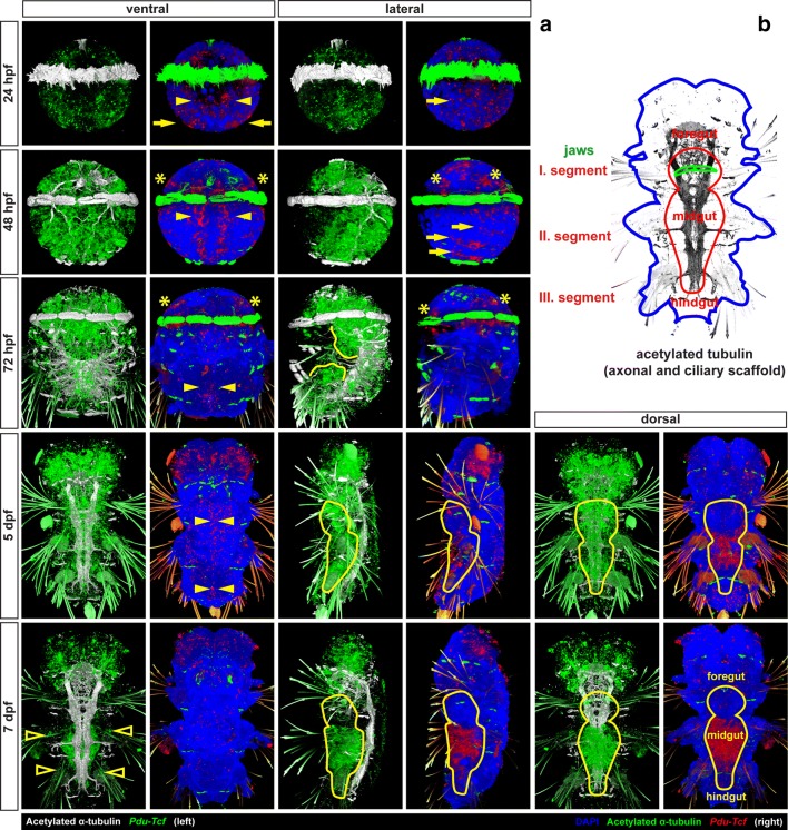
Fig. 3.
Expression of Pdu-Tcf throughout development of Platynereis dumerilii. a The expression of Pdu-Tcf during development (left: Tcf green, acetylated tubulin white; right: Tcf red, DAPI blue, acetylated tubulin green). At 24 hpf, Pdu-Tcf is broadly expressed at low levels in both the episphere and hyposphere. At 48 hpf in the hyposphere, Pdu-Tcf is present in ectodermal cells along the midline (yellow arrowheads) and in the segmental pattern (yellow arrows). It persists in the episphere, e.g. in future larval (ventral) and adult (dorsal) eye regions (yellow asterisks). At 72 hpf stage, Pdu-Tcf is expressed mainly in the episphere and stomodeal region, whereas it becomes more scarce in a majority of the hyposphere. This trend continues throughout 5 dpf, where expression mainly restricted to the brain ganglia of the head lobes. At 7 dpf, Pdu-Tcf is still expressed in the brain; however, a new strong expression is observed in the midgut and hindgut. There is also a small patch of weaker expression at the base of each parapodium (empty arrowheads). The expression patterns are described in greater detail in the text. Approximate size of a 48 hpf larva is around 130 μm, and all images are to scale. Stage and orientation are indicated; anterior up; in lateral view ventral to the right

