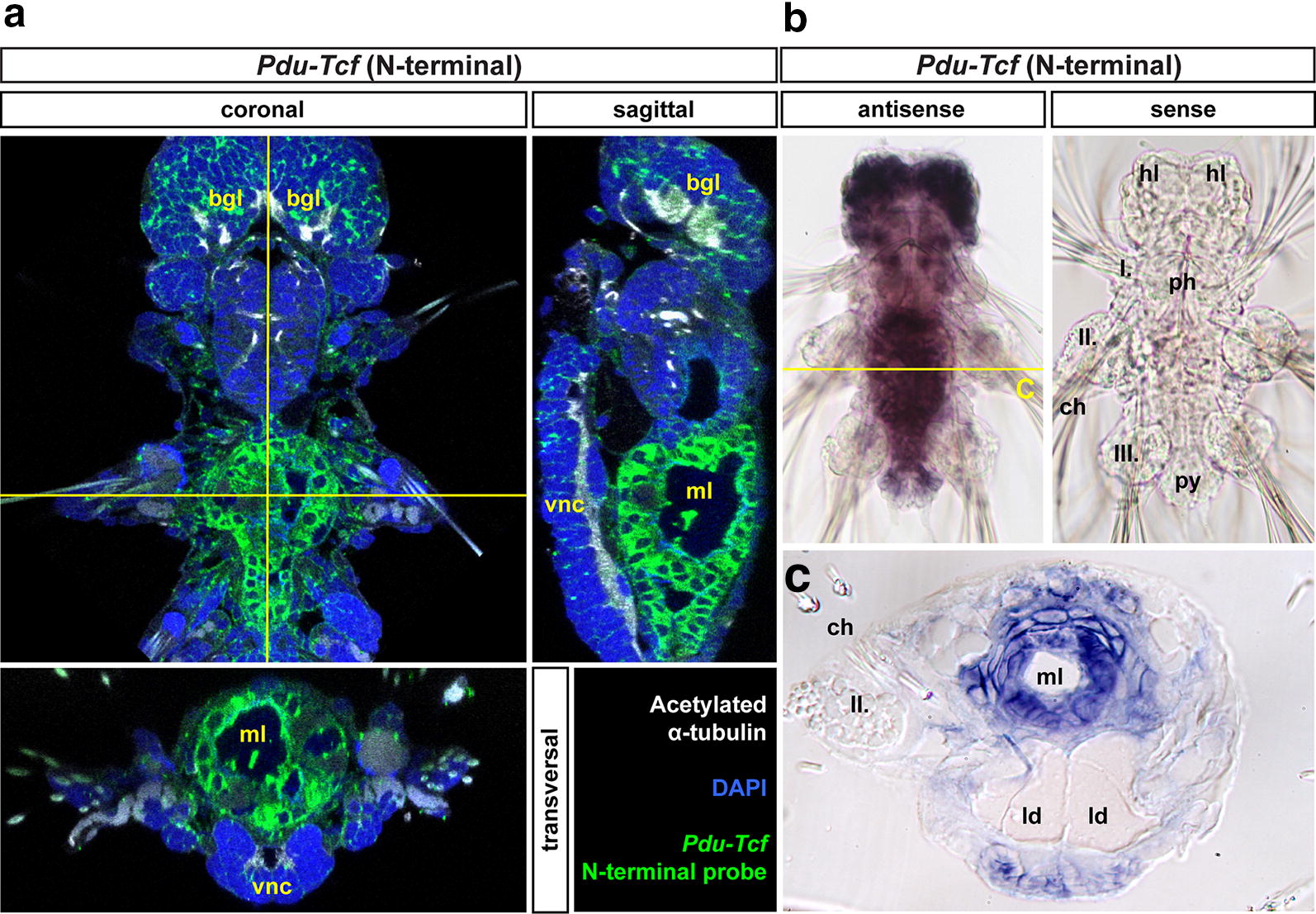
Fig. 5.

Pdu-Tcf is specifically detected in the gut of 7 dpf larva by N-terminal probe. a Virtual orthogonal sections through a confocal z-stack of a 7 dpf Platynereis larva after prolonged staining of fluorescent in situ hybridizaton with a probe recognizing the N-terminal part of Pdu-Tcf show the expression of this gene in the midgut and hindgut. b Pdu-Tcf N-terminal antisense (left) and sense (right) probe NBT/BCIP stainings show that the antisense probe specifically detects Pdu-Tcf in the brain and in the gut. Also treatment with levamisole did not abolish the antisense probe staining demonstrating that the observed signal was not due to persisting endogenous alkaline phosphatase activity (which was thus successfully inactivated by hybridization temperature). The same fact is also demonstrated by the lack of staining with sense probe (right) or after hybridization without a probe (not shown). The absence of signal with sense probe shows that the observed signal is not due to unspecific binding of digoxigenin-labelled probe. c Physical coronal section through the body of 7 dpf larva after in situ hybridization with antisense probe against N terminus of Pdu-Tcf confirms its presence in the gut. The section is approximately on the level of the second segment, i.e. through midgut. I., II., III.—first, second and third body segments’ parapodia, bgl brain ganglia, hl head lobes, ld lipid droplets, ml midgut lumen, ph pharynx (foregut), py pygidium, vnc ventral nerve cord. For schematics of 7 dpf larval gut morphology, see Fig. 2
