Abstract
A 60-year-old female with no significant medical history presented with hematuria. A computed tomography (CT) scan revealed extensive lymphadenopathy with hypodensities in the liver and spleen, and she was referred for an 18F-fluorodeoxyglucose (18F-FDG) positron emission tomography/CT (PET/CT) study to assess for malignancy of unknown primary. PET/CT revealed extensive 18F-FDG avid lymphadenopathy as well as innumerable intensely 18F-FDG avid lung, liver and splenic nodules, highly concerning for malignancy. A PET-guided bone marrow biopsy of the posterior superior iliac spine revealed several non-necrotizing, well-formed granulomas, consistent with sarcoidosis. The patient was managed conservatively and remained clinically well over the subsequent 9 years of follow-up.
Keywords: Sarcoidosis, artifact, mimic, lymphadenopathy, metastases, 18F-fluorodeoxyglucose, positron emission tomography
Abstract
Tıbbi anamnezinde özellik olmayan 60 yaşında bir kadın hematüri ile başvurdu. Bilgisayarlı tomografide (BT) yaygın lenfadenopatiyle birlikte karaciğer ve dalakta hipodens alanlar saptanması üzerine primeri bilinmeyen malignite değerlendirilmesi amacıyla 18F-fluorodeoksiglukoz (18F-FDG) pozitron emisyon tomografisi/BT (PET/BT) için yönlendirildi. PET/BT’de 18F-FDG tutan yaygın lenfadenopatiler ve ölçülemeyecek kadar fazla yoğun 18F-FDG tutan akciğer, karaciğer ve dalak nodülleri saptandı, malignite açısından şüpheli bulundu. PET-kılavuzluğunda posterior superior spina iliacadan yapılan kemik iliği biyopsisinde sarkoidoz ile uyumlu non-kazeifiye granülomlar saptandı. Hasta konservatif olarak takip edildi ve 9 yıllık takip süresinde klinik sorun oluşmadı.
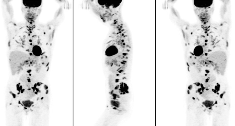
Figure 1. A 60-year-old woman, non-smoker with no significant medical history presented with recurrent hematuria. Computed tomography (CT) abdomen/pelvis identified intra-abdominal, retroperitoneal and inguinal lymphadenopathy, and small hepatic/splenic hypodensities, prompting referral for positron emission tomography/CT (PET/CT) to assess for malignancy of unknown origin. Maximum intensity projection images revealed widespread foci of intense 18F-FDG uptake throughout the skeleton and soft-tissues.

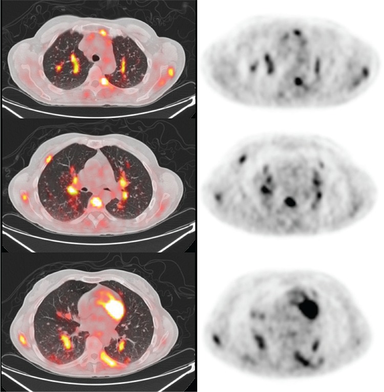
Figure 2. Transaxial (A) PET/CT fusion, (B) PET, and (C) CT images revealed extensive bone involvement of the skull base, right clavicle, spine, multiple ribs, sternum, proximal left humerus, right femur and extensively throughout the pelvis with maximum standardized uptake value (SUVmax) 8.3.

Figure 3. Intense 18F-FDG uptake was also noted in numerous peribronchovascular and subpleural nodules in both lungs (largest was 1.0 cm in diameter with SUVmax 3.2).

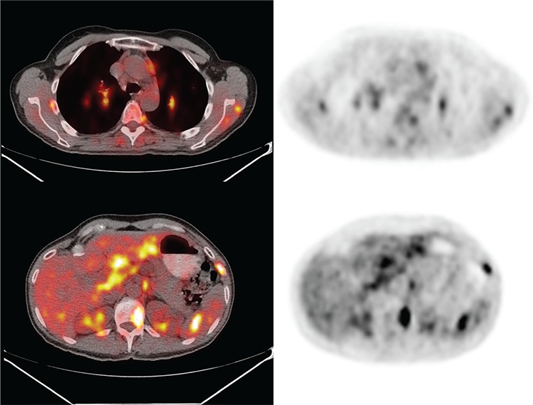
Figure 4. There was widespread adenopathy including left retromandibular, mediastinal, hilar, abdominal, retroperitoneal and inguinal nodes (largest mediastinal node measured 1.4 cm, and the most metabolically active was a right perihilar lymph node with SUVmax 5.2). The innumerable liver and spleen hypodensities identified on CT were intensely 18F-FDG avid with SUVmax 4.3 in the liver and 5.9 in the spleen. No primary malignancy was identified, but findings were interpreted as highly concerning for disseminated metastatic disease. The patient remained asymptomatic. PET-guided bone marrow biopsy of the left posterior superior iliac spine revealed several non-necrotizing, well-formed granulomas. These granulomas were paratrabecular in distribution and were composed of tightly apposed epithelioid histiocytes, with occasional multinucleated giant cells, consistent with sarcoidosis.

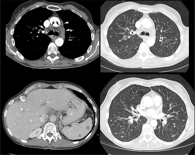
Figure 5. Follow-up diagnostic CT of chest/abdomen/pelvis performed 6 months later, again revealed extensive poorly marginated lung nodules, thoracic and abdominal lymphadenopathy, and splenic and hepatic hypodensities, unchanged as compared to prior PET/CT and consistent with stable granulomatous disease. The patient was managed conservatively with observation and remained malignancy-free over the subsequent 9 years of clinical and imaging follow-up. Sarcoidosis is a chronic multisystem granulomatous disorder of unknown etiology, which is characterized pathologically by non-caseating granulomas that can present almost anywhere in the body (1,2,3,4). 18F-FDG uptake in the granulomas of sarcoidosis can be very intense, likely due to metabolic activity of activated macrophages (2,3,5). 18F-FDG-avid lesions of skeletal sarcoidosis cannot be reliably differentiated from metastases or other benign bone processes (Paget’s disease, fibrous dysplasia, giant cell tumors, osteomyelitis) on the basis of semi-quantitative (SUV), visual or other analysis, and therefore remain a pitfall of oncologic PET/CT interpretation (6,7,8,9,10). 18F-FDG uptake in pulmonary sarcoidosis lesions has been described and the severity of pulmonary involvement has been shown to be associated with 18F-FDG activity in persistently symptomatic sarcoidosis patients (11). CT studies show presence of hepatic and splenic nodules in approximately 5-15% of sarcoidosis patients and splenic nodules tend to be larger than hepatic nodules (12). Intense 18F-FDG uptake in hepatic and splenic sarcoidosis lesions has been previously described (13). This case shows an uncommon presentation of disseminated sarcoidosis in the skeleton, lymph nodes, as well as organs such as the lungs, liver and spleen. This impressive pattern of disseminated 18F-FDG uptake can be easily mistaken for extensive metastatic disease when interpreting oncologic PET/CT studies.

Footnotes
Ethics
Informed Consent: All subjects in the study gave written informed consent or the institutional review board waived the need to obtain informed consent.
Peer-review: Externally and internally peer-reviewed.
Authorship Contributions
Surgical and Medical Practices: W.M., M.P., C.R., S.P., Concept: W.M., M.P., C.R., S.P., Design: W.M., M.P., C.R., S.P., Data Collection or Processing: W.M., M.P., C.R., S.P., Analysis or Interpretation: W.M., M.P., C.R., S.P., Literature Search: W.M., M.P., C.R., S.P., Writing: W.M., M.P., C.R., S.P.
Conflict of Interest: William Makis, Mark Palayew, Christopher Rush and Stephan Probst declare that they have no conflicts of interest.
Financial Disclosure: The authors declare that this study has received no financial support.
References
- 1.Judson MA. Sarcoidosis: clinical presentation, diagnosis, and approach to treatment. Am J Med Sci. 2008;335:26–33. doi: 10.1097/MAJ.0b013e31815d8276. [DOI] [PubMed] [Google Scholar]
- 2.Aberg C, Ponzo F, Raphael B, Amorosi E, Moran V, Kramer E. FDG positron emission tomography of bone involvement in sarcoidosis. AJR Am J Roentgenol. 2004;182:975–977. doi: 10.2214/ajr.182.4.1820975. [DOI] [PubMed] [Google Scholar]
- 3.Zhuang H, Alavi A. 18-fluorodeoxyglucose positron emission tomographic imaging in the detection and monitoring of infection and inflammation. Semin Nucl Med. 2002;32:47–59. doi: 10.1053/snuc.2002.29278. [DOI] [PubMed] [Google Scholar]
- 4.Ludwig V, Fordice S, Lamar R, Martin WH, Delbeke D. Unsuspected skeletal sarcoidosis mimicking metastatic disease on FDG positron emission tomography and bone scintigraphy. Clin Nucl Med. 2003;28:176–179. doi: 10.1097/01.RLU.0000053528.35645.70. [DOI] [PubMed] [Google Scholar]
- 5.Cook GJ, Fogelman I, Maisey MN. Normal physiological and benign pathological variants of 18-fluoro-2-deoxyglucose positron-emission tomography scanning: potential for error in interpretation. Semin Nucl Med. 1996;26:308–314. doi: 10.1016/s0001-2998(96)80006-7. [DOI] [PubMed] [Google Scholar]
- 6.Teirstein AS, Machac J, Almeida O, Lu P, Padilla ML, Iannuzzi MC. Results of 188 Whole Body FDG PET Scans in 137 Patients with Sarcoidosis. Chest. 2007;132:1949–1953. doi: 10.1378/chest.07-1178. [DOI] [PubMed] [Google Scholar]
- 7.Makis W, Probst S. Extensive polyostotic fibrous dysplasia evaluated for malignant transformation with 99mTc-MDP bone scan and 18F-FDG PET/CT. BJR Case Rep. 2016;2:1–4. doi: 10.1259/bjrcr.20150440. [DOI] [PMC free article] [PubMed] [Google Scholar]
- 8.Makis W, Stern J. Chronic vascular graft infection with fistula to bone causing vertebral osteomyelitis, imaged with F-18 FDG PET/CT. Clin Nucl Med. 2010;35:794–796. doi: 10.1097/RLU.0b013e3181ef099a. [DOI] [PubMed] [Google Scholar]
- 9.Aoki J, Watanabe H, Shinozaki T, Takagishi K, Ishijima H, Oya N, Sato N, Inoue T, Endo K. FDG PET of primary benign and malignant bone tumors: standardized uptake value in 52 lesions. Radiology. 2001;219:774–777. doi: 10.1148/radiology.219.3.r01ma08774. [DOI] [PubMed] [Google Scholar]
- 10.Kobayashi A, Shinozaki T, Shinjyo Y, Kato K, Oriuchi N, Watanabe H, Takagishi K. FDG PET in the clinical evaluation of sarcoidosis with bone lesions. Ann Nucl Med. 2000;14:311–313. doi: 10.1007/BF02988216. [DOI] [PubMed] [Google Scholar]
- 11.Sobic-Saranovic D, Artiko V, Obradovic V. FDG PET imaging in sarcoidosis. Semin Nucl Med. 2013;43:404–411. doi: 10.1053/j.semnuclmed.2013.06.007. [DOI] [PubMed] [Google Scholar]
- 12.Warshauer DM, Molina PL, Hamman SM, Koehler RE, Paulson EK, Bechtold RE, Perlmutter ML, Hiken JN, Francis IR, Cooper CJ, et al. Nodular sarcoidosis of the liver and spleen: analysis of 32 cases. Radiology. 1995;195:757–762. doi: 10.1148/radiology.195.3.7754007. [DOI] [PubMed] [Google Scholar]
- 13.Soussan M, Augier A, Brillet PY, Weinmann P, Valeyre D. Functional imaging in extrapulmonary sarcoidosis: FDG-PET/CT and MR features. Clin Nucl Med. 2014;39:e146–159. doi: 10.1097/RLU.0b013e318279f264. [DOI] [PubMed] [Google Scholar]


