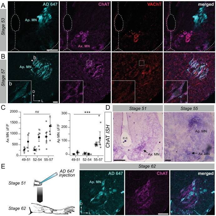
Figure 2. Developmental emergence of the molecular ACh phenotype in appendicular MNs.
(A, B) Examples of fluorescence immunolabeling against ChAT (magenta) and VAChT (red) in appendicular MNs (Ap. MN) labeled with retrograde Alexa Fluor dextran 647 (AD 647, cyan) in stages 53 (A) and 57 (B) tadpoles. Insets (b) in B show x60 magnification of stage 57 appendicular MNs. (C) Variation of fluorescence (∆F/F) of axial (left plot) and appendicular (right plot) MNs for ChAT and VAChT immuno-signals at stages 49–51 (n = 4), stages 52–54 (n = 7) and stage 55–57 (n = 8). Grey dots represent the averaged ∆F/F values for ChAT (squares) and VAChT (circles) in each preparation, and the black dots are ∆F/F grand means ± SEM for all preparations in a given developmental group. ns non significant, ***p<0.001, Kruskall-Wallis test. (D) Examples of in situ hybridization (ISH) labeling for ChAT mRNA in the appendicular spinal column at stages 51 and 55. (E) Example of fluorescence immunolabeling against ChAT in stage 62 appendicular MNs that were previously labeled with AD 647 injected into the hindlimb at stage 51 (see schematic at left). All scale bars = 50 µm; D, dorsal; L, lateral.

