Abstract
We described an 88-year-old woman presented with large aneurysm on the carotid siphon of the right internal carotid artery (ICA). Digital subtraction angiography showed extravasation from the distal cervical segment of the right ICA due to positioning a guiding catheter for intra-aneurysmal coil embolization. Transoral carotid ultrasonography (TOCU) showed arrested bleeding and a pseudolumen in the distal cervical segment of the right ICA. We originally described that TOCU was useful for evaluating iatrogenic extravasation and extracranial ICA dissection during neurointervention.
INTRODUCTION
The incidence of iatrogenic internal carotid artery (ICA) dissection during neurointerventional procedures is 0.26%–0.4% [1, 2]. Transoral carotid ultrasonography (TOCU) is useful for detecting ICA dissection during acute stroke because a high portion of the extracranial ICA can be visualized [3–5]. Here, we evaluated iatrogenic vessel injury in the distal ICA using TOCU during neurointervention.
CASE REPORT
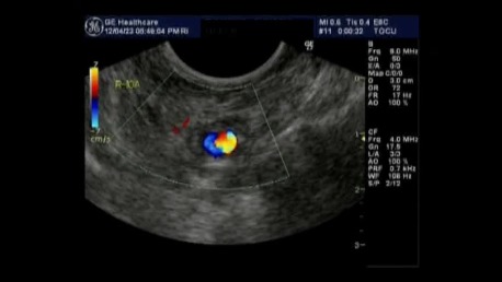
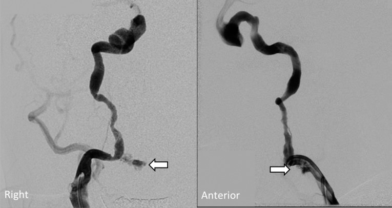
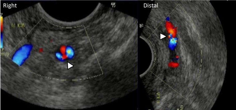
An 88-year-old woman presented with worsening right hemifacial pain, oculomotor, and abducens nerve palsy. Digital subtraction angiography (DSA) showed an aneurysm with a maximal diameter of 17 mm on a horizontal segment of the carotid siphon of the right ICA. We planned coil embolization using an Enterprise vascular remodeling device (Codman, Miami, FL, USA) under general anesthesia to meet her request for relieving hemifacial pain. We struggled to position a 7 Fr guiding catheter using a coaxial system with a 5 Fr catheter and a stiff 0.035-inch guidewire after anticoagulation with intravenous unfractionated heparin. DSA showed contrast medium extravasation from the distal cervical segment of the right ICA (Figure 1). Minor extravasation persisted at 40 min after immediate reversal of anticoagulation with protamine sulfate. Twenty minutes after final DSA, we tried to evaluate extravasation and the inner contour of the ICA by ultrasonography during catheterization to minimize the amount of contrast medium for repeated DSA. TOCU with color Doppler imaging using a convex array transducer (6 MHz), which was located beside intubated tracheal tube, showed a pseudolumen and no further extravasation in the distal cervical segment of the right ICA (Figure 2 and Movie). Computed tomography showed a small hematoma behind the pharynx. The patient had no respiratory signs or additional neurological deficits despite the absence of additional therapy. Follow up magnetic resonance angiography 7 days after DSA did not detect the pseudoaneurysm on the right ICA.
Figure 1. DSA shows extravasation of contrast medium from distal cervical segment of right ICA (white arrows). (Left and right) Anteroposterior and lateral views, respectively.
Figure 2. TOCU with color Doppler image shows arrested bleeding and pseudolumen in distal cervical segment of right ICA (white arrow head). (Left and right) Axial and longitudinal images, respectively.
Movie. TOCU with color Doppler axial image scanned the cervical segment of right ICA from the far distal segment to the proximal segment.

DISCUSSION
Paramasivam and colleagues [1] reported that most iatrogenic dissections arising during neurointerventional procedures are minimal intimal tears (67%). The incidence of extravasation from iatrogenic dissection might be extremely rare. To verify the arrest of extravasation from an injured vessel during neurointerventions requires frequent DSA, which would lead to an excess of contrast medium. On the other hand, ultrasonography is less invasive and easily repeatable. TOCU can assess the far distal segment of the ICA more effectively than conventional carotid ultrasonography [3–5]. We originally described that TOCU was useful for evaluating iatrogenic extravasation and extracranial ICA dissection due to positioning a guiding catheter. Ultrasonography, including TOCU should be widely applied to evaluate iatrogenic vessel injury during neurointerventional procedures.
REFERENCES
- Paramasivam S, et al. Iatrogenic dissection during neurointerventional procedures: a retrospective analysis. J NeuroIntervent Surg. 2012;4(5):331–335. doi: 10.1136/neurintsurg-2011-010103. [DOI] [PubMed] [Google Scholar]
- Cloft HJ, et al. Arterial dissections complicating cerebral angiography and cerebrovascular interventions. AJNR Am J Neuroradiol. 2000;21(3):541–545. [PMC free article] [PubMed] [Google Scholar]
- Yasaka M, et al. Transoral carotid ultrasonography. Stroke. 1998;29(7):1383–1388. doi: 10.1161/01.str.29.7.1383. [DOI] [PubMed] [Google Scholar]
- Nagasawa H, et al. Acute morphological change in an extracranial carotid artery dissection on transoral carotid ultrasonography. Circulation. 2008;118(10):1064–1065. doi: 10.1161/CIRCULATIONAHA.108.770693. [DOI] [PubMed] [Google Scholar]
- Suzuki R, et al. Identification of internal carotid artery dissection by transoral carotid ultrasonography. Cerebrovasc Dis. 2012;33(4):369–377. doi: 10.1159/000336121. [DOI] [PubMed] [Google Scholar]


