. 2018 Jun 13;7:e34574. doi: 10.7554/eLife.34574

Figure 4. Polymerase elements required for preferential plus-end localization of Stu2.

Polymerase assays were carried out using wild-type tubulin (0.8 μM) and a panel of polymerase mutants (200 nM each). Representative kymographs are shown for Stu2 variants. Plus-end localization requires at least two tubulin-binding TOGs (WT, TOG1*-TOG2, ∆cc-2xBasic) and is not sensitive to how they are linked (compare TOG1*-TOG2 to ∆cc-2xBasic). Unexpectedly, polymerases with 0 (TOG1*-TOG2*) or one active TOGs (TOG1*-TOG2-∆cc-2xBasic) robustly coat the body of the microtubule. See also Figure 4—figure supplement 1.

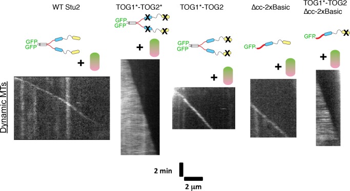
Figure 4.

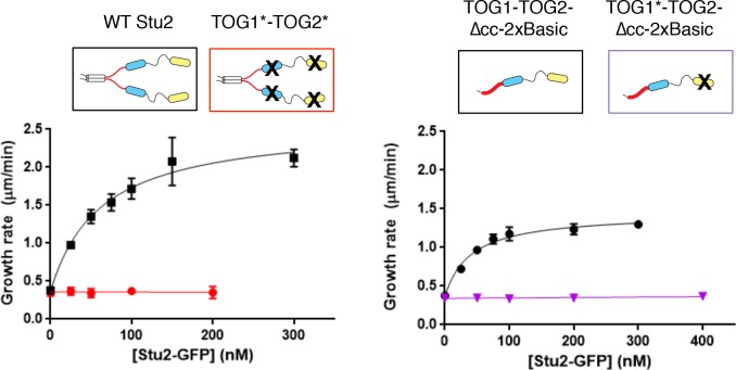
Figure 4—figure supplement 1. Additional data from assays using mutated TOGs.

Figure 4—figure supplement 1.

‘Double dead’ Stu2 (TOG1*-TOG2*) (left) with both TOGs inactivated does not show polymerase activity (red). Data for wild-type Stu2 (black) are reproduced from Figure 1. Stu2(TOG1*-TOG2)-∆cc-2xBasic (right), which contains a single active TOG domain, does not show polymerase activity (purple). Data for Stu2(TOG1*-TOG2)-∆cc-2xBasic (black; this construct has two active TOGs) are reproduced from Figure 1. Black: N = 45 from three independent chambers; Red and Purple: N = 20 measurements. All error bars are SEM. Smooth curves indicate a hyperbolic fit to the data.
Figure 4—figure supplement 1—source data 1. Numerical data associated with Figure 4—figure supplement 1.
DOI: 10.7554/eLife.34574.014