Abstract
We report the development of macrocyclic melanocortin derivatives of MT-II and SHU-9119, achieved by modifying the cycle dimension and incorporating constrained amino acids in ring-closing. This study culminated in the discovery of novel agonists/antagonists with an unprecedented activity profile by adding pieces to the puzzle of the melanocortin receptor selectivity. Finally, the resulting 19- and 20-membered rings represent a suitable frame for the design of further therapeutic ligands as selective modulators of the melanocortin system.
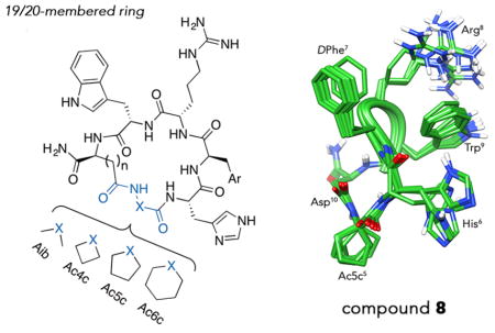
Graphical Abstract
INTRODUCTION
The proopiomelanocortin (POMC) gene, mostly expressed in the central nervous system (CNS) of humans, encodes its corresponding precursor polypeptide of 241 amino acids, which is subject to post-translational proteolytic cleavages.1 From these processes the melanocortin peptides, α-, β-, and γ-melanocyte stimulating hormones (MSH) and the adrenocorticotropin hormone (ACTH), arise.1 Such peptide hormones are the endogenous bioactive ligands for the human melanocortin receptors (hMCRs), and along with the melanocortin inverse agonists agouti signaling protein (ASiP) and agouti related protein (AgRP), all comprise the melanocortin system.1,2 To date, five G-protein-coupled receptor (GPCR) subtypes have been discovered and investigated for their ability to mediate several functions in the human body.3,4 Although the melanocortin system is well-known machinery involved in the regulation of several physiopathological functions,1 structure–activity relationships (SARs) knowledge is still lacking in terms of structural requirements to reach fine modulation of hMCRs. In fact, extensive studies have been carried out,3,5,6 unveiling the core tetrapeptide His-Phe-Arg-Trp as a conserved and fundamental sequence for receptor activation.7 Furthermore, modifications of the α-MSH sequence have turned out to be successful for the development of other representative compounds, both linear and cyclic derivatives, such as the lead agonist MT-II (1, Chart 1) and antagonist SHU-9119 (2, Chart 1),8–12 albeit nonselective.
Chart 1.
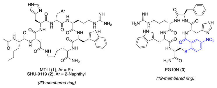
Chemical Structures and Ring Cycle Dimension of MT-II (1), SHU-9119 (2), and PG10N (3)
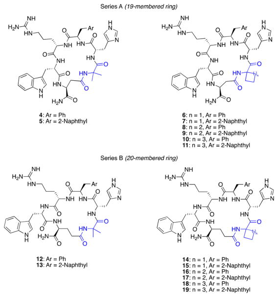
We have previously designed macrocyclic melanocortin analogues by a reduction of the ring cycle, by means of an alkylthioaryl bridge, from typically 23 members of 1 and 2 to 19 members (e.g., compound PG10N,13 3 in Chart 1). In particular, these previously reported compounds have implied key features to attain improved selectivity, such as (i) reduced ring size and (ii) stabilized β-turn conformation. Therefore, herein we describe the design and synthesis of novel derivatives achieved by modifications of the cycle dimension and the incorporation of constrained amino acids in ring-closing. For this approach, we present the synthesis of side-chain-to-tail cyclic 1 and 2 analogues containing 2-aminoisobutyric acid (Aib), 1-aminocyclobutane-1-carboxylic acid (Ac4c), 1-amino-cyclopentane-1-carboxylic acid (Ac5c), and 1-aminocyclohexane-1-carboxylic acid (Ac6c) residues, all involved in a side-chain-to-tail cyclization with Asp and Glu acidic residues (4–19, Chart 2). This strategy has been pursued to investigate the influence of the cycle structure and to understand the effects of incorporation of constrained Aib-derived amino acids on the conformation and interaction with melanocortin receptors. This study has led to the discovery of compounds with unprecedented profiles of activity and selectivity and has improved SAR knowledge regarding the melanocortin system.
Chart 2.
Chemical Structures and Ring Cycle Dimension of Synthesized Compounds 4–19 Divided as Series A and B (19- and 20-Membered Rings, Respectively)
RESULTS AND DISCUSSION
Design
Extensive SAR studies performed on melanocortin ligands pointed out that conformational restriction can contribute to receptor selectivity.14–16 Starting from the exciting results obtained with 3 (Chart 1) and with the aim to discover new potent and selective ligands, we have designed novel macrocyclic compounds in which an α,α-disubstituted amino acid has replaced the alkylthioaryl bridge at the N-terminus (Chart 2). With α-hydrogen exchanged with a methyl group, the Aib residue is almost invariably restricted to ϕ, ψ values of either −60° (±20°) and −30° (±20°) or 60° (±20°) and 30° (±20°).17 Ac4c–Ac6c not only adopt similar ϕ, ψ angle constraints of Aib but have additional constraints to the N–Cα–C′ (τ) bond angle. For example, Ac4c was shown to have a wider than normal τ angle of 114.7°.18 It is expected to have a decreasing trend for the τ bond angle for Ac4c–Ac6c. These structural constraints have been introduced to further restrict the conformation of the macrocyclic compounds, which in turn may improve potency and selectivity. Then, ring closure is obtained by a lactam bridge formed by the carboxyl group of side chain of a Glu or Asp residue at position 10 and the amino group of residue 5. Following this strategy, we have designed and synthesized two series of macrocyclic compounds containing Asp (series A, 4–11) or Glu (series B, 12–19). The resulting macrocyclic peptidomimetics, possessing a 19- or 20-membered ring, have conserved the melanocortin core sequence His-DPhe/DNal(2′)-Arg-Trp (Chart 2).
Chemistry
Peptides 4–19 have been manually synthesized by solid-phase peptide synthesis (SPPS) using conventional 9-fluorenylmethoxycarbonyl (Fmoc) chemistry.19 The synthetic steps of selective allyl ester hydrolysis and intramolecular coupling have been performed by microwave (μW)-assisted SPPS, following slightly modified protocols elsewhere reported.20,21 Upon cyclization and cleavage from the solid support, crude peptides have been purified by reverse-phase high pressure liquid chromatography (RP-HPLC).
Biological Evaluation
All synthesized compounds (4–19) have been evaluated for their binding affinities to the hMCRs 1, 3, 4, and 5 in competitive binding assays using the radiolabeled ligand [125I]-NDP-α-MSH and for their agonist potency in cAMP assays employing the HEK293 cells expressing those receptors. Data of the most representative analogues are collected in Table 1 (for the whole library of compounds see Table S2 in Supporting Information).
Table 1.
Biological Activities of the More Representative Macrocyclic Peptides at the hMC1R and 3–5 hMCRs
| hMC1R | hMC3R | hMC4R | hMC5R | |||||||||
|---|---|---|---|---|---|---|---|---|---|---|---|---|
|
|
|
|
|
|||||||||
| IC50a (nM) | EC50b (nM) | Act %c | IC50a (nM) | EC50b (nM) | Act %c | IC50a (nM) | EC50b (nM) | Act %c | IC50a (nM) | EC50b (nM) | Act %c | |
| 1 | 1.2 ± 0.2 | 1.8 ± 0.2 | 100 | 1.2 ± 0.2 | 1.5 ± 0.2 | 100 | 1.1 ± 0.2 | 2.8 ± 0.4 | 100 | 7.5 ± 0.5 | 3.3 ± 0.6 | 100 |
| 8 | 440 ± 6 | 28 ± 4 | 100 | 10 ± 1 | 30 ± 4 | 100 | 260 ± 23 | 280 ± 7 | 100 | NBe | NAd | 0 |
| 10 | 240 ± 10 | 13 ± 2 | 100 | 11 ± 1 | 33 ± 3 | 100 | 160 ± 9 | 10 ± 1 | 96 | >1000 | NAd | 0 |
| 12 | 320 ± 32 | 15 ± 4 | 100 | 57 ± 6 | 12 ± 4 | 100 | 74 ± 3 | 5.3 ± 0.4 | 100 | 440 ± 115 | 14 ± 1 | 92 |
| 13 | 470 ± 10 | 13 ± 2 | 100 | 27 ± 5 | 19 ± 2 | 60 | 370 ± 41 | 18 ± 1 | 100 | 12 ± 1 | NAd | 0 |
| 15 | 120 ± 6 | 41 ± 4 | 100 | 2.2 ± 0.2 | NAd | 0 | 71 ± 2 | NAd | 0 | 21 ± 1 | NAd | 0 |
| 19 | 100 ± 12 | 631 ± 25 | 100 | 4.4 ± 0.1 | NAd | 0 | 59 ± 4 | NAd | 0 | 20 ± 3 | NAd | 0 |
IC50 = concentration of peptide at 50% specific binding (N = 4).
EC50 = effective concentration of peptide that was able to generate 50% maximal intracellular cAMP accumulation (N = 4).
Act % max is ratio of the highest cAMP level triggered by peptides over the highest cAMP level triggered by MT-II (1). The peptides were tested at a range of concentrations from 10−10 to 10− M.
NA: 0% cAMP accumulation at 10−5 M.
NB: no binding at 10−5 M.
Overall, binding data indicate that affinities of the new compounds toward hMCRs are dependent on macrocycle ring size. In particular, the compounds of series A (4–11) show medium/high nM range or null affinity for all hMCRs (IC50 > 70 nM) except for the hMC3R. In fact, 4, 5, 7–11 strongly bind to the hMC3R (IC50 < 20 nM). This leads to some selective ligands (7–11) at the hMC3R. Exceptions to the above observation are compound 6, which does not bind to any hMCR, and compounds 4 and 5, which have high and moderate affinity also for hMC4R. Increasing the ring size to 20 atoms (series B) leads to 12–19, which show low affinities for hMC1R (IC50 > 40 nM). In regard to hMC3R, DPhe7 containing compounds generally show lower affinity compared to their corresponding 19-membered cycles (12 vs 4, 16 vs 8, 18 vs 10). Conversely, DNal7 derivatives (13, 15, 17, 19) retain similar or improved affinity for hMC3R compared to their lower homologs. For hMC4R, compounds 14, 16, and 18 show no binding at this receptor. Finally, considering hMC5R, some DNal7 derivatives (13, 15, 19) show good affinities (IC50 = 12–21 nM), significantly improved compared to the series A homologs.
Considering the functional activity measured in terms of cAMP levels, all the compounds are full agonists at hMC1R as their parents 1 and 2, but their potency is always lower than 1 (EC50 > 8 nM). For the hMC3R, all the analogs containing a DPhe7 are full agonists as the parent 1. Potencies on this receptor depend on the ring size, with series B derivatives (12, 14, 16, 18) being generally more potent than the corresponding compounds belonging to series A. Among those, 14 and 16 have low nM EC50 values (about 1 nM) similar to 1. In particular, 14 is found to be an interesting agonist at hMC3R but without selectivity with respect to the hMC1R (1:1) and with only modest selectivity when compared to hMC5R (1:7). Interestingly, 8 and 16 are selective agonists for the hMC3R in terms of binding and activity when compared to hMC4R and hMC5R. In particular, 16 shows a nM binding at hMC1R and hMC3R and no binding at the 4 and 5 hMCRs. This compound represents a highly potent full agonist at the hMC1R and hMC3R with EC50 values of 8.6 and 1.0 nM, respectively. The analogs containing DNal7 are often partial agonists at hMC3R with the notable exceptions of 15 and 19, which behave as full antagonists. In this context, 13 and 17, which differ from 15 and 19 only due to the α,α-amino acid at position 5, are potent partial agonists at hMC3R (EC50 of 19 and 5 nM, respectively). Thus, the constrained amino acid at position 5 influences the activity profile of the 2-derived (i.e., DNal containing) analogs and similar results are also observed for the hMC4R. In fact, 15 and 19 are full antagonists at hMC4R while 17 is a partial agonist and 5 and 13 are full agonists at hMC4R.
Considering the activity on hMC5R, many compounds that show some affinity for this receptor have full agonist activity resembling the parent peptides 1 and 2. In this context, 12 is a potent agonist (EC50 < 15 nM) at all the hMCRs. Notable exceptions are 13, 15, and 19, which are ligands with high affinity and full antagonism at hMC5R. In particular, 13 is a potent antagonist at hMCR5, a full agonist at hMC1R and hMC4R, and a partial agonist at hMC3R, which is a unique activity profile for a melanocortin agent. Moreover, 15 and 19 containing DNal(2′) residue in position 7 showed an unexpected but exceptional profile of activity resulting in potent antagonists at all hMCRs except for the hMC1R. These compounds represent the first macrocyclic peptides endowed with full antagonist activity at the 3, 4, and 5 hMCRs.
According to the bioassay results, α,α-diakylated amino acids played an important role for modulating selectivity. In series A, the Ac6c contained peptides 10 and 11 are 4–7 times more potent at hMC1R than the Aib containing compounds 4 and 5. The Ac4c containing peptides 6 and 7, on the other hand, are 40–107 times less potent at hMC4R than the Aib containing compounds 4 and 5. In series B, the Ac6c containing peptides 18 and 19 become 4–49 times less potent at hMC1R than the Aib containing compounds 12 and 13. More importantly, Ac4c and Ac6c have enhanced the antagonist activity of the DNal(2′) containing peptides 15 and 19 at hMC3R and hMC4R. These modulations on potency are probably due to different presentations of the His-DPhe/DNal(2′)-Arg-Trp pharmacophore as a result of the ϕ, ψ, and τ angle constrains.
NMR Analysis
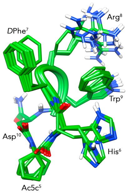
Detailed conformational analysis by solution NMR was performed on 8. Compound 8 was chosen because it is a high affinity (IC50 = 10 nM) ligand at hMC3R with a selectivity of at least 25 times over all the other hMCRs. It is also a potent hMC3R full agonist (EC50 = 30 nM) with high functional selectivity vs the other central hMCRs (4 and 5). Moreover, belonging to the series A, 8 was designed to be also highly conformationally constrained. A whole set of one-dimensional (1D) and two-dimensional (2D) NMR spectra in a 200 mM aqueous solution of dodecylphosphocholine (DPC) were collected for 8. DPC micelle solutions were used since they are membrane mimetic environments and are largely used for conformational studies of peptide neurotransmitters and hormones.22,23 All NMR parameters are reported in Table S3, Supporting Information. NMR-derived constraints obtained for 8 were used as the input data for a simulated annealing structure calculation. NOESY spectra of 8 showed diagnostic NOEs consistent with turn/helical structures (Table S4, Supporting Information). The restrained simulated annealing calculation gave an ensemble of structures showing a type II′ β-turn about residues DNal(2′)7-Arg8, followed by a short 310-helix along residues Trp9 and Glu10 (Figure 1). The complete ensemble of structures fulfilled the NOE restraints, with no violations exceeding 0.5 Å. Considering the side chains, DPhe7, Arg8, and Trp9 show a clear preference for trans, g+, and g− and orientations, respectively. His6 is less defined, but there is a predominance of the g− orientation (about 60% of the calculated structures). Spatial proximity of the Arg8 side chain to the indole of Trp9 is in accordance with the significant upfield shift of the proton signals of the Arg8 residue.
Figure 1.
Lowest energy conformers of compound 8 (PDB code 6FCE).
Docking
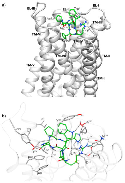
8 was also docked against an hMC3R model,22 using the program AUTODOCK.24 This gave a high populated low energy cluster for the 8/hMC3R complex, whose best scored pose is shown in Figure 2. The predicted binding site is placed among TM3–7, EL1–3 (Figure 2a). Main interactions between the peptide and hMC3R are shown in Figure 2b. For comparison purpose, NMR structure of 125 was also docked to the hMC3R model. Docking results are reported in Figure S1 of the Supporting Information. 1 location in the docking best pose is very similar to 8. The main difference is the position of Trp9 side chain in the two complexes, which could be responsible of the different activity profiles of the two compounds. This issue requires further investigation.
Figure 2.
(a) hMC3R model22 complexed with 8. Heavy atoms of 8 (carbon, green; nitrogen, blue; oxygen, red; sulfur, yellow). Receptor backbone is represented in gray ribbon. (b) 8 within the binding pocket of hMC3R. Hydrogen bonds are represented with dashed lines.
CONCLUSIONS
We have developed novel melanocortin ligands by reducing the cycle dimension of known hMCRs agonist and antagonist and introducing α,α-disubstituted constrained amino acids. Among the novel compounds, we have obtained (i) highly potent agonists at the hMC3R (8, 10, 12, 14, 16, 18), some of which were also highly selective toward this receptor (8 and 10); (ii) a potent agonist at all the hMCRs (12); (iii) a potent antagonist at hMC5R, full agonist at hMC1R and hMC4R, and partial agonist at hMC3R (13), which is a unique activity profile for a melanocortin agent; (iv) potent antagonists at the 3, 4, and 5 hMCRs and agonists at hMC1R (15 and 19) which is again an unprecedented activity profile for hMCRs. All these compounds enlarge our structure–activity relationships knowledge for the melanocortin ligands and offer new tools for pharmacological studies. Moreover, the activities shown by these compounds demonstrate that the new cyclic scaffolds with 19- and 20-membered ring are suitable for the development of hMCRs ligands and can be employed for further development of melanocortins. Finally, NMR-derived structure of 8 and its docking model against hMC3R were calculated. Overall, the results will help to develop novel peptide and non-peptide analogues acting on this target which is of overwhelming importance in different pathological conditions in which melanocortin system play an important role.
EXPERIMENTAL SECTION
General Procedures
Microwave irradiation was performed on a Biotage Initiator+ apparatus on the high-absorption level; temperature was monitored automatically. LC–MS analyses were performed on a LC–MS instrument from Agilent Technologies equipped with an analytical C18 column and 6110 quadrupole, in positive electrospray ionization (ESI) mode, to confirm that difficult couplings had achieved >90% conversion. HRMS measurements were recorded on a LTQ Orbitrap mass spectrometer in positive ESI mode, and proton adducts [M + H]+ were used for empirical formula confirmation.
Purification of peptides 4–19 was performed by RP-HPLC (Shimadzu preparative liquid chromatograph LC-8A) equipped with a preparative column (Phenomenex Kinetex C18 column, 150 mm × 21.2 mm, 5 μm, 100 Å) using linear gradients of MeCN (0.1% TFA) in water (0.1% TFA), from 10% to 90% over 20 min, with a flow rate of 10 mL/min and UV detection at 220 nm. Final products were obtained by lyophilization of the appropriate fractions after removal of the MeCN by rotary evaporation. Peptides 4–19 were analyzed by analytical RP-UHPLC (Shimadzu Nexera liquid chromatograph LC-30AD) equipped with a C18-bonded reverse-phased column (Phenomenex Kinetex, 150 mm × 4.6 mm, 2.6 μm, 100 Å) using gradient elution of two different solvent systems [gradient 1 of MeCN (0.1% TFA) in water (0.1% TFA) from 10% to 90% over 15 min; gradient 2 of MeOH (0.1% TFA) in water (0.1% TFA) from 10% to 90% over 15 min; flow rate = 1 mL/min; diode array UV–vis detector; see Supporting Information]. Such analytical methods assessed for purity of peptides 4–19 > 99% (see Table S1, Supporting Information), and their correct molecular weights were also confirmed by HRMS spectrometer.
Peptide Synthesis
The synthesis of macrocylic peptides 4–19 was performed in a stepwise fashion via solid-phase peptide synthesis (SPPS), as elsewhere reported.19 In particular, each peptide sequence was assembled on a Rink amide resin (0.2 mmol from 0.74 mmol/g of loading substitution) as solid support placed into a plastic syringe tube equipped with Teflon filter, stopper, and stopcock. The resin, stored as Fmoc-protected, was preswollen in DMF for 20 min, then treated with 20% piperidine in DMF solution (5 min × 1, 25 min × 1) to remove the Fmoc group. The first peptide bond formation by the coupling with the first residue [Fmoc-Asp(OAll) or Fmoc-Glu(OAll)] (3 equiv) was achieved by adding 3-fold excess of 2-(1H-benzotriazole-1-yl)-1,1,3,3-tetramethyluronium hexafluorophosphate (HBTU) and 1-hydroxybenzotriazole (HOBt) in the presence of a 6-fold excess of N,N-diisopropylethylamine (DIEA). The Fmoc deprotection was carried out as described above. After each coupling and Fmoc-deprotection step, the peptide resins were washed with DMF (2 mL × 3), DCM (2 mL × 3), and DMF (2 mL × 3), and reactions were monitored by the colorimetric Kaiser test for the detection of solid-phase bound primary amines.26
The peptide resins carrying the allyl esters of aspartic and glutamic residues in C-terminal were hydrolyzed by a slightly modified procedure elsewhere described.20,21 In particular, the resins were washed with DCM (2 mL × 3), suspended in a solution of Pd(PPh3)4 (0.15 equiv) and N,N′-dimethylbarbituric acid (NDMBA) (7 equiv) in 3:2 dry DCM/DMF (v/v), and placed in a 20 mL μW reaction vessel. The vessel was sealed and heated to 40 °C using μW irradiation for 5 min. The vessel was opened, and the reaction mixture was transferred to a plastic syringe tube. The resins were filtered, washed with DMF (2 mL × 3) and DCM (2 mL × 3), and the allyl-deprotection procedure was repeated under the same conditions. The resins were filtered, washed with DMF (2 mL × 3), a 0.5% solution of sodium N,N-diethyldithiocarbamate in DMF (30 min ×2), and DCM (2 mL × 3). After complete removal of the allyl group from the peptide was ascertained by LC–MS of the residue from cleavage of an aliquot of resin (5 mg), the N-terminal Fmoc group was finally removed, as above-described. The released amines were thus coupled to acids by using PyAOP (2 equiv) and HOAt (2 equiv), which are known effective coupling reagents for the intramolecular amide bond formation,27,28 in the presence of DIEA (4 equiv). The reaction was carried out in a sealed 20 mL μW reaction vessel, heated to 45 °C under μW irradiation for 5 min. The vessel was opened, and the reaction mixture was transferred to the plastic syringe tube. The peptide-resin was thoroughly washed with DCM (5 × 2 mL) and dried under argon.
Peptides 4–19 were released from the resin and the protecting groups cleaved simultaneously by means of a reaction cocktail consisting of 95:2.5:2.5 TFA/TIS/H2O (v/v/v) at rt for 3 h. The resin was removed by filtration and crude peptides were recovered by precipitation with chilled anhydrous Et2O as white to pale beige- colored amorphous solids.
Biological Activity Assays
Competitive binding assays with [125I]-NDP-MSH and cAMP assays were performed on HEK293 cells stably expressed human melanocortin receptors (hMC1R, hMC3R, hMC4R, hMC5R). The methods used previously described protocols.29–34
NMR Spectroscopy
The samples for NMR spectroscopy were prepared by dissolving the appropriate amount of 8 in 0.54 mL of 1H2O (pH 5.5), 0.06 mL of 2H2O to obtain 2 mM peptide and 200 mM DPC-d38. NMR spectra were recorded on a Varian INOVA 700 MHz spectrometer equipped with a z-gradient 5 mm triple-resonance probe head. 1D and 2D NMR spectra were recorded and processed as described in the Supporting Information.
Structure Calculation
The NOE-based distance restraints were obtained from NOESY spectrum of 8 collected with a mixing time of 100 ms. The NOE cross peaks were integrated with the XEASY35 program and were converted into upper distance bounds using the CALIBA program incorporated into the program package DYANA.36 Only NOE derived constraints were considered in the annealing procedures (Table S4, Supporting Information). Restrained simulated annealing was performed using the Discover module of the InsightII program. More details are reported in the Supporting Information.
Receptor Models and Docking
Three-dimensional structure model of hMC3R was generated by I-TASSER37 server for protein structure and function prediction, as previously reported.22 The initial pose for the hMC3R/8 or hMC3R/1 complexes was generated by docking the lowest energy conformers of 8 and 1 obtained by NMR to the hMC3R model using the program AUTODOCK 4.0.24 The side chains of His6, DPhe7, Arg8, and Trp9 of 8 and 1 were considered flexible in the docking procedure. Refinement of lowest energy pose of hMC3R/8 and hMC3R/1 complexes was achieved by in vacuo energy minimization with the Discover algorithm using the steepest descent and conjugate gradient methods until a rmsd of 0.05 kcal/mol per Å was reached. The backbone atoms of the TM and IL domains of the hMC3R were held in their position; the ligands and ELs were free to relax.
Supplementary Material
Acknowledgments
We thank the “Finanziamento della Ricerca di Ateneo, Università degli Studi di Napoli Federico II, Annualità 2016 (Prot. N. 0016503)” to A.C. and for support of this research. The study was also supported by NIH Grant GM108040 to M.C.
ABBREVIATIONS USED
- 1D
one-dimensional
- 2D
two-dimensional
- Ac4c
1-aminocyclobutanecarboxylic acid
- Ac5c
1-aminocyclopentane-carboxylic acid
- Ac6c
1-aminocyclohexanecarboxylic acid
- Aib
2-aminoisobutyrric acid
- DIEA
N,N-diisopropylethylamine
- DPC
dodecylphosphocholine
- EL
extracellular loop
- HBTU
2-(1H-benzotriazole-1-yl)-1,1,3,3-tetramethyluronium hexa-fluorophosphate
- hMCR
human melanocortin receptor
- HOBt
1-hydroxybenzotriazole
- MSH
melanocyte stimulating hormone
- Nal(2′)
2-naphtylalanine
- NDMBA
N,N′-dimethyl-barbituric acid
- POMC
proopiomelanocortin
- RP-HPLC
reversed-phase high performance liquid chromatography
- TM
transmembrane domain
Footnotes
Accession Codes
The PDB code for the NMR structure of 8 in DPC solution is 6FCE. Authors will release the atomic coordinates and experimental data upon article publication.
Notes
The authors declare no competing financial interest.
DEDICATION
Dedicated to Prof. Victor J. Hruby on the occasion of his birthday.
The Supporting Information is available free of charge on the ACS Publications website at DOI: 10.1021/acs.jmed-chem.8b00488.
Materials, analytical data and biological activity of 4–19; 1H NMR resonance assignments of 8 in DPC solution; NOE derived upper limit constraints of 8; docking result of 1/hMC3R complex (PDF)
Molecular formula strings and some data (CSV)
PDB coordinates of 1/hMC3R (PDB)
PDB coordinated of 8/hMC3R (PDB)
References
- 1.Cone RD. Studies on the Physiological Functions of the Melanocortin System. Endocr Rev. 2006;27:736–749. doi: 10.1210/er.2006-0034. [DOI] [PubMed] [Google Scholar]
- 2.Voisey J, Carroll L, van Daal A. Melanocortins and Their Receptors and Antagonists. Curr Drug Targets. 2003;4:586–597. doi: 10.2174/1389450033490858. [DOI] [PubMed] [Google Scholar]
- 3.Zhou Y, Cai M. Novel Approaches to the Design of Bioavailable Melanotropins. Expert Opin Drug Discovery. 2017;12:1023–1030. doi: 10.1080/17460441.2017.1351940. [DOI] [PMC free article] [PubMed] [Google Scholar]
- 4.Grieco P, Carotenuto A, Auriemma L, Limatola A, Di Maro S, Merlino F, Mangoni ML, Luca V, Di Grazia A, Gatti S, Campiglia P, Gomez-Monterrey I, Novellino E, Catania A. Novel α-MSH Peptide Analogues with Broad Spectrum Antimicrobial Activity. PLoS One. 2013;8:e61614. doi: 10.1371/journal.pone.0061614. [DOI] [PMC free article] [PubMed] [Google Scholar]
- 5.Cai M, Hruby VJ. Design of Cyclized Selective Melanotropins. Biopolymers. 2016;106:876–883. doi: 10.1002/bip.22976. [DOI] [PMC free article] [PubMed] [Google Scholar]
- 6.Hruby VJ, Cai M, Nyberg J, Muthu D. Approaches to the Rational Design of Selective Melanocortin Receptor Antagonists. Expert Opin Drug Discovery. 2011;6:543–557. doi: 10.1517/17460441.2011.565743. [DOI] [PMC free article] [PubMed] [Google Scholar]
- 7.Abdel-Malek Z. Melanocortin Receptors: Their Functions and Regulation by Physiological Agonists and Antagonists. Cell Mol Life Sci. 2001;58:434–441. doi: 10.1007/PL00000868. [DOI] [PMC free article] [PubMed] [Google Scholar]
- 8.Sawyer TK, Sanfilippo PJ, Hruby VJ, Engel MH, Heward CB, Burnett JB, Hadley ME. 4-Norleucine, 7-D-Phenylalanine-Alpha-Melanocyte-Stimulating Hormone: a Highly Potent Alpha-Melanotropin with Ultralong Biological Activity. Proc Natl Acad Sci U S A. 1980;77:5754–5758. doi: 10.1073/pnas.77.10.5754. [DOI] [PMC free article] [PubMed] [Google Scholar]
- 9.Grieco P, Balse PM, Weinberg D, Macneil T, Hruby VJ. D-Amino Acid Scan of Gamma-Melanocyte-Stimulating Hormone: Importance of Trp(8) on Human MC3 Receptor Selectivity. J Med Chem. 2000;43:4998–5002. doi: 10.1021/jm000211e. [DOI] [PubMed] [Google Scholar]
- 10.Hadley ME, Marwan MM, Al-Obeidi F, Hruby VJ, Castrucci AL. Linear and Cyclic Alpha-Melanotropin [4–10]-Fragment Analogues that Exhibit Superpotency and Residual Activity. Pigm Cell Res. 1989;2:478–484. doi: 10.1111/j.1600-0749.1989.tb00242.x. [DOI] [PubMed] [Google Scholar]
- 11.Al-Obeidi F, Castrucci AMDL, Hadley ME, Hruby VJ. Potent and Prolonged-acting Cyclic Lactam Analogs of Alpha-Melanotropin: Design Based on Molecular Dynamics. J Med Chem. 1989;32:2555–2561. doi: 10.1021/jm00132a010. [DOI] [PubMed] [Google Scholar]
- 12.Hruby VJ, Lu D, Sharma SD, de L, Castrucci A, Kesterson RA, Al-Obeidi FA, Hadley ME, Cone RD. Cyclic lactam Alpha-Melanotropin Analogs of Ac-Nle4-Cyclo[Asp5,D-Phe7,Lys10]-Alpha-Melanocyte-Stimulating Hormone-(4-10)-NH2 with Bulky Aromatic Amino Acids at Position 7 Show High Antagonist Potency and Selectivity at Specific Melanocortin Receptors. J Med Chem. 1995;38:3454–3461. doi: 10.1021/jm00018a005. [DOI] [PubMed] [Google Scholar]
- 13.Grieco P, Cai M, Liu L, Mayorov A, Chandler K, Trivedi D, Lin G, Campiglia P, Novellino E, Hruby VJ. Design and Microwave-Assisted Synthesis of Novel Macrocyclic Peptides Active at Melanocortin Receptors: Discovery of Potent and Selective hMC5R Receptor Antagonists. J Med Chem. 2008;51:2701–2707. doi: 10.1021/jm701181n. [DOI] [PMC free article] [PubMed] [Google Scholar]
- 14.Doedens L, Opperer F, Cai M, Beck JG, Dedek M, Palmer E, Hruby VJ, Kessler H. Multiple N-Methylation of MT-II Backbone Amide Bonds Leads to Melanocortin Receptor Subtype hMC1R Selectivity: Pharmacological and Conformational Studies. J Am Chem Soc. 2010;132:8115–8128. doi: 10.1021/ja101428m. [DOI] [PMC free article] [PubMed] [Google Scholar]
- 15.Cai M, Marelli UK, Bao J, Beck JG, Opperer F, Rechenmacher F, Mcleod KR, Zingsheim MR, Doedens L, Kessler H, Hruby VJ. Systematic Backbone Conformational Constraints on a Cyclic Melanotropin Ligand Leads to Highly Selective Ligands for Multiple Melanocortin Receptors. J Med Chem. 2015;58:6359–6367. doi: 10.1021/acs.jmedchem.5b00102. [DOI] [PMC free article] [PubMed] [Google Scholar]
- 16.Grieco P, Cai M, Han G, Trivedi D, Campiglia P, Novellino E, Hruby VJ. Further Structure-Activity Studies of Lactam Derivatives of MT-II and SHU-9119: Their Activity and Selectivity at Human Melanocortin Receptors 3, 4, and 5. Peptides. 2007;28:1191–1196. doi: 10.1016/j.peptides.2007.02.012. [DOI] [PMC free article] [PubMed] [Google Scholar]
- 17.Venkatraman J, Shankaramma SC, Balaram P. Design of Folded Peptides. Chem Rev. 2001;101:3131–3152. doi: 10.1021/cr000053z. [DOI] [PubMed] [Google Scholar]
- 18.Gatos M, Formaggio F, Crisma M, Toniolo C, Bonora GM, Benedetti Z, Di Blasio B, Iacovino R, Santini A, Saviano M, Kamphuis J. Conformational Characterization of the 1-Amino-cyclobutane-1-Carboxylic Acid Residue in Model Peptides. J Pept Sci. 1997;3:110–122. doi: 10.1002/(SICI)1099-1387(199703)3:2%3C110::AID-PSC88%3E3.0.CO;2-6. [DOI] [PubMed] [Google Scholar]
- 19.Lubell WD, Blankenship JW, Fridkin G, Kaul R. Peptides. Thieme; Stuttgart, Germany: 2005. Science of Synthesis 21.11; pp. 713–809. [Google Scholar]
- 20.Tala SR, Schnell SM, Haskell-Luevano C. Microwave-Assisted Solid-Phase Synthesis of Side-Chain to Side-Chain Lactam-Bridge Cyclic Peptides. Bioorg Med Chem Lett. 2015;25:5708–5711. doi: 10.1016/j.bmcl.2015.10.095. [DOI] [PMC free article] [PubMed] [Google Scholar]
- 21.Merlino F, Yousif AM, Billard É, Dufour-Gallant J, Turcotte S, Grieco P, Chatenet D, Lubell WD. Urotensin II(4–11) Azasulfuryl Peptides: Synthesis and Biological Activity. J Med Chem. 2016;59:4740–4752. doi: 10.1021/acs.jmedchem.6b00108. [DOI] [PubMed] [Google Scholar]
- 22.Carotenuto A, Merlino F, Cai M, Brancaccio D, Yousif AM, Novellino E, Hruby VJ, Grieco P. Discovery of Novel Potent and Selective Agonists at the Melanocortin-3 Receptor. J Med Chem. 2015;58:9773–9778. doi: 10.1021/acs.jmedchem.5b01285. [DOI] [PMC free article] [PubMed] [Google Scholar]
- 23.Cai M, Stankova M, Muthu D, Mayorov A, Yang Z, Trivedi D, Cabello C, Hruby VJ. An Unusual Conformation of γ-Melanocyte-Stimulating Hormone Analogues Leads to a Selective Human Melanocortin 1 Receptor Antagonist for Targeting Melanoma Cells. Biochemistry. 2013;52:752–764. doi: 10.1021/bi300723f. [DOI] [PMC free article] [PubMed] [Google Scholar]
- 24.Goodsell DS, Morris GM, Olson AJ. Automated Docking of Flexible Ligands: Applications of AutoDock. J Mol Recognit. 1996;9:1–5. doi: 10.1002/(sici)1099-1352(199601)9:1<1::aid-jmr241>3.0.co;2-6. [DOI] [PubMed] [Google Scholar]
- 25.Grieco P, Brancaccio D, Novellino E, Hruby VJ, Carotenuto A. Conformational Study on Cyclic Melanocortin Ligands and New Insight into Their Binding Mode at the MC4 Receptor. Eur J Med Chem. 2011;46:3721–3733. doi: 10.1016/j.ejmech.2011.05.038. [DOI] [PMC free article] [PubMed] [Google Scholar]
- 26.Gaggini F, Porcheddu A, Reginato G, Rodriquez M, Taddei M. Colorimetric Tools for Solid-Phase Organic Synthesis. J Comb Chem. 2004;6:805–810. doi: 10.1021/cc049963a. [DOI] [PubMed] [Google Scholar]
- 27.Han SY, Kim YA. Recent Development of Peptide Coupling Reagents in Organic Synthesis. Tetrahedron. 2004;60:2447–2467. [Google Scholar]
- 28.Generoso SF, Giustiniano M, La Regina G, Bottone S, Passacantilli S, Di Maro S, Cassese H, Bruno A, Mallardo M, Dentice M, Silvestri R, Marinelli L, Sarnataro D, Bonatti S, Novellino E, Stornaiuolo M. Pharmacological Folding Chaperones Act as Allosteric Ligands of Frizzled4. Nat Chem Biol. 2015;11:280–286. doi: 10.1038/nchembio.1770. [DOI] [PubMed] [Google Scholar]
- 29.Zhou Y, Mowlazadeh Haghighi S, Zoi I, Sawyer JR, Hruby VJ, Cai M. Design of MC1R Selective Gamma-MSH Analogues with Canonical Amino Acids Leads to Potency and Pigmentation. J Med Chem. 2017;60:9320–9329. doi: 10.1021/acs.jmedchem.7b01295. [DOI] [PMC free article] [PubMed] [Google Scholar]
- 30.Cai M, Marelli UK, Mertz B, Beck JG, Opperer F, Rechenmacher F, Kessler H, Hruby VJ. Structural Insights into Selective Ligand-Receptor Interactions Leading to Receptor Inactivation Utilizing Selective Melanocortin 3 Receptor Antagonists. Biochemistry. 2017;56:4201–4209. doi: 10.1021/acs.biochem.7b00407. [DOI] [PMC free article] [PubMed] [Google Scholar]
- 31.Cai M, Mayorov AV, Ying J, Stankova M, Trivedi D, Cabello C, Hruby VJ. Design of Novel Melanotropin Agonists and Antagonists with High Potency and Selectivity for Human Melanocortin Receptors. Peptides. 2005;26:1481–1485. doi: 10.1016/j.peptides.2005.03.020. [DOI] [PubMed] [Google Scholar]
- 32.Cai M, Mayorov AV, Cabello C, Stankova M, Trivedi D, Hruby VJ. Novel 3D Pharmacophore of Alpha-MSH/Gamma-MSH Hybrids Leads to Selective Human MC1R and MC3R Analogues. J Med Chem. 2005;48:1839–1848. doi: 10.1021/jm049579s. [DOI] [PubMed] [Google Scholar]
- 33.Cai M, Stankova M, Pond SJ, Mayorov AV, Perry JW, Yamamura HI, Trivedi D, Hruby VJ. Real Time Differentiation of G-protein Coupled Receptor (GPCR) Agonist and Antagonist by two Photon Fluorescence Laser Microscopy. J Am Chem Soc. 2004;126:7160–7161. doi: 10.1021/ja049473m. [DOI] [PubMed] [Google Scholar]
- 34.Cai M, Cai C, Mayorov AV, Xiong C, Cabello CM, Soloshonok VA, Swift JR, Trivedi D, Hruby VJ. Biological and Conformational Study of Beta-substituted Prolines in MT-II Template: Steric Effects Leading to Human MC5 Receptor Selectivity. J Pept Res. 2004;63:116–131. doi: 10.1111/j.1399-3011.2003.00105.x. [DOI] [PubMed] [Google Scholar]
- 35.Bartels C, Xia TH, Billeter M, Güntert P, Wüthrich K. The Program XEASY for Computer-Supported NMR Spectral Analysis of Biological Macromolecules. J Biomol NMR. 1995;6:1–10. doi: 10.1007/BF00417486. [DOI] [PubMed] [Google Scholar]
- 36.Güntert P, Mumenthaler C, Wüthrich K. Torsion Angle Dynamics for NMR Structure Calculation with the New Program Dyana. J Mol Biol. 1997;273:283–298. doi: 10.1006/jmbi.1997.1284. [DOI] [PubMed] [Google Scholar]
- 37.Roy A, Kucukural A, Zhang Y. I-TASSER: a Unified Platform for Automated Protein Structure and Function Prediction. Nat Protoc. 2010;5:725–738. doi: 10.1038/nprot.2010.5. [DOI] [PMC free article] [PubMed] [Google Scholar]
Associated Data
This section collects any data citations, data availability statements, or supplementary materials included in this article.


