Abstract
Leiomyoma of uterine origin is a common histologically benign neoplasm in women; however, growth intravenously with intracardiac extension is a rare phenomenon. This is a diagnostic challenge that can present with varied clinical manifestations and multiple differential diagnosis. This is a case of a 45-year-old female patient with chest heaviness and an intracardiac mass on 2-dimensional (2D) echocardiogram. Previous history of hysterectomy was likewise noted. Imaging workup, including 2D echocardiogram and contrast-enhanced chest and abdomen computed tomography scans, was performed which demonstrated a large, heterogeneous, elongated filling defect in the right atrium and right ventricle extending to the inferior vena cava, left renal vein, and left gonadal vein. The diagnosis was made after resection of the tumor in a single-stage operation. The histopathologic and immunoprofile of the resected tumor were consistent with leiomyoma. The use of multiple imaging modalities such as 2D echocardiogram and computed tomography are essential in the investigation of the intracaval masses with intracardiac extension. Although intravenous leiomyoma with intracardiac extension is a rare phenomenon, radiologists and clinicians alike should be mindful of this differential diagnosis.
Keywords: Leiomyoma, Intravascular extension, Intracardiac extension
Introduction
The most frequent neoplasm in the female genital tract is leiomyoma characterized by histologically benign-looking smooth muscle cells. Intravenous leiomyoma is a rare manifestation and even more so with intracardiac extension [1], [2]. Only 300 cases of intravenous leiomyoma have been reported in the English literature [3]. Although it is histologically benign, it can take multiple patterns of venous spread [4]. The symptom of intravenous leiomyoma is nonspecific and is dependent on the extent of the tumor [5]. Intravenous leiomyoma has been reported in women with concurrent leiomyoma and/or with a history of hysterectomy [6], [7]. Multiple imaging modalities such as echocardiography, computed tomography (CT), and magnetic resonance (MR) imaging can be used to establish the diagnosis and the operative plan [2], [8]. In this case report, an intravenous leiomyoma with intracardiac extension was treated with a single-stage operation.
Case report
A 45-year-old female patient was admitted due to chest heaviness and was initially managed as a case of acute coronary syndrome. The patient also had occasional palpitations and syncopal attacks for the past 3 months. Troponin I and echocardiographic findings were normal. Physical examination revealed a grade 2/6 systolic murmur, which was audible along the right upper sternal border along with a widely split second heart sound. Other physical findings were unremarkable. Upon investigation, it was found that the patient previously underwent total abdominal hysterectomy with left salpingo-oophorectomy secondary to myoma uteri with cystic degeneration and left ovary with corpus luteum. The operation was done 2 years before the onset of the cardiac symptoms.
Echocardiography showed a right atrial mass, which prolapses into the right ventricle during diastole. There was resultant dilation of the right atrium and right ventricle, and moderate tricuspid regurgitation with dilated tricuspid valve annulus.
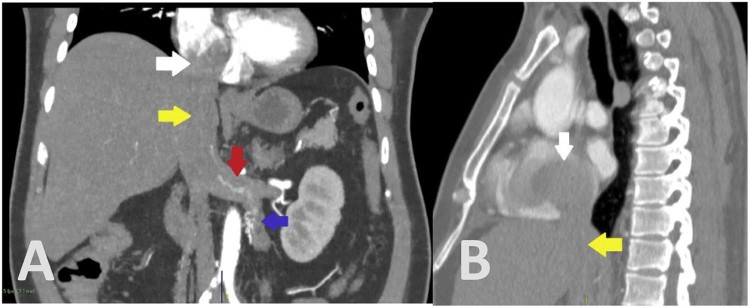
Enhanced CT scan of the chest and abdomen demonstrated a large, heterogeneous, elongated filling defect in the right atrium and right ventricle (Fig. 1) extending to the inferior vena cava (IVC), left renal vein, and left gonadal vein (Fig. 2). Liver is normal in size with heterogeneous parenchyma in venous phase, likely due to congestion caused by the filling defect within the IVC.
Fig. 1.
(A, B) Coronal and axial views showing the right atrial mass (white arrows).
Fig. 2.
(A, B) Coronal and sagittal views showing the extent of the filling defect, from the right atrium (white arrow) to the inferior vena cava (yellow arrow), left renal vein (red arrow), and left gonadal vein (blue arrow).
The patient was started on enoxaparin on an initial presumption of hypercoagulability. Laboratory workup for hypercoagulability, thrombosis, and myeloproliferative disorder, and metastatic workup for ovarian cancer were done; findings were within normal limits.
The patient underwent excision of the IVC and right atrial mass via a midline sternotomy incision. Cardiopulmonary bypass was instituted with cannulations at the aorta, right atrium-superior vena cava, and IVC.
The excised mass consisted of 2 pieces of elongated and tan-cream mass, with glistening and smooth surface, each measuring 7.5 × 4.0 × 3.5 cm and 7.5 × 3.0 × 2.5 cm. Each piece of mass has a resection end measuring 3.8 and 2.9 cm, respectively. Histomorphologic features and immunoprofile were consistent with leiomyoma. Neither mitosis nor necrosis was appreciated in the obtained specimen.
The patient had an uneventful postoperative course with no demonstrable residual mass within the right atrial cavity and visualized proximal IVC on transesophageal echocardiography.
Discussion
Leiomyoma is the most frequent neoplasm in the female genital tract; however, leiomyoma located intravenously with intracardiac extension is a rare phenomenon [1], [2]. This case is rare presentation of a common neoplasm causing a diagnostic dilemma.
Intravenous leiomyoma lesions were first described in 1897 by Birch-Hirschfield from an autopsy case, and the first case of intracardiac extension was then reported by Durck in 1907 [9], [10]. Only 300 cases of intravenous leiomyoma have been reported in the English literature as cited by Matos et al., dated 2013 [3]. Further, approximately 50% of the 68 cases studied by Lam et al. had intracardiac involvement [4].
There has been no established pathogenesis of this tumor. Fourteen cases have been studied by Norris and Parmley, and 2 main theories have been proposed. The first one shows that it originates directly from the vein walls, whereas the second may be due to intravascular projections into adjacent venous channels from a primary uterine leiomyoma [2].
Reported cases of intravenous leiomyoma had an age range of 20-70 years, with a median age of 45 years [2], [11]. Concurrent benign uterine leiomyoma or with a history of hysterectomy were noted [6], [7]. Lam et al. studied 68 reported cases of intravenous leiomyoma with intracardiac extension and found that 38 patients (55.9%) had a history of hysterectomy [4]. The correlation with race, fertility, or parity has yet to be proven [6], [7].
The clinical presentation of intravenous leiomyoma is varied, with the symptoms dependent on the extent of the tumor [12]. The obstruction of the venous return causes the clinical manifestations of this tumor [5]. Other symptoms that may occur include syncope, dyspnea, easy fatigability, chest pain, ascites, and hepatomegaly [12]. According to Wu et al., the most common presentation is heart failure; however, approximately 13% of patients with intracardiac extension may be asymptomatic [13].
The histopathologic features of intravenous leiomyoma are similar to a uterine leiomyoma. Under microscopy, intravenous leiomyoma consists of whorled, anastomosing fascicles of uniform, spindle-shaped smooth muscles with none or minimal amount of nuclear fission [14]. On immunohistochemical staining, intravenous leiomyoma are positive for muscle actin when evaluated with the monoclonal antibody HHF-35 or with antibodies to alpha-SMA [15]. In our case, it was consistent with the histomorphologic and immunoprofile features of leiomyoma.
Intravenous leiomyoma can take several pathways of venous spread with possible intracardiac extension. Lam et al. described 2 routes of spread into the systemic venous circulation. First possible pathway is via the uterine vein which can extend into the internal iliac veins, common iliac veins, and then the inferior vena cava. Second, growth in the ovarian vein may extend into the subphrenic segment of the inferior vena cava and circumvent the iliac veins [4], as seen in this case.
Echocardiography is the initial imaging of choice that is used to evaluate intracardiac lesions, providing high-resolution and real-time images [16]. CT and MR imaging are then used for their multiplanar abilities and large fields of view [2]. These modalities can provide supplementary information on the extension of the lesion to detect associated uterine leiomyoma and help in establishing the operative plan [8].
Diagnostic investigation of an intracardiac mass generally starts with the exclusion of thrombi. The presence of structural heart diseases, atrial fibrillation, and low cardiac output state are factors in thrombus formation [17], [18]. The patient had no demonstrable structural heart disease and the only related history was hypertension. Laboratory workups were likewise negative for hypercoagulability.
Other differential diagnosis of the right atrial mass includes right atrial myxoma and metastasis from tumors with caval extension, such as renal cell carcinoma, adrenal cortical carcinoma, lymphoma, leiomyosarcoma, and hepatocellular carcinoma [19], [20], [21], [22], [23].
Surgical treatment is required for complete removal of the intravenous leiomyoma [20], [21], [24], [25]. The first successful resection of the intracardiac extension of this lesion was reported by Timmis et al. [26]. As intravenous leiomyoma does not generally invade blood vessels, it can be simply removed by downward traction from the involved vein [27], [28]. An important feature is the lack of adhesion to the wall of the cardiac chambers and venous structures [29], as seen in this case.
Long-term follow-up is recommended for intravenous leiomyoma because of the high possibility of recurrence [21], [30]. Multiple studies have reported recurrence rates of up to 30%, with a follow-up range of 7 months to 17 years [21], [31], [32]. It is suggested by Matos et al. that MR imaging is preferable for follow-up due to its superior soft tissue contrast resolution, being nonradiating, and with a higher safety profile of the intravenous contrast media [3].
Footnotes
Acknowledgments: The author greatly appreciates the guidance of Dr. Marvin Tamana, the supervising consultant of this paper. Dr. Arnel Co for the assistance in finding this rare case. Dr. Neilson Tino for the critical remarks that helped in improving the construction of this case report. The rest of the residents and fellows of the Department of Radiology in the Philippine Heart Center have likewise helped in this paper.
Competing Interests: The authors have declared that no competing interests exist.
References
- 1.Nam M.S., Jeon M.J., Kim Y.T., Kim J.W., Park K.H., Hong Y.S. Pelvic leiomyomatosis with intracaval and intracardiac extension: a case report and review of the literature. Gynecol Oncol. 2003;89(1):175–180. doi: 10.1016/s0090-8258(02)00138-5. [DOI] [PubMed] [Google Scholar]
- 2.Norris H.J., Parmley T. Mesenchymal tumors of the uterus. V. Intravenous leiomyomatosis. A clinical and pathologic study of 14 cases. Cancer. 1975;36(6):2164–2178. doi: 10.1002/cncr.2820360935. [DOI] [PubMed] [Google Scholar]
- 3.Matos A.P., Ramalho M., Palas J., Heredia V. Heart extension of an intravenous leiomyomatosis. Clin Imaging. 2013;37(2):369–373. doi: 10.1016/j.clinimag.2012.04.022. [DOI] [PubMed] [Google Scholar]
- 4.Lam P.M., Lo K.W., Yu M.Y., Wong W.S., Lau J.Y., Arifi A.A. Intravenous leiomyomatosis: two cases with different routes of tumor extension. J Vasc Surg. 2004;39(2):465–469. doi: 10.1016/j.jvs.2003.08.012. [DOI] [PubMed] [Google Scholar]
- 5.Li R., Shen Y., Sun Y., Zhang C., Yang Y., Yang J. Intravenous leiomyomatosis with intracardiac extension: echocardiographic study and literature review. Tex Heart Inst J. 2014;41:502–506. doi: 10.14503/THIJ-13-3533. [DOI] [PMC free article] [PubMed] [Google Scholar]
- 6.Clement P.B. Intravenous leiomyomatosis of the uterus. Pathol Annu. 1988;23:152–183. [PubMed] [Google Scholar]
- 7.Andrade L., Torresan R., Sales J., Jr, Vicentini R., De Souza G.A. Intravenous leiomyomatosis of the uterus: a report of three cases. Pathol Oncol Res. 1998;4:44–47. doi: 10.1007/BF02904695. [DOI] [PubMed] [Google Scholar]
- 8.Fasih N., Shanbhogue A., Macdonald D., Fraser-Hill M.A., Papadatos D., Kielar A.Z. Leiomyomas beyond the uterus: unusual locations, rare manifestations. Radiographics. 2008;28:1931–1946. doi: 10.1148/rg.287085095. [DOI] [PubMed] [Google Scholar]
- 9.Birch-Hirschfeld F.V. 5th ed. vol. 1. Vogel; Leipzig (Germany): 1896. p. 226. (Lehrbuch der Pathologischen Anatomie). [Google Scholar]
- 10.Durck H. Ueber ien Kontinvierlich durch die entere Holhlvene in das Herz vorwachsendes: fibromyom des uterus. Munch Med Wochenschr. 1907;54:1154. [Google Scholar]
- 11.Kaszar-Seibert D.J., Gauvin G.P., Rogoff P.A., Vittimberga F.J., Margolis S., Hilgenberg A.D. Intracardiac extension of intravenous leiomyomatosis. Radiology. 1988;168:409–410. doi: 10.1148/radiology.168.2.3393658. [DOI] [PubMed] [Google Scholar]
- 12.Li B., Chen X., Chu Y.D., Li R.Y., Li W.D., Ni Y.M. Intracardiac leiomyomatosis: a comprehensive analysis of 194 cases. Interact Cardiovasc Thorac Surg. 2013;17(1):132–138. doi: 10.1093/icvts/ivt117. [DOI] [PMC free article] [PubMed] [Google Scholar]
- 13.Wu C.K., Luo J.L., Yang C.Y., Huang Y.T., Wu X.M., Cheng C.L. Intravenous leiomyomatosis with intracardiac extension. Intern Med. 2009;48:997–1001. doi: 10.2169/internalmedicine.48.1780. [DOI] [PubMed] [Google Scholar]
- 14.Vanni R., Lynch A., Morton C. Uterus: leiomyoma [Internet] 2007. http://atlasgeneticsoncology.org/Tumors/leiomyomID5031.html Atlasgeneticsoncology.org. accessed 19.10.17.
- 15.Schurch W., Skalli O., Seemayer T.A., Gabbiani G. Intermediate filament proteins and actin isoforms as markers for soft tissue tumor differentiation and origin. I. Smooth muscle tumors. Am J Pathol. 1987;128:91–103. [PMC free article] [PubMed] [Google Scholar]
- 16.Oliveira R., Branco L., Galrinho A., Abreu A., Abreu J., Fiarresga A. Cardiac myxoma: a 13-year experience in echocardiographic diagnosis. Rev Port Cardiol. 2010;29:1087–1100. [PubMed] [Google Scholar]
- 17.Alam M. Pitfalls in the echocardiographic diagnosis of intracardiac and extracardiac masses. Echocardiography. 1993;10:181–191. doi: 10.1111/j.1540-8175.1993.tb00029.x. [DOI] [PubMed] [Google Scholar]
- 18.Restrepo C.S., Largoza A., Lemos D.F., Diethelm L., Koshy P., Castillo P. MR imaging findings of benign cardiac tumors. Curr Probl Diagn Radiol. 2005;34:12–21. doi: 10.1067/j.cpradiol.2004.10.002. [DOI] [PubMed] [Google Scholar]
- 19.Sun C., Wang X.M., Liu C., Xv Z.D., Wang D.P., Sun X.L. Intravenous leiomyomatosis: diagnosis and follow-up with multislice computed tomography. Am J Surg. 2010;200(3):e41–e43. doi: 10.1016/j.amjsurg.2009.09.027. [DOI] [PubMed] [Google Scholar]
- 20.Fang B.-R., Ng Y.-T., Yeh C.-H. Intravenous leiomyomatosis with extension to the heart: echocardiographic features: a case report. Angiology. 2007;58(3):376–379. doi: 10.1177/0003319707302504. [DOI] [PubMed] [Google Scholar]
- 21.Clay T.D., Dimitriou J., McNally O.M., Russell P.A., Newcomb A.E., Wilson A.M. Intravenous leiomyomatosis with intracardiac extension—a review of diagnosis and management with an illustrative case. Surg Oncol. 2013;22(3):e44–e52. doi: 10.1016/j.suronc.2013.03.004. [DOI] [PubMed] [Google Scholar]
- 22.Grebenc M.L., Rosado-de-Christenson M.L., Green C.E., Burke A.P., Galvin J.R. Cardiac myxoma: imaging features in 83 patients. Radiographics. 2002;22:673–689. doi: 10.1148/radiographics.22.3.g02ma02673. [DOI] [PubMed] [Google Scholar]
- 23.Buckley O., Madan R., Kwong R., Rybicki F.J., Hunsaker A. Cardiac masses, Part 2: key imaging features for diagnosis and surgical planning. AJR Am J Roentgenol. 2011;197(5):W842–W851. doi: 10.2214/AJR.11.6903. [DOI] [PMC free article] [PubMed] [Google Scholar]
- 24.Sogabe M., Kawahito K., Aizawa K., Sato H., Misawa Y. Uterine intravenous leiomyomatosis with right ventricular extension. Ann Thorac Cardiovasc Surg. 2014;20(Suppl.):933–936. doi: 10.5761/atcs.cr.13-00309. [DOI] [PubMed] [Google Scholar]
- 25.Kang L.Q., Zhang B., Liu B.G., Liu F.H. Diagnosis of intravenous leiomyomatosis extending to heart with emphasis on magnetic resonance imaging. Chin Med J. 2012;125(1):33–37. [PubMed] [Google Scholar]
- 26.Timmis A.D., Smallpeice C., Davies A.C., Macarthur A.M., Gishen P., Jackson G. Intracardiac spread of intravenous leiomyomatosis with successful surgical excision. N Engl J Med. 1980;303:1043–1044. doi: 10.1056/NEJM198010303031806. [DOI] [PubMed] [Google Scholar]
- 27.Lou Y.F., Shi X.P., Song Z.Z. Intravenous leiomyomatosis of the uterus with extension to the right heart. Cardiovasc Ultrasound. 2011;9:25. doi: 10.1186/1476-7120-9-25. [DOI] [PMC free article] [PubMed] [Google Scholar]
- 28.Liu B., Liu C., Guan H., Li Y., Song X., Shen K. Intravenous leiomyomatosis with inferior vena cava and heart extension. J Vasc Surg. 2009;50(4):897–902. doi: 10.1016/j.jvs.2009.04.037. [DOI] [PubMed] [Google Scholar]
- 29.Xu Z.-F. Uterine intravenous leiomyomatosis with cardiac extension: imaging characteristics and literature review. World J Clin Oncol. 2013;4(1) doi: 10.5306/wjco.v4.i1.25. article 25. [DOI] [PMC free article] [PubMed] [Google Scholar]
- 30.Mariyappa N., Manikyam U.K., Krishnamurthy D., Preeti K., Agarwal Y., Prakar U. Intravenous leiomyomatosis. Niger J Surg. 2012;18(2):105–106. doi: 10.4103/1117-6806.103122. [DOI] [PMC free article] [PubMed] [Google Scholar]
- 31.Moniaga N.C., Randall L.M. Uterine leiomyomatosis with intra-caval and intra-cardiac extension. Gynecol Oncol Case Rep. 2012;2(4):130–132. doi: 10.1016/j.gynor.2012.08.003. [DOI] [PMC free article] [PubMed] [Google Scholar]
- 32.Ahmed M., Zangos S., Bechstein W.O. Intravenous leiomyomatosis. Eur Radiol. 2004;14:1316–1317. doi: 10.1007/s00330-003-2186-z. [DOI] [PubMed] [Google Scholar]


