Abstract
Aim: In this study, we clarified changes of the surface dose to a low-density material on a carbon couch and verified whether a novel rigid couch (HM couch) could reduce the surface dose. Materials and Methods: We measured the surface dose using only a carbon couch (iBeam Couchtop STANDARD; BrainLab), a low-density material (Styrofoam board) on the carbon couch, and an HM couch for 6 and 10 MV photon beams. Results: A 5-cm styrofoam board placed on the carbon couch reduced the surface dose by approximately 7-9%, while it had no impact on the depth dose profile; however, in use, such a thickness may cause collision of the patient with the gantry head. The HM couch reduced the surface dose by approximately 7-9% and shifted the depth dose profile by approximately 0.4 cm in the depth direction compared to the carbon couch. Conclusion: The HM couch has the potential to reduce skin toxicity and is expected to be useful in clinical practice instead of carbon couches.
Keywords: Skin toxicity, surface dose, carbon couch, HM couch
Many kinds of carbon couches are commonly used in megavoltage radiotherapy to support patients. The carbon fiber composition minimizes imaging artifacts in image-guided radiotherapy (1). Its characteristics are suitable for use with posterior beams (2). However, the surface dose close to the couch tends to be increased, shifting the depth dose curve to the surface of the patient, i.e. the skin (1,3-6). Smith et al. reported a five- to six-fold increase in surface dose when using a carbon couch for 6 and 10 MV photon beams, respectively (6). Certain studies have shown that immobilization devices can increase the risk of skin toxicity (7-10). Use of both a carbon couch top and immobilization device was associated with a grade 2 or higher skin toxicity in stereotactic body radiation therapy (11,12).
To solve these problems, the development of the tennis racket table has provided an advantage for reduction of skin toxicity, however, it can exhibit sagging in excess of 5.0 mm, which can cause a systematic error in accuracy and isocentric reproducibility (5). Gray et al. reported that the large air gap created by a patient positioning device reduces the surface dose (13), therefore placing a low-density material on the carbon couch may prevent the creation of air gaps, thereby reducing the surface dose for treatment. An ideal couch material needs to have high permeability and low potential for sagging and should adequately support a patient, allowing the surface dose to be reduced. The HM couch (Toppan Printing Co., Ltd., Tokyo, Japan) has been developed for such a purpose and has the characteristics of being light, strong, rigid, and solid.
In this study, we evaluated the surface doses to a thick low-density material on a carbon couch top and HM couch top to investigate whether the HM couch was able to reduce the surface dose and be useful in clinical application instead of the carbon couch top.
Materials and Methods
Carbon couch, styrofoam board, and HM couch. We employed a carbon couch (iBeam Couchtop STANDARD, BrainLab, Heimstetten, Germany) 200 cm long and 53 cm wide in this study. It was constructed from a plastic foam material with a thickness of 4.6 cm, sandwiched between two layers of carbon fiber, each with a thickness of 0.2 cm. Its maximum load was 185 kg (14). Styrofoam board (E-Board C; ESFORM, Nagano, Japan, 1.5, 3.0, and 5.0 cm thick) was employed as a low-density material. The HM couch (Toppan Printing Co., Ltd., Tokyo, Japan) is shown in Figure 1a. It is constructed from polycarbonate foam sandwiched between two thin layers of glass fiber, measuring 5.0 cm in thickness. The polycarbonate is very light and has one of the highest weight resistances of plastics, and a density of 0.1 g/cm3. The components of the glass fiber (wt%) was 53.0% SiO2, 15.0% Al2O3, 21.0% CaO, 2.0% MgO, 8.0% B2O3, and 0.3% Na2O and K2O, and with density of 2.55 g/cm3.
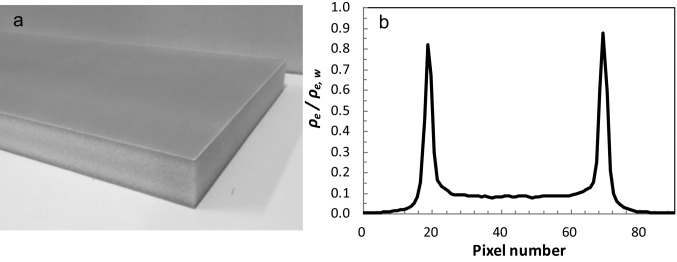
Figure 1. Photograph of HM couch (a) and relative water electron density profile (b). The pixel size was 0.976 mm. The HM couch is composed of thin glass fiber sandwiching a polycarbonate foam core with a total thickness of 5.0 cm.
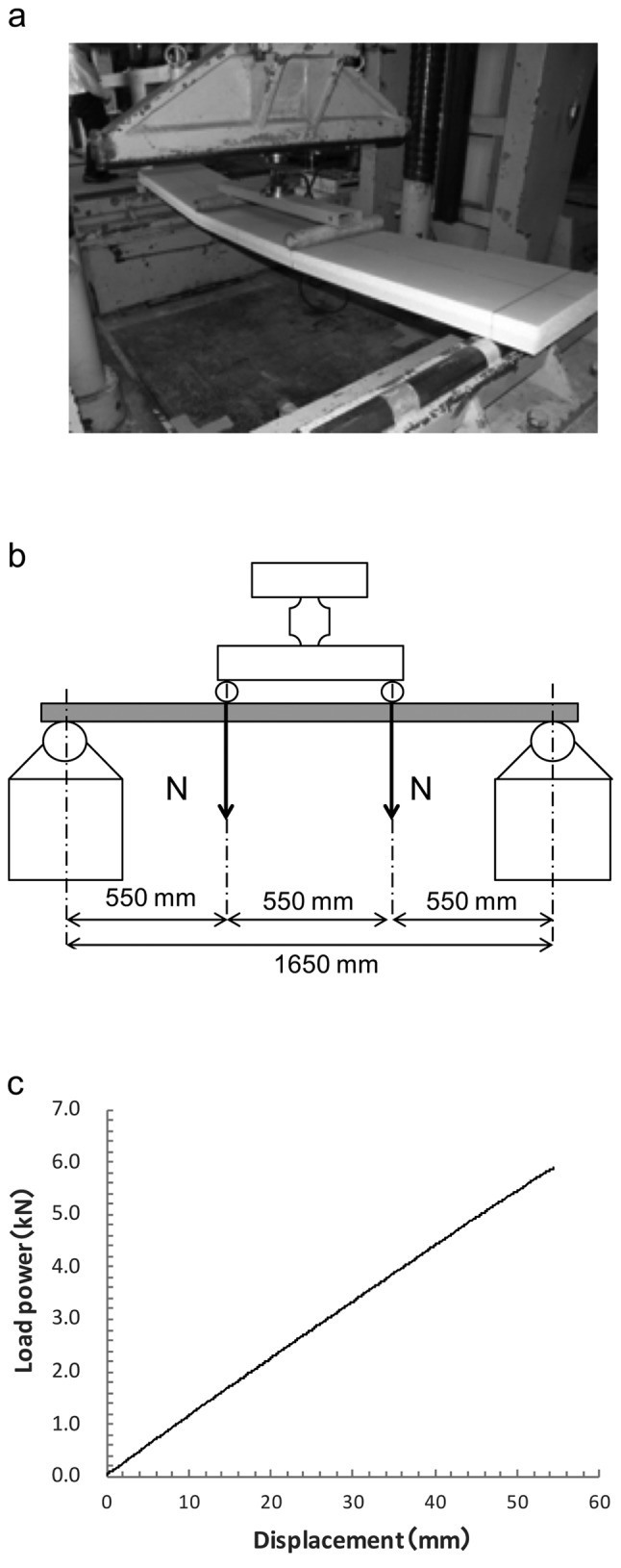
To assess the structure of the HM couch, computed tomography (CT) (Optima, GE Healthcare, Little Chalfont, UK) was employed, using a section thickness of 0.2 cm, FOV=50 cm, and tube voltage of 120 kV. The relative electron densities were calculated from CT values and the electron density table in the treatment planning system. Figure 1b shows the CT images and the relative electron density profile for the HM couch. Additionally, we performed a load test on the HM couch to verify its load capacity of 185 kg (the same as that for the carbon couch). Figure 2 shows the schema of geometry for the load test. The size of the HM couch was 200×53×5 cm. Figure 2a shows the photograph of a broken HM couch. The load power was 2N as shown in Figure 2b. Figure 2c shows the result of the load test. The maximum load of the HM couch, 2N, was 5.88 kN (600 kg). Therefore, the HM couch can hold a large load adequately.
Figure 2. Photograph (a) and schema (b) of the geometry for the load test of the HM couch, and the results of the test (c). The maximum load value of the HM couch, defined as the breaking point (shown in the photograph), 2N, was 5.88 kN (600 kg).
Surface dose measurements. We used a solid water phantom (Gammex RMI, Miccleton WI, USA), and a plane parallel ionization chamber (PPIC) (Murkus Ion Chamber; PTW, Freiburg, Germany) to measure the absorbed surface doses for 6 and 10 MV photon beams from a clinical linear accelerator (TrueBeam; Varian Medical Systems, Palo Alto CA, USA). The dose of 200 MU was delivered with a field size of 10×10 cm2 at source to surface distance (SSD) of 100 cm.
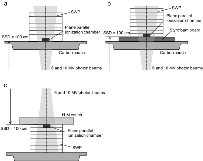
In measurements with the PPIC, Figure 3 shows the experimental geometry for measurements of surface doses with the carbon couch (Figure 3a) and with the styrofoam board on the carbon couch (Figure 3b). Firstly, we measured the surface doses without the carbon couch at a gantry angle of 0˚. Then we measured the surface doses for the carbon couch (Figure 3a) and styrofoam board of 1.5, 3.0, and 5.0 cm thickness on the carbon couch (Figure 3b). Figure 3c shows the schema of geometry for measurements of surface doses for the HM couch. We compared the mean surface dose from three irradiations for the surface dose measurements.
Figure 3. Schematic views of measurements of absorbed surface doses and depth dose profiles for carbon couch (a), carbon couch with styrofoam boards of 1.5, 3.0 or 5.0 cm thicknesses (b), and HM couch (c) using a plane parallel ionization chamber. SSD: Source to surface distance; SWP: solid water phantom .
In the surface dose measurements, a charged particle equilibrium was not established because the ionization chamber was located in the build-up region, which can cause perturbation effects in PPIC by scattered radiation, mostly from the chamber sidewall (15). The perturbation effects cause overestimation of surface dose (16). Therefore, we measured the depth of maximum dose (dmax) for 6 and 10 MV photon beams and set them as reference doses, and corrected the perturbation effects (15).
Depth dose profiles for carbon couch, styrofoam board on the carbon couch, and HM couch. We obtained the depth dose profiles with the carbon couch, styrofoam board of each thickness (1.5, 3.0, and 5.0 cm) on the carbon couch, and HM couches as shown in Figure 3a, b, and c, respectively, using the solid water phantom and PPIC for 6 and 10 MV photon beams. Measurements were taken with 100 MU irradiation with a field size of 10×10 cm2 at an SSD of 100 cm. The depth dose profiles were normalized to the doses at a depth of 5.0 cm in the solid water phantom in which a charged particle equilibrium was established. In the build-up region, the doses were corrected for perturbation effect (15).
Results
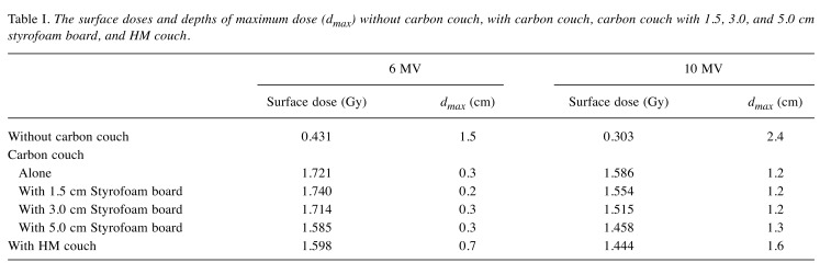
Surface dose. Table I shows the surface doses and the values of dmax with and without the carbon couch, and styrofoam board on the carbon couch and HM couch for the 6 and 10 MV photon beams. The standard deviations of measurements by PPIC were within 0.5%. The surface doses for the 5.0 cm styrofoam board on the carbon couch and HM couch were approximately 7-9% less than that for the carbon couch alone.
Table I. The surface doses and depths of maximum dose (dmax) without carbon couch, with carbon couch, carbon couch with 1.5, 3.0, and 5.0 cm styrofoam board, and HM couch.
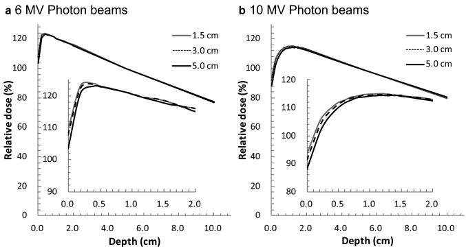
Depth dose profiles for carbon couch, styrofoam board on the carbon couch, and HM couch. Figure 4 shows the depth dose profiles for the styrofoam boards of different thicknesses on the carbon couch for the 6 and 10 MV photon beams. The values of dmax were almost equal without and with styrofoam boards, while the relative surface doses decreased as the thickness of the styrofoam board increased. With the 6 MV photon beams, the reduction in surface dose compared to the carbon couch alone were 0.0%, 1.3%, and 5.1% for styrofoam boards of 1.5, 3.0, and 5.0 cm thickness, respectively (Figure 4a). For the 10 MV photon beams, the corresponding reduction of surface dose compared with the carbon couch alone were 2.1%, 4.3%, and 8.0%, respectively (Figure 4b).
Figure 4. Depth dose profiles for the carbon couch with styrofoam boards with thicknesses of 1.5, 3.0, and 5.0 cm for 6 (a) and 10 (b) MV photon beams, which were normalized to the dose at a depth of 5.0 cm in a solid water phantom .
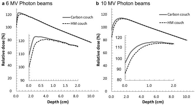
Figure 5 shows the depth dose profiles for the carbon couch and HM couch with the 6 and 10 MV photon beams. The HM couch reduced the surface dose by 7.9% and shifted the dmax value 0.4 cm in the depth direction compared to the carbon couch for 6 MV photon beams (Figure 5a). For the 10 MV photon beams, the HM couch reduced the surface dose by 9.9% and shifted the dmax value 0.4 cm in the depth direction compared to the carbon couch (Figure 5b). The surface doses for the HM couch were less than those using the 5.0 cm styrofoam board on the carbon couch by approximately 2-3%. In the region where charged particle equilibrium was established, the gradients of depth dose profiles for the carbon couch and HM couch were equal for both the 6 and 10 MV photon beams. The depth dose profiles for the HM couch were shifted approximately 0.4 cm in the depth direction compared to the carbon couch, which indicates the water-equivalent thickness of the HM couch was approximately 0.4 cm less than that of the carbon couch.
Figure 5. Depth dose profiles for the carbon couch and HM couch with 6 (a) and 10 (b) MV photon beams, which were normalized to the doses at a depth of 5.0 cm in a solid water phantom.
Discussion
In this study, we investigated the surface doses for a carbon couch and styrofoam board as a low-density material on a carbon couch. The surface dose with carbon couch was increased four- to five-fold compared to without that of a carbon couch alone since the carbon couch shifted the depth dose profile to the surface by approximately 1.2 cm (Table I), that accorded the nominal value of water-equivalent thickness. However, the styrofoam board, especially of 5.0 cm thickness, reduced the surface dose as shown in Figure 4, since build-down might be caused in the styrofoam board and secondary build-up was established (13). The combination of both a material of low absorption and a large gap was effective in reducing the surface dose. However, the large gap of 5.0 cm might not allow its use in clinical practice due to the increased possibility of a collision between the patient and the gantry head.
We also verified the dosimetric characteristics for the HM couch. Used instead of the carbon couch, the HM couch reduced the surface dose by an additional 7-9% (Table I), since the depth dose profile for HM couch was shifted from the surface (Figure 5). The differences in water-equivalent thickness for the carbon couch and HM couch caused this shift of depth dose profile. The water-equivalent thickness of HM couch was approximately 0.76±0.02 cm, as determined from the relative electron density profiles shown in Figure 1b (17), while that of the carbon couch was approximately 1.2 cm. The surface dose for the HM couch was almost equal to that of the 5.0 cm styrofoam board on the carbon couch where the irradiation MU was equal. However, in the depth dose profile normalized at an arbitrary depth at which a charged particle equilibrium was established, the surface dose for the HM couch was approximately 2-3% less than that of the 5.0 cm styrofoam board on the carbon couch since the HM couch had the characteristic of low attenuation for radiation compared with the 5.0 cm styrofoam board on the carbon couch. This indicates that the lower irradiation rate was enough to deliver a prescribed dose using the HM couch compared with the 5.0 cm styrofoam board on the carbon couch. A low irradiation rate is particularly more important for stereotactic body radiation therapy, which can be associated with significant skin toxicity (11).
Some reports have described methods for reduction of skin toxicity. The accurate calculation of skin dose with radiation treatment planning is important for predicting the influence of the carbon couch (3,5,6,9) and another recommendation is the use of multiple beams (11). The HM couch appears to reduce the surface dose associated with skin toxicity more simply than other methods. Its lower cost also recommends its use instead of the carbon couch.
Conclusion
Skin toxicity can be reduced by placing a low-density material of 5.0 cm or thicker on the carbon couch, although it may cause a collision between the patient and gantry head. The HM couch has also the potential to reduce skin toxicity to the same extent with reduction of the likelihood of collision. The HM couch is expected to be useful in clinical practice instead of carbon couches.
Conflicts of Interest
Hajime Monzen has a consultancy agreement with, and financial interest in, TOPPAN PRINTING CO., LTD, Tokyo.
Acknowledgements
This work was supported by the JSPS KAKENHI [grant numbers 17K09071 and 16K09027]. We would like to thank Mr. Masaru Hayakawa for his valuable support.
References
- 1.Sepälä JKH, Kulmala JAJ. Increased beam attenuation and surface dose by different couch inserts of treatment tables used in megavoltage radiotherapy. J Appl Clin Med Phys. 2011;12:15–23. doi: 10.1120/jacmp.v12i4.3554. [DOI] [PMC free article] [PubMed] [Google Scholar]
- 2.McCormack S, Diffey J, Morgan A. The effect of gantry angle on megavoltage photon beam attenuation by a carbon fiber couch insert. Med Phys. 2005;32:483–487. doi: 10.1118/1.1852792. [DOI] [PubMed] [Google Scholar]
- 3.Meydanci TP, Kemikler G. Effect of a carbon fiber tabletop on the surface dose and attenuation for high-energy photon beams. Radiat Med. 2008;26:539–544. doi: 10.1007/s11604-008-0271-6. [DOI] [PubMed] [Google Scholar]
- 4.Spezi E, Ferri A. Dosimetric characteristics of the SIEMENS IGRT carbon fiber tabletop. Med Dosim. 2007;32:295–298. doi: 10.1016/j.meddos.2006.11.006. [DOI] [PubMed] [Google Scholar]
- 5.Higgins DM, Whitehurst P, Morgan AM. The effect of carbon fiber couch inserts on surface dose with beam size variation. Med Dosim. 2001;26:251–254. doi: 10.1016/s0958-3947(01)00071-1. [DOI] [PubMed] [Google Scholar]
- 6.Smith DW, Christophides D, Dean C, Naisbit M, Mason J, Morgan A. Dosimetric characterization of the iBEAM evo carbon fiber couch for radiotherapy. Med Phys. 2010;37:3595–3606. doi: 10.1118/1.3451114. [DOI] [PubMed] [Google Scholar]
- 7.Lee KW, Wu JK, Jeng SC, Hsueh YW, Cheng JCH. Skin dose impact from vacuum immobilization device and carbon fiber couch in intensity-modulated radiation therapy for prostate cancer. Med Dosim. 2009;34:228–232. doi: 10.1016/j.meddos.2008.10.001. [DOI] [PubMed] [Google Scholar]
- 8.Vieira SC, Kaarten RSJP, Dirkx MLP, Heijmen BJM. Two-dimensional measurement of photon beam attenuation by the treatment couch and immobilization devices using an electronic portal imaging device. Med Phys. 2003;30:2981–2987. doi: 10.1118/1.1620491. [DOI] [PubMed] [Google Scholar]
- 9.Lee N, Chuang C, Quivey JM, Phillipse TL, Akazawa P, Verhey LJ, Xia P. Skin toxicity due to intensity-modulated radiotherapy for head-and-neck carcinoma. Int J Radiat Oncol Biol Phys. 2002;53:630–637. doi: 10.1016/s0360-3016(02)02756-6. [DOI] [PubMed] [Google Scholar]
- 10.Carl J, Vestergaard A. Skin damage probabilities using fixation materials in high-energy photon beams. Radiother Oncol. 2000;55:191–198. doi: 10.1016/s0167-8140(00)00177-8. [DOI] [PubMed] [Google Scholar]
- 11.Hoppe BS, Laser B, Kowalski AV, Fontenla SC, Pena-Greenberg E, Yorke ED, Lovelock DM, Hunt MA, Rosenzweig KE. Acute skin toxicity following stereotactic body radiation therapy for stage I non-small-cell lung cancer: Who at risk. Int J Radiat Oncol Biol Phys. 2008;72:1281–1286. doi: 10.1016/j.ijrobp.2008.08.036. [DOI] [PubMed] [Google Scholar]
- 12.Arthur JO, Lee G, Heng L, Ivaylo M, Andrew M. Dosimetric effects caused by couch tops and immobilization devices: Report of AAPM Task Group 176. Med Phys. 2014;41:061501. doi: 10.1118/1.4876299. [DOI] [PubMed] [Google Scholar]
- 13.Gray A, Oliver LD, Johnston PN. The accuracy of the pencil beam convolution and anisotropic analytical algorithms in predicting the dose effects due to attenuation from immobilization devices and large air gap. Med Phys. 2009;36:3181–3191. doi: 10.1118/1.3147204. [DOI] [PubMed] [Google Scholar]
- 14.Njeh CF, Parker J, Spurgin J, Rhoe E. A validation of carbon fiber imaging couch top modeling in two radiation therapy treatment planning systems: Philips Pinnacle3 and BrainLAB iPlan RT Dose. Radiat Oncol. 2012;7:190–200. doi: 10.1186/1748-717X-7-190. [DOI] [PMC free article] [PubMed] [Google Scholar]
- 15.Gerbi BJ, Khan FM. Measurement of dose in the buildup region using fixed-separation plane-parallel ionization chambers. Med Phys. 1990;17:17–26. doi: 10.1118/1.596522. [DOI] [PubMed] [Google Scholar]
- 16.Das IJ, Cheng CW, Watts RJ, Ahnesjö A, Gibbons J, Li XA, Lowenstein J, Mitra RK, Simon WE, Zhu TC. Accelerator beam data commissioning equipment and procedures: Report of the TG-106 of the Therapy Physics Committee of the AAPM. Med Phys. 2014;35:4186–4215. doi: 10.1118/1.2969070. [DOI] [PubMed] [Google Scholar]
- 17.Gerig LH, Niedbala M, Nyiri BJ. Dose perturbations by two carbon fiber treatment couches and the ability of a commercial treatment planning system to predict these effects. Med Phys. 2010;37:322–328. doi: 10.1118/1.3271364. [DOI] [PubMed] [Google Scholar]


