Abstract
The effects of neoadjuvant chemotherapy on the minimum alveolar concentration (MAC) values of sevoflurane and desflurane in patients with hepatocellular carcinoma (HCC) complicated with jaundice were investigated. Eighty patients with HCC complicated with jaundice were selected. Forty patients underwent the neoadjuvant chemotherapy and were grouped into the desflurane group (Group D) and the sevoflurane group (Group S). Patients in all chemotherapy groups received 2 cycles of chemotherapy prior to surgery and underwent surgical treatment 3 weeks after chemotherapy. The remaining 40 patients in the control group were divided into the desflurane group (Group C1) and the sevoflurane group (Group C2). Changes in MAP, HR and BIS at different time points before and after anesthesia induction and skin incision were compared among the groups. Results showed that there were no significant differences in MAP, HR and BIS before anesthesia induction (T0) (P>0.05); at each time point from T1 to T6, MAP, HR and BIS of Group D were significantly lower than those of Group C1 (P>0.05). Furthermore, MAP, HR and BIS of Group S were significantly lower than those of Group C2 (P>0.05). The MACMean of sevoflurane and desflurane were compared among all patient groups using the mean method. MACMean values of Group D were significantly lower than those of Group C1 (P<0.05). Notably, MACDixon values of sevoflurane and desflurane were compared among all patient groups using the Dixon method and the differences were statistically significant (P<0.05). Logistic regression analyses were conducted, respectively, which revealed that the MAC of sevoflurane and desflurane were associated with whether patients received the neoadjuvant chemotherapy. MACLog of sevoflurane and desflurane were decreased in patients receiving the neoadjuvant chemotherapy. The results suggested that neoadjuvant chemotherapy can reduce MAC values of sevoflurane and desflurane in HCC patients complicated with jaundice and may improve these patients' sensitivity to sevoflurane and desflurane.
Keywords: anti-tumor combined chemotherapy regimen, anesthetic, inhalation, minimum alveolar effective concentration
Introduction
Hepatic lesions of hepatocellular carcinoma (HCC) in the late stage can cause extensive hepatic failure and the compression of bile duct tumor, and jaundice occurs in about 10 to 40% of the patients. This change is often referred to as jaundice or cholestatic hepatocytes. Neoadjuvant chemotherapy not only narrows the scope of tumor surgeries, but also can significantly improve the survival rate of patients (1–3). Unlike other cancer patients, the central nervous system and peripheral nervous system of patients are damaged after neoadjuvant chemotherapy (4–6). At present, neoadjuvant chemotherapy has been clinically promoted, and one of the key issues of postoperative survival rate of patients is the depth of anesthesia, so exploring the impact of chemotherapy on patients is an important factor for safe anesthesia in patients. The target effect-site concentration EC50 of patients receiving neoadjuvant chemotherapy before operation is reduced compared with that of patients receiving no chemotherapy (7,8). Sevoflurane and desflurane are relatively new types of inhaled anesthetics that have been widely used clinically (9,10).
Desflurane has been widely used for the maintenance of general anesthesia for ambulatory surgery in adults and a type of inhalational agent with the least blood gas solubility coefficient and fastest recovery. However, desflurane has not been widely used in the pediatric population because of its two disadvantages: Its pungent smell and irritant nature, which makes it unsuitable for its use for induction of general anesthesia; and it association with inducing airway complications, such as a laryngospasm, breath holding, and cough (11). Because of the low blood-gas and blood-tissue solubility, sevoflurane has increasingly become more popular, and may provide rapid recovery after general anesthesia (12). Notably, sevoflurane is well known to cause emergence agitation (EA). A recent Cochrane review revealed that compared to sevoflurane, desflurane has a relative risk of EA of 1.46 with 95% confidence interval of 0.92–2.31 (13).
Due to changes in neuropathology caused by chemotherapy drugs, whether the neoadjuvant chemotherapy for patients will affect the minimum alveolar concentration (MAC) values of sevoflurane and desflurane during anesthesia needs to be further studied. Therefore, in this study, the effects of neoadjuvant chemotherapy drugs on sevoflurane and desflurane were investigated by the further study on the MAC values of sevoflurane and desflurane used to anesthetize HCC patients complicated with jaundice after neoadjuvant chemotherapy, to develop a much safer and more effective surgical program for HCC patients complicated with jaundice after neoadjuvant chemotherapy and provide a theoretical basis.
Materials and methods
General data
80 HCC patients complicated with jaundice were selected, in which 40 patients received neoadjuvant chemotherapy. The present study was approved by the ethics committee of Xiangyang No. 1 People's Hospital, Hubei University of Medicine and informed consents were signed by the patients and/or guardians. The chemotherapy regimen was oxaliplatin combined with tegafur: 150 mg/m2 oxaliplatin was intravenously instilled for 3 h on the 1st day; patients orally took tegafur after a meal for consecutive 14 days, and the initial dose was adjusted according to the body surface area of patients (the dose was adjusted to 40 mg/m2 for patients with less than 1.25 m2 body surface area; the dose was adjusted to 50 mg/m2 for patients with 1.25~1.5 m2 body surface area; the dose was adjusted to 60 mg/m2 for patients with more than 1.5 m2 body surface area). 21 days formed 1 cycle. The sevoflurane and desflurane treatment time for each patient was once. The concentration was from 0 to 2 times the concentration of the basis of MAC. The concentration was adjusted continuously by a special device.
Inclusion criteria: Patients whose American Society of Anesthesiologists (ASA) Score reached Grade I–II; HCC patients complicated with jaundice; patients with basically normal results in preoperative routine examinations; patients whose body mass index (BMI) was 22–23; patients aged 30–62 years old.
Exclusion criteria: Patients with a history of severe cardiovascular system or respiratory system disease, or renal dysfunction; patients with hepatic encephalopathy; patients with a history of mental or neurological disease; patients with a long history of taking psychiatric drugs or alcohol dependence; patients whose heart rate (HR) were less than 50 bpm before skin incision; patients whose mean arterial blood pressure (MAP) was less than 50mmHg; patients receiving drug intervention; patients whose intubation was not successfully conducted for the first time.
Grouping of subjects: A total of 40 patients receiving chemotherapy were randomly divided into the desflurane group (Group D, n=20) and the sevoflurane group (Group S, n=20). Patients in all the chemotherapy groups were treated with chemotherapy for 2 cycles (14 days for 1 chemotherapy cycle), and received surgical treatment 3 weeks after chemotherapy. A total of 40 patients receiving no neoadjuvant chemotherapy were selected as the control group, and were randomly divided into the desflurane group (Group C1, n=20) and the sevoflurane group (Group C2, n=20).
Study methods
Anesthesia methods
All patients did not undergo preoperative medication. Patients were infused with sodium chloride by injection via the peripheral venous route at a rate of 10 ml kg−1 h−1. Electrocardiography, pulse, oxygen saturation and bispectral index (BIS) were monitored, and radial artery catheterization was performed under local anesthesia as an invasive method to measure arterial blood pressure. Anesthesia was induced by target-controlled infusion of propofol (Approval no.: National Medicine Permission no. H20123318; manufacturing enterprise: Xi'an Libang Pharmaceutical Co., Ltd.; target plasma concentration: 2 µg/ml) and remifentanil [Approval no.: National Medicine Permission no. H20123422; manufacturing enterprise: China National Pharmaceutical Industrial Corporation Ltd., (Langfang Branch); target plasma concentration: 4 ng/ml], and changes in consciousness of patients were carefully observed. Endotracheal intubation was carried out for mechanical ventilation. After that, patients began to inhale sevoflurane (Approval no.: National Medicine Permission no. H20070172; manufacturing enterprise: Shanghai Hengrui Pharmaceutical Co., Ltd.) and desflurane to maintain anesthesia, respectively, and the infusion of propofol and remifentanil was stopped. The volatilization pot was adjusted according to the end tidal concentrations of sevoflurane and desflurane so that the end tidal concentrations of them reached the target values and lasted for 15 min or longer. HR, MAP and BIS before anesthesia induction (T0), at 2 min before skin incision (T1), at 1 min before skin incision (T2), immediately before skin incision (T3), immediately after skin incision (T4), at 1 min after skin incision (T5) and at 2 min after skin incision were recorded, after which 0.15 mg kg−1 cisatracurium (Approval no.: National Medicine Permission no. H20090202; manufacturing enterprise: Zhejiang Xianju Pharmaceutical Co., Ltd.) was used according to the operation condition.
Test methods
The mean values of HR, MAP and BIS measured at from T0 to T6 were calculated, and changes in HR, MAP and BIS at each time point were compared, which was recorded by Multifunctional monitor. If the amplitude of changes in the mean values of HR, MAP and BIS was greater than or equal to 15%, it was considered to be positive; if the amplitude was less than 15%, it was considered to be negative. According to the principle of sequential allocation method, the test would be terminated when more than or equal to 6 waveforms alternating from a positive direction to a negative direction appeared.
The mean values of the end tidal concentrations of sevoflurane and desflurane were selected at each pair of crossings, which were the concentrations when half of the patients had no response to plantar acupuncture. According to the principle of sequential allocation method, MAC values of sevoflurane and desflurane and 95% confidence interval (CI) were calculated.
If the MAP value was less than 50 mmHg before the skin incision, 6 mg ephedrine (Approval no.: National Medicine Permission no. H42021159; manufacturing enterprise: Hubei Kelun Pharmaceutical Co., Ltd.) was intravenously injected, and if the HR was less than 50 beats/min, 0.5 mg atropine (Approval no.: National Medicine Approval no. H42021159; manufacturing enterprise: Hubei Kelun Pharmaceutical Co., Ltd.) was intravenously injected.
Statistical treatments
Statistical analysis of the data was carried out using SPSS v22.0 software (IBM Corp., Armonk, NY, USA). Measurement data were expressed as mean ± standard deviation (x±s), and the one-way analysis of variance was used to reveal the age, height, weight, preoperative fasting time and preoperative infused fluid volume, and to detect the intergroup differences in HR and MAP values at each time point among various groups. The repeated measures analysis of variance was used to analyze the intragroup differences in HR and MAP values in each group. The pairwise comparisons were detected using Student-Newman-Keuls-q (SNK-q) test. The logistic regression analysis, Dixon method and mean method were used to determine MAC values of sevoflurane and desflurane, respectively. The changes in MAC values of sevoflurane and desflurane were detected via linear regression analysis and correlation analysis so as to determine the sex distribution ratio. P<0.05 was considered to indicate a statistically significant difference.
Results
Comparisons of general data of four groups of patients
80 patients completed the test, and intraoperative and postoperative accidents did not occur in the follow-ups. The general conditions of four groups of patients were generally similar (P>0.05), and there were no significant differences in all indicators during the operation (Table I).
Table I.
Comparisons of baseline data of four groups of patients.
| Sex | |||||||
|---|---|---|---|---|---|---|---|
| Group | n | Male | Female | Age (years) | Weight (kg) | Height (cm) | BMI (kg/m2) |
| Group D | 20 | 12 | 8 | 50.21±8.17 | 64.45±11.78 | 165.52±6.93 | 22.45±2.31 |
| Group S | 20 | 10 | 10 | 52.32±7.55 | 66.12±13.01 | 168.37±6.29 | 22.39±2.45 |
| Group C1 | 20 | 6 | 14 | 51.75±7.33 | 63.33±14.08 | 167.06±6.42 | 22.57±2.60 |
| Group C2 | 20 | 12 | 8 | 52.51±7.11 | 63.22±14.14 | 166.72±6.31 | 22.49±2.98 |
| χ2 or t-value | 0.480 | 6.457 | 4.251 | 5.085 | 2.059 | ||
| P-value | 0.187 | 0.207 | 0.235 | 0.591 | 0.436 | ||
BMI, body mass index.
Changes in MAP values of four groups of patients at different time points before anesthesia induction and before and after skin incision
There was no significant difference in the comparison of MAP value at T0 among four groups of patients (P>0.05). At each time point from T1 to T6, the MAP values of Group D were significantly lower than those of Group C1, and the differences were statistically different (P<0.05). The MAP values of Group S were significantly lower than those of Group C2, and the differences were statistically different (P<0.05). The differences in MAP values among the four groups were statistically significant (P<0.05; Table II).
Table II.
Changes in MAP of four groups of patients at different time points before anesthesia induction and before and after skin incision.
| Group | n | T0 | T1 | T2 | T3 | T4 | T5 | T6 |
|---|---|---|---|---|---|---|---|---|
| Group D | 20 | 90.25±5.13 | 60.88±5.22a | 62.50±5.29a | 63.37±5.61a | 64.59±5.87a | 66.32±6.03a | 67.06±6.33a |
| Group S | 20 | 91.33±5.59 | 62.02±5.31b | 63.15±5.12b | 64.37±5.50b | 65.45±5.99b | 67.09±5.25b | 68.18±5.27b |
| Group C1 | 20 | 88.71±5.01 | 66.21±6.01 | 67.30±6.26 | 67.54±6.54 | 70.07±6.72 | 73.92±6.88 | 76.39±6.99 |
| Group C2 | 20 | 88.92±5.34 | 67.56±6.21 | 69.59±6.77 | 70.32±6.79 | 72.55±6.98 | 76.90±7.21 | 77.99±7.45 |
| F-value | 1.083 | 6.371 | 6.593 | 5.427 | 6.486 | 8.443 | 14.477 | |
| P-value | 0.362 | 0.0007 | 0.0005 | 0.0011 | 0.0006 | 0.0004 | 0.0000 |
P<0.05 Group D vs. Group C1
P<0.05 Group S vs. Group C2. MAP, minimum alveolar concentration.
Changes in HR values of four groups of patients at different time points before anesthesia induction and before and after skin incision
There was no significant difference in the comparison of HR value at T0 among four groups of patients (P>0.05). The HR values of Group D were significantly lower than those of Group C1, and the differences were statistically significant (P<0.05). The MAP values of Group S were significantly lower than those of Group C2, and the differences were statistically different (P<0.05). The differences in MAP values among the four groups were statistically significant (P<0.05; Table III).
Table III.
Changes in HR of four groups of patients at different time points before anesthesia induction and before and after skin incision.
| Group | n | T0 | T1 | T2 | T3 | T4 | T5 | T6 |
|---|---|---|---|---|---|---|---|---|
| Group D | 20 | 87.52±5.96 | 73.07±6.88a | 70.22±6.53a | 68.19±6.73a | 75.72±6.90a | 78.14±7.52a | 78.99±7.83a |
| Group S | 20 | 89.59±6.10 | 74.01±6.21b | 71.05±6.31b | 69.97±6.81b | 74.99±6.93b | 79.83±6.99b | 80.07±7.92b |
| Group C1 | 20 | 85.23±5.66 | 78.15±7.03 | 75.32±7.01 | 73.81±6.82 | 78.15±7.03 | 81.33±7.95 | 88.45±8.52 |
| Group C2 | 20 | 85.91±5.83 | 79.31±7.42 | 75.07±6.98 | 72.91±6.52 | 80.56±7.69 | 85.68±7.92 | 90.39±9.21 |
| F-value | 2.622 | 3.927 | 3.128 | 2.978 | 3.342 | 4.127 | 4.595 | |
| P-value | 0.0567 | 0.0116 | 0.0305 | 0.0365 | 0.0213 | 0.0186 | 0.0133 |
P<0.05 Group D vs. Group C1
P<0.05 Group S vs. Group C2. HR, heart rate.
Changes in BIS values of four groups of patients at different time points before anesthesia induction and before and after skin incision
There was no significant difference in the comparison of BIS value at T0 among four groups of patients (P>0.05). The BIS values of Group D were significantly lower than those of Group C1, and the differences were statistically significant (P<0.05). The BIS values of Group S were significantly lower than those of Group C2, and the differences were statistically different (P<0.05). The differences in MAP values among the four groups were statistically significant (P<0.05; Table IV).
Table IV.
Changes in bispectral index values of four groups of patients at different time points before anesthesia induction and before and after skin incision.
| Group | n | T0 | T1 | T2 | T3 | T4 | T5 | T6 |
|---|---|---|---|---|---|---|---|---|
| Group D | 20 | 93.23±6.90 | 81.21±6.01a | 67.30±6.26a | 61.54±6.54a | 54.07±6.72a | 48.92±6.88a | 46.39±6.99a |
| Group S | 20 | 91.56±6.21 | 80.56±6.21b | 66.59±6.77b | 60.32±6.79b | 52.55±6.98b | 46.90±6.21b | 47.99±7.45b |
| Group C1 | 20 | 93.52±6.77 | 85.08±5.22 | 72.50±5.29 | 66.37±5.61 | 58.59±5.87 | 53.32±6.03 | 57.06±6.33 |
| Group C2 | 20 | 93.84±6.82 | 87.34±5.31 | 73.15±5.12 | 67.37±5.50 | 57.45±5.99 | 55.09±5.25 | 58.18±5.27 |
| F-value | 0.46 | 3.927 | 3.247 | 4.957 | 3.683 | 4.325 | 4.568 | |
| P-value | 0.709 | 0.0129 | 0.0322 | 0.0065 | 0.0232 | 0.0166 | 0.0147 |
P<0.05 Group D vs. Group C1
P<0.05 Group S vs. Group C2.
Determination of MAC values of sevoflurane and desflurane by mean method
The MAC values of sevoflurane and desflurane were calculated using the mean method. The MACMean value was 2.17±0.13% (95% CI: 2.00–2.31%) in Group D, 2.09±0.17% (95% CI: 2.01–2.32%) in Group S, 3.13±0.11% (95% CI: 3.01–3.35%) in Group C1 and 3.15±0.12% (95% CI: 3.05–3.57%) in Group C2. Comparisons of MACMean values of sevoflurane and desflurane among four groups of patients showed that MACMean values of Group D were significantly lower than those of Group C1, and the differences were statistically significant (P<0.05); MACMean values of Group S were significantly lower than those of Group C2, and the differences were statistically significant (P<0.05). The differences in MACMean values among four groups were statistically significant (P<0.05; Table V).
Table V.
Determination of MAC values of sevoflurane and desflurane by mean method.
| Group | n | MACMean (%) |
|---|---|---|
| Group D | 20 | 2.17±0.13a |
| Group S | 20 | 2.09±0.17b |
| Group C1 | 20 | 3.13±0.11 |
| Group C2 | 20 | 3.15±0.12 |
| F-value | 377.547 | |
| P-value | 0.000 | |
P<0.05 Group D vs. Group C1
P<0.05 Group S vs. Group C2. MAC, minimum alveolar concentration.
Determination of MAC values of sevoflurane and desflurane by Dixon method
The MAC values of sevoflurane and desflurane were calculated using the Dixon method. The MACDixon value was 2.18% (95% CI: 2.11–2.42%) in Group D, 2.03% (95% CI: 2.01–2.53%) in Group S, 3.08% (95% CI: 3.04–3.52%) in Group C1 and 3.109% (95% CI: 3.04–3.59%) in Group C2. Comparisons of MACDixon values of sevoflurane and desflurane among four groups of patients showed that MACDixon values of Group D were significantly lower than those of Group C1, and the differences were statistically significant (P<0.05); MACDixon values of Group S were significantly lower than those of Group C2, and the differences were statistically significant (P<0.05). The differences in MACDixon values among four groups were statistically significant (P<0.05; Table VI).
Table VI.
Determination of MAC values of sevoflurane and desflurane by the Dixon method (n=20).
| Group | n | MACDixon (%) |
|---|---|---|
| Group D | 20 | 2.18 |
| Group S | 20 | 2.03 |
| Group C1 | 20 | 3.08 |
| Group C2 | 20 | 3.09 |
| F-value | 118.322 | |
| P-value | 0.000 | |
MAC, minimum alveolar concentration.
Logistic regression analyses of MAC values of sevoflurane and desflurane
Logistic regression analyses showed that the MAC values of sevoflurane and desflurane were closely related to whether patients received neoadjuvant chemotherapy, and MACLog values of sevoflurane and desflurane were decreased in patients receiving neoadjuvant chemotherapy. The MACLog value was 2.19% (95% CI: 2.10–2.51%) in Group D, 2.05% (95% CI: 2.00–2.47%) in Group S, 3.08% (95% CI: 3.03–3.55%) in group C1 and 3.09% (95% CI: 3.04–3.59%) in Group C2. The differences in MACLog values among four groups of patients were significant (P<0.05), and the comparisons between Group D and Group C1 and between Group S and Group C2 showed P<0.05 (Table VII).
Table VII.
Determination of MAC values of sevoflurane and desflurane by logistic regression analyses.
| Group | n | MACLog (%) |
|---|---|---|
| Group D | 20 | 2.19 |
| Group S | 20 | 2.05 |
| Group C1 | 20 | 3.08 |
| Group C2 | 20 | 3.09 |
| F-value | 19.564 | |
| P-value | 0.003 |
MAC, minimum alveolar concentration.
Variation trend of sevoflurane and desflurane concentrations to stimulus responses of skin incision
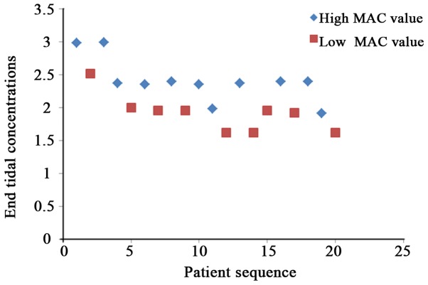
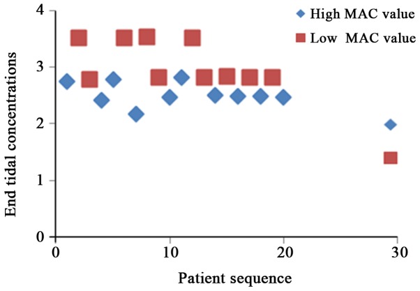
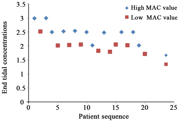
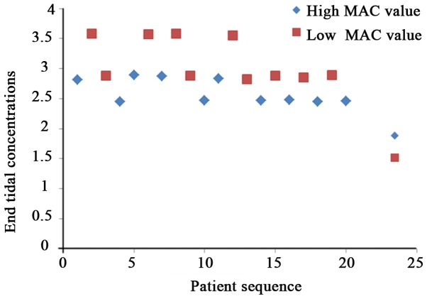
According to the principle of sequential allocation method, the variation trend graphs of different end tidal concentrations of sevoflurane and desflurane to stimulus responses in four groups of patients were obtained (Figs. 1–4).
Figure 1.
Variation trend graph of different end tidal concentrations of desflurane to stimulus responses of skin incision in patients of Group D Among patients receiving neoadjuvant chemotherapy drugs, the inhibitory effect of patients with high MAC value of desflurane on stimulus responses of skin incision is better than those with low MAC value of desflurane.
Figure 4.
Variation trend graph of different end tidal concentrations of sevoflurane to stimulus responses of skin incision in patients of Group C2. It indicates that among patients receiving neoadjuvant chemotherapy drugs, the inhibitory effect of patients with low MAC value of sevoflurane on stimulus responses of skin incision is better than those with high MAC value of sevoflurane.
Discussion
HCC complicated with jaundice rarely occurs, and lesions often occur in the late tumor phase with a relatively lower surgical resection rate and a relatively higher incidence rate of complications. Neoadjuvant chemotherapy is a systemic chemotherapy method prior to operation or radiotherapy based on the local treatment of malignant tumors (14,15). At present, the study results of many researchers have gradually confirmed the effect of neoadjuvant chemotherapy in the treatment of malignant tumors, which is significant in patients with malignant tumors, so in practical clinical applications, neoadjuvant chemotherapy has been gradually applied in patients with malignant tumors prior to operation or radiotherapy. However, there is a need for further studies on the relationship between chemotherapy drugs and anesthetics, so in this study, a reasonable treatment regimen was selected before operation or radiotherapy. Besides, HCC patients complicated with jaundice with same treatment courses inhaled sevoflurane and desflurane for anesthesia together with patients receiving no neoadjuvant chemotherapy. MAC values were detected so as to explore whether neoadjuvant chemotherapy could improve the sensitivity of HCC patients complicated with jaundice to sevoflurane and desflurane.
There were no significant differences in the age, sex, height, BMI and other conditions among the three groups of patients. The surgical treatment regimen was to perform preoperative chemotherapy for patients in the neoadjuvant chemotherapy group. The specific action dose of oxaliplatin was 150 mg/qd and that of tegafur was 60 mg/bid. After the chemotherapy, the computed tomography (CT) was used to assess the tumor changes, and patients were further examined. Operation patients underwent the surgical treatment at the end of 3-week 2 cycles of chemotherapy, thus ensuring the homogeneity within the chemotherapy group.
The test results also revealed that compared with those of patients in Group C, MAC values of patients in Group S and Group D were reduced, confirming that neoadjuvant chemotherapy could reduce MAC values of HCC patients complicated with jaundice. Chemotherapy leads to the emergence of many adverse reactions in patients, and as the basic principle of selecting neoadjuvant chemotherapy drugs and regimens is the high efficiency and low toxicity, the regimen selected for the neoadjuvant chemotherapy group was oxaliplatin combined with tegafur (16,17). Most of inhaled anesthetics are discharged from the body through the lung, and a small part of them can be discharged from the human body through the skin and urine, but they are not discharged through the liver, so despite of the impact of chemotherapy drugs, the liver function of patients receiving neoadjuvant chemotherapy may be damaged to a certain degree. However, in the body's metabolic process, significant changes will not occur in sevoflurane and desflurane, so that the potency of sevoflurane and desflurane will not be affected. The reason for the decrease in MAC values of halothane and desflurane is likely to the changes in the efficacy of inhaled anesthetics. As the inhalation of anesthetics calms patients down and hypnotize them, it hinders the normal brain function of patients (18–20). Therefore, changes in the brain functional status of patients after neoadjuvant chemotherapy result in decreased MAC values of sevoflurane and desflurane titers (21–23). The MAC value of sevoflurane of patients receiving no chemotherapy detected in the test was 3.08%, which was consistent with that in other studies (24,25).
In summary, preoperative neoadjuvant chemotherapy can reduce MAC values of sevoflurane and desflurane of HCC patients complicated with jaundice. Therefore, from the perspective of improving the safety of anesthesia for patients, the dose of anesthetics needs to be appropriately reduced for patients receiving neoadjuvant chemotherapy. Neoadjuvant chemotherapy can increase the sensitivity of patients to sevoflurane and desflurane, but the specific mechanism of action still needs to be further studied.
Figure 2.
Variation trend graph of different end tidal concentrations of sevoflurane to stimulus responses of skin incision in patients of Group S. Among patients receiving neoadjuvant chemotherapy drugs, the inhibitory effect of patients with high MAC value of sevoflurane on stimulus responses of skin incision is better than those with low MAC value of sevoflurane.
Figure 3.
Variation trend graph of different end tidal concentrations of desflurane to stimulus responses of skin incision in patients of Group C1. Among patients receiving neoadjuvant chemotherapy drugs, the inhibitory effect of patients with low MAC value of desflurane on stimulus responses of skin incision is better than those with high MAC value of desflurane.
Acknowledgements
Not applicable.
Funding
No funding was received.
Availability of data and materials
The datasets used and/or analyzed during the present study are available from the corresponding author on reasonable request.
Authors' contributions
LZ drafted this manuscript. LZ and XM were responsible for the conception and design of the study. MZ collected the patient data and revised the manuscript critically for important intellectual content. MZ and YD analyzed and interpreted the data. All authors read and approved the final manuscript.
Ethics approval and consent to participate
The study was approved by the Ethics Committee of Xiangyang No. 1 People's Hospital, Hubei University of Medicine. Signed written informed consents were obtained from the patients and/or guardians.
Consent for publication
Not applicable.
Competing interests
The authors declare that they have no competing interests.
References
- 1.Ronellenfitsch U, Schwarzbach M, Hofheinz R, Kienle P, Nowak K, Kieser M, Slanger TE, Burmeister B, Kelsen D, Niedzwiecki D, et al. Predictors of overall and recurrence-free survival after neoadjuvant chemotherapy for gastroesophageal adenocarcinoma: Pooled analysis of individual patient data (IPD) from randomized controlled trials (RCTs) Eur J Surg Oncol. 2017;43:1550–1558. doi: 10.1016/j.ejso.2017.05.005. [DOI] [PubMed] [Google Scholar]
- 2.Kümmel S, Paepke S, Huober J, Schem C, Untch M, Blohmer JU, Eiermann W, Gerber B, Hanusch C, Hilfrich J, et al. Randomised, open-label, phase II study comparing the efficacy and the safety of cabazitaxel versus weekly paclitaxel given as neoadjuvant treatment in patients with operable triple-negative or luminal B/HER2-negative breast cancer (GENEVIEVE) Eur J Cancer. 2017;84:1–8. doi: 10.1016/j.ejca.2017.06.037. [DOI] [PubMed] [Google Scholar]
- 3.Feng X, Chen A, Zhang Y, Wang J, Shao L, Wei L. Central nervous system toxicity of metallic nanoparticles. Int J Nanomedicine. 2015;10:4321–4340. doi: 10.2147/IJN.S78308. [DOI] [PMC free article] [PubMed] [Google Scholar]
- 4.Winocur G, Henkelman M, Wojtowicz JM, Zhang H, Binns MA, Tannock IF. The effects of chemotherapy on cognitive function in a mouse model: A prospective study. Clin Cancer Res. 2012;18:3112–3121. doi: 10.1158/1078-0432.CCR-12-0060. [DOI] [PubMed] [Google Scholar]
- 5.Sipocz I, Pinter T, Skaliczky Z, Kullmann T. Effective systemic palliative chemotherapy for intracranial metastases of breast cancer. Orv Hetil. 2016;157:1809–1813. doi: 10.1556/650.2016.30557. (In Hungarian) [DOI] [PubMed] [Google Scholar]
- 6.Nudelman KN, McDonald BC, Wang Y, Smith DJ, West JD, O'Neill DP, Zanville NR, Champion VL, Schneider BP, Saykin AJ. Cerebral perfusion and gray matter changes associated with chemotherapy-induced peripheral neuropathy. J Clin Oncol. 2016;34:677–683. doi: 10.1200/JCO.2015.62.1276. [DOI] [PMC free article] [PubMed] [Google Scholar]
- 7.He ZJ, Hu YH, Fan ZY. Median effective effect-site concentration of intravenous anesthetics for loss of consciousness in neoadjuvant chemotherapy patients. Chin Med J (Engl) 2011;124:504–508. [PubMed] [Google Scholar]
- 8.Trenerry C, Peters MDJ, Corsini N, Damarell RA, Wilson C, Flight I. Patient-reported outcomes following neoadjuvant chemotherapy or chemoradiotherapy treatment for esophageal cancer: A scoping review protocol. JBI Database System Rev Implement Rep. 2017;15:1499–1507. doi: 10.11124/JBISRIR-2016-003240. [DOI] [PubMed] [Google Scholar]
- 9.Weng Y, Yang L, Corringer PJ, Sonner JM. Anesthetic sensitivity of the Gloeobacter violaceus proton-gated ion channel. Anesth Analg. 2010;110:59–63. doi: 10.1213/ANE.0b013e3181c4bc69. [DOI] [PMC free article] [PubMed] [Google Scholar]
- 10.Liang Q, Anderson WD, Jones ST, Souza CS, Hosoume JM, Treptow W, Covarrubias M. Positive allosteric modulation of Kv channels by sevoflurane: Insights into the structural basis of inhaled anesthetic action. PLoS One. 2015;10:e0143363. doi: 10.1371/journal.pone.0143363. [DOI] [PMC free article] [PubMed] [Google Scholar]
- 11.Klock PA, Jr, Czeslick EG, Klafta JM, Ovassapian A, Moss J. The effect of sevoflurane and desflurane on upper airway reactivity. Anesthesiology. 2001;94:963–967. doi: 10.1097/00000542-200106000-00008. [DOI] [PubMed] [Google Scholar]
- 12.Kotwani MB, Malde AD. Comparison of maintenance, emergence and recovery characteristics of sevoflurane and desflurane in pediatric ambulatory surgery. J Anaesthesiol Clin Pharmacol. 2017;33:503–508. doi: 10.4103/joacp.JOACP_194_16. [DOI] [PMC free article] [PubMed] [Google Scholar]
- 13.Costi D, Cyna AM, Ahmed S, Stephens K, Strickland P, Ellwood J, Larsson JN, Chooi C, Burgoyne LL, Middleton P. Effects of sevoflurane versus other general anaesthesia on emergence agitation in children. Cochrane Database Syst Rev. 2014;12:CD007084. doi: 10.1002/14651858.CD007084.pub2. [DOI] [PMC free article] [PubMed] [Google Scholar]
- 14.Chen VE, Gillespie EF, Zakeri K, Murphy JD, Yashar CM, Lu S, Einck JP. Pathologic response after neoadjuvant chemotherapy predicts locoregional control in patients with triple negative breast cancer. Adv Radiat Oncol. 2017;2:105–109. doi: 10.1016/j.adro.2017.01.012. [DOI] [PMC free article] [PubMed] [Google Scholar]
- 15.Mirkin KA, Luke FE, Gangi A, Pimiento JM, Jeong D, Hollenbeak CS, Wong J. Sarcopenia related to neoadjuvant chemotherapy and perioperative outcomes in resected gastric cancer: A multi-institutional analysis. J Gastrointest Oncol. 2017;8:589–595. doi: 10.21037/jgo.2017.03.02. [DOI] [PMC free article] [PubMed] [Google Scholar]
- 16.Hentic O, Safi D, Jerome C, Magaly Z, Vinciane R, Sebastien G, Frederique M, Anne C, Philippe L, Philippe R, et al. Neoadjuvant gemcitabine-oxaliplatin (GemOx) combination followed by chemoradiotherapy (CRT) in borderline pancreatic adenocarcinoma (BPC): A promising management. Pancreatology. 2015;15(Suppl):S112. doi: 10.1016/j.pan.2015.05.397. [DOI] [Google Scholar]
- 17.Seigers R, Loos M, Van Tellingen O, Boogerd W, Smit AB, Schagen SB. Cognitive impact of cytotoxic agents in mice. Psychopharmacology (Berl) 2015;232:17–37. doi: 10.1007/s00213-014-3636-9. [DOI] [PubMed] [Google Scholar]
- 18.Zhou C, Liang P, Liu J, Ke B, Wang X, Li F, Li T, Bayliss DA, Chen X. HCN1 channels contribute to the effects of amnesia and hypnosis but not immobility of volatile anesthetics. Anesth Analg. 2015;121:661–666. doi: 10.1213/ANE.0000000000000830. [DOI] [PMC free article] [PubMed] [Google Scholar]
- 19.Sun X, Liu J, Wang Y, Bai X, Chen Y, Qian J, Zhu H, Liu F, Qiu X, Sun S, et al. Methotrexate-cytarabine-dexamethasone combination chemotherapy with or without rituximab in patients with primary central nervous system lymphoma. Oncotarget. 2017;8:49156–49164. doi: 10.18632/oncotarget.17101. [DOI] [PMC free article] [PubMed] [Google Scholar]
- 20.Lutterbeck CA, Kern DI, Machado ÊL, Kümmerer K. Evaluation of the toxic effects of four anti-cancer drugs in plant bioassays and its potency for screening in the context of waste water reuse for irrigation. Chemosphere. 2015;135:403–410. doi: 10.1016/j.chemosphere.2015.05.019. [DOI] [PubMed] [Google Scholar]
- 21.Liu X, Dingley J, Elstad M, Scull-Brown E, Steen PA, Thoresen M. Minimum alveolar concentration (MAC) for sevoflurane and xenon at normothermia and hypothermia in newborn pigs. Acta Anaesthesiol Scand. 2013;57:646–653. doi: 10.1111/aas.12055. [DOI] [PubMed] [Google Scholar]
- 22.Mishra RK, Mahajan C, Prabhakar H, Kapoor I, Bithal PK. Effect of nitrous oxide on bispectral index values at equi-minimum alveolar concentrations of sevoflurane and desflurane. Indian J Anaesth. 2017;61:482–485. doi: 10.4103/ija.IJA_363_16. [DOI] [PMC free article] [PubMed] [Google Scholar]
- 23.Du W, Li C, Wang H, Zhao A, Shen J, Yong F, Jia H. Effect of neoadjuvant chemotherapy on sevoflurane MAC-BAR value of patients undergoing radical stomach carcinoma surgery. Int J Clin Exp Med. 2015;8:5649–5657. [PMC free article] [PubMed] [Google Scholar]
- 24.Voulgaris DA, Egger CM, Seddighi MR, Rohrbach BW, Love LC, Doherty TJ. The effect of nitrous oxide on the minimum alveolar concentration (MAC) and MAC derivatives of isoflurane in dogs. Can J Vet Res. 2013;77:131–135. [PMC free article] [PubMed] [Google Scholar]
- 25.Suarez MA, Seddighi R, Egger CM, Rohrbach BW, Cox SK, KuKanich BK, Doherty TJ. Effect of fentanyl and lidocaine on the end-tidal sevoflurane concentration preventing motor movement in dogs. Am J Vet Res. 2017;78:12–16. doi: 10.2460/ajvr.78.1.12. [DOI] [PubMed] [Google Scholar]
Associated Data
This section collects any data citations, data availability statements, or supplementary materials included in this article.
Data Availability Statement
The datasets used and/or analyzed during the present study are available from the corresponding author on reasonable request.


