Abstract
Although it is difficult to detect αvβ6 integrin (αvβ6) in normal epithelia cells, its expression is upregulated during wound healing and carcinogenesis. Overexpression of αvβ6 has been demonstrated in epithelial cell carcinomas, such as adenocarcinoma of the colon and ovary. However, the expression of αvβ6 has not been reported in hepatocellular carcinoma (HCC). We previously indicated that LPA may induce αvβ6-mediated TGF-β1 signaling mechanisms during the pathogenesis of lung injury and fibrosis. In addition, transforming growth factor-β1 (TGF-β1) and lysophosphatidic acid (LPA) have been demonstrated to participate in the progression of HCC. In the present study, we hypothesized that TGF-β1 and LPA would serve a key role in the subunit integrin β6 (Itgβ6) transcriptional regulatory mechanism in HCC. It was identified that human HCC tissues and Hep-3B cells expressed Itgβ6. Treatment of Hep-3B with TGF-β1 or LPA increased the expression of Itgβ6. Furthermore, truncation experiments indicated a positive regulatory region at −326 to −157 bp of the Itgβ6 promoter. TGF-β1 and LPA increased transcriptional activation at this regulatory region. To the best of our knowledge, the present study was the first to demonstrate Itgβ6 expression in HCC, and the data indicate that TGF-β1 and LPA regulate Itgβ6 expression through the Itgβ6 gene promoter, which is an important factor in the development of HCC.
Keywords: gene promoter, hepatocellular carcinoma, integrin β6, transforming growth factor-β1, lysophosphatidic acid
Introduction
Primary liver cancers (PLC), including cholangiocarcinoma (CC) and hepatocellular carcinoma (HCC), are the third and sixth most common causes of cancer-associated mortalities for men and women, respectively. Worldwide, >560,000 people are diagnosed with PLC annually (1,2). Of these, 55% of the cases are in China, where PLC was the second most common cause of cancer mortality in 2002 (1–3). HCC accounts for 85–90% of all PLC worldwide (4); HCC and PLC often are used interchangeably. The median survival length in unresectable cases is >4 months, and >1 year for untreated patients with less advanced disease, which demonstrates the poor prognosis of this type of cancer (1,4,5). Therefore, it is crucial to understand the mechanisms that regulate the progression of HCC for the development of novel and effective therapeutic approaches.
Integrins mediate the adhesion between the cellular cytoskeleton and extracellular matrix. They serve a key role in outside-in and inside-out signaling, and also control a variety of vital cell functions including: Adhesion; differentiation; migration; cell division; and apoptosis (6–8). αvβ6 integrin (αvβ6) is an important member of the integrin family, which is unique as it is expressed exclusively in epithelial cells (9). During embryogenesis, αvβ6 is expressed at high levels in the developing lung, skin and kidney epithelia cells, while its expression is downregulated in healthy adult epithelia cells (10–12). Previously, αvβ6 integrin was demonstrated to be highly upregulated in carcinomas of the breast, cholangiocarcinoma, pancreatic ductal, colon, stomach, ovary and endometrium, and also in liver metastases derived from colorectal and pancreatic carcinomas (13–19). However, the expression and role of αvβ6 has not been thoroughly investigated in HCC.
We previously indicated that Lysophosphatidic acid (LPA) induces αvβ6-mediated transforming growth factor-β1 (TGF-β1) activation (20), and Geng et al (21) demonstrated similar results. TGF-β1 is involved in the development of tumor-initiating cells, contributes to angiogenesis and promotes liver cancer development (22–24). There is significant evidence that also indicates its important role for Autotaxin-LPA signaling in human HCC (25–27). Furthermore, β6 only partners with αv, forming a single heterodimer, and its synthesis is rate-limiting for αvβ6 expression (9). Based on results from these previous studies, we hypothesized that the integrin β6 subunit (Itgβ6) is upregulated in HCC, and that there is an association between TGF-β1 and LPA in the expression of Itgβ6. In the present study, the expression levels for Itgβ6 in HCC tissue samples and tumor-derived cell lines were evaluated, and the role of LPA and TGF-β1 in the regulation of Itgβ6 expression and transcriptional activation of Itgβ6 gene promoter was investigated in Hep-3B.
Materials and methods
Cell culture
Cell lines were purchased from the Shanghai Institute of Biochemistry and Cell Biology, Shanghai Institutes of Biological Sciences, Chinese Academy of Sciences (Shanghai, China). Human hepatic cancer Hep-3B cells were cultured in Dulbecco's modified Eagle's medium (DMEM; Gibco; Thermo Fisher Scientific, Inc., Waltham, MA, USA) supplemented with 10% fetal bovine serum (FBS; Gibco; Thermo Fisher Scientific, Inc.). Human hepatocyte HL-7702 cells were maintained in RPMI-1640 (RPMI-1640; Gibco; Thermo Fisher Scientific, Inc.) supplemented with 20% FBS containing penicillin (100 U/ml) and streptomycin sulfate (100 µg/ml). Cell lines were maintained at 37°C with 5% CO2 in a humid incubator.
Human samples
Paraffin-embedded human liver and colorectal carcinoma tissues were obtained from the Second Affiliated Hospital of Shantou Medical College (Shantou, China). Samples were collected from consecutive patients undergoing surgical resection for HCC (n=23), hepatic metastatic adenoma [n=2; 1 male, 1 female, age range 50–52 years (mean, 51.00±1.41)] and colorectal carcinoma [n=5; 4 males, 1 female, age range 54–78 years (mean, 64.80±11.30)]. Histologically normal liver tissues were collected from patients undergoing resection for other carcinomas [n=4; 3 males, 1 female, age range 54–78 years (mean, 62.00±10.86)], and served as controls. The population of patients with HCC included 21 men and 2 women ranging in age between 37–78 years (mean, 50.04±2.45). HCC was histologically confirmed, and tumor staging was performed using the Cancer of the Liver Italian Program (CLIP; which incorporates Child stage, tumor morphology, AFP levels, and the presence of portal vein thrombosis) system (28). Patient characteristics are summarized in Table I; no patients had received preoperative chemotherapy. The Ethical Committees of the Second Affiliated Hospital of Shantou Medical College and the Medical College of Shantou University approved the present study, and all patients provided written informed consent.
Table I.
Characteristics of patients with HCC.
| Variables | HCC (n=23) |
|---|---|
| Age, years | |
| Mean ± standard error of the mean | 50.04±2.445 |
| Sex | |
| Male | 21 |
| Female | 2 |
| CLIP stages (0–6) | |
| 0 | 5 |
| 1 | 3 |
| 2 | 11 |
| 3 | 4 |
CLIP, cancer of the liver Italian program; HCC, hepatocellular carcinoma.
Immunohistochemistry
The histological sections were de-paraffinized in 100% xylene for 30 min at room temperature, and then incubated in graded ethanol (100% ethanol for 15 min, 95% ethanol for 5 min, 90% ethanol for 5 min, then 70% ethanol for 5 min, successively) for 30 min at room temperature. Subsequent to blocking endogenous peroxidase activity in 3% H2O2 for 30 min at room temperature, the sections were incubated at 4°C overnight with the primary anti-Itgβ6 mouse monoclonal antibody (Merck KGaA, Darmstadt, Germany) applied at a dilution rate of 1:50 in antibody diluents (Dako; Agilent Technologies, Inc., Santa Clara, CA, USA), followed by PV9000 reagent (ZSGB-BIO; OriGene Technologies, Inc., Beijing, China) according to the manufacturer's protocol. A 3-amino-9-ethylcarbazole kit (ZSGB-BIO; OriGene Technologies, Inc.) was used to visualize positive staining according to the manufacturer's protocol. Nuclei were stained with 0.5% hematoxylin for 10 sec at room temperature and covered with glycerogelatin. The primary antibodies were replaced by antibody diluent to serve as controls (50:1; DAKO; Agilent Technologies, Inc., Santa Clara, CA, USA). The anti-Itgβ6 mouse monoclonal antibody (mAb) immunoreactivity colorectal carcinoma specimen that is known to be positive for β6 was used as a positive control. Staining was scored using the following scale: Negative (−, 0% of cytoplasmic stain), weak (+, <20%), moderate (++, 20–50%), strong (+++, >50%). A total of 10 random views were taken of each section and the proportions of cytoplasmic stain were calculated in these views. Representative images were captured at ×200 original magnification using a light microscope (BX-51TRF; Olympus Corporation, Tokyo, Japan).
Reverse transcription-quantitative polymerase chain reaction (RT-qPCR)
Hep-3B cells were stimulated with either: i) TGF-β1 (5 ng/ml) for 8, 12 and 24 h; ii) LPA (10 µM) for 12, 24, 48 and 72 h; iii) DMEM/0.1% bovine serum albumin (BSA) alone (serving as a negative control) in 6 well plates. Hep-3B cells were stimulated with either TGF-β (5 ng/ml) for 8 h, LPA (10 µM) for 24 h, TGF-β (5 ng/ml) for 8 h + LPA (10 µM) for 24 h, or DMEM/0.1% BSA alone (serving as a negative control) in 6 well plates. Total RNA was extracted using universal RNA Pure reagent (Guangzhou Dongsheng Biotech, Guangzhou, China) according to the manufacturer's protocol. PCR conditions were: 94°C for 2 min, followed by 35–40 cycles of 94°C for 30 sec, 58°C for 30 sec and 72°C for 1 min, finished at 72°C for 5–10 min. Template cDNA was obtained using reverse transcription of 1 µg of the total RNA with EasyScript First-Strand cDNA Synthesis SuperMix (Beijing TransGen Biotech Co., Ltd., Beijing, China), and relative transcript levels were quantified using qPCR on an ABI PRISM® 7300 Fast Real-Time PCR System (Applied Biosystems; Thermo Fisher Scientific, Inc.), according to the manufacturer's protocol. The expression of Itgβ6 and the housekeeping gene human 18s rRNA (18s) gene were determined using the primer sequences: Itgβ6 forward primer 5′-GCAAGCTGCTGTGTGTAAGGAA-3′ and reverse primer 5′-CTTGGGTTACAGCGAAGATCAA-3′; 18s forward primer 5′-TTGGTGGAGCGATTTGTCTG-3′ and reverse primer 5′-AATGGGGTTCAACGGGTTAC-3′. Itgβ6 and 18s transcripts were amplified using the SYBR® Premix Ex Taq™ kit (Takara Biotechnology Co., Ltd., Dalian, China), according to manufacturer's protocol. The thermocycler conditions were as follows: Initial denaturation at 94°C for 30 sec, followed by 40 cycles of 95°C for 5 sec and 60°C for 30 sec. The relative expression levels of Itgβ6 mRNAs were determined using the comparative cycle threshold (Cq) method (29) that utilized 18s as the endogenous control. Quantity One 4.4 (Bio-Rad Laboratories, Inc., Hercules, CA, USA) was used for analysis.
Western blot assays
Hep-3B cells were seeded in 25 cm2 flasks (Corning Incorporated, NY, USA) containing DMEM supplemented with 10% FBS. Cells were grown to 60–70% confluence and the medium was changed to DMEM with 0.1% BSA for 30 h in the absence (control) or presence of TGF-β1 (5 ng/ml) for 14 h, LPA (10 µM) for 30 h, or TGF-β1 (5 ng/ml) for 14 h + LPA (10 µM) for 30 h. Cells were then washed with ice-cold PBS 3 times and lysed in radioimmunoprecipitation assay buffer (50 mM Tris-HCl, pH 7.4, 1% Nonidet P-40, 150 mM NaCl, 1 mM EDTA, 1 mM phenylmethylsulfonyl fluoride, 1 µg/ml aprotinin and leupeptin, 1 mM sodium orthovanadate and 1 mM NaF). Following centrifugation (13,400 × g at 4°C for 15 min), the supernatant was collected for protein analysis. Protein concentrations were determined using a BCA Protein Assay kit (Merck KGaA) according to the manufacturer's protocol. Protein samples (80 µg) were separated by 10% SDS-PAGE and transferred onto nitrocellulose membranes (Bio TraceTMNT; Pall Corporation, Port Washington, NY, USA), and incubated with anti-integrin β6 mouse mAb (cat. no., 442.5C4; Merck KGaA; dilution, 1:200) in TBST at 4°C overnight prior to washing and incubation at room temperature for 1 h with horseradish peroxidase-conjugated goat anti-mouse secondary antibody (sc-2031; Santa Cruz Biotechnology, Inc., Hercules, CA, USA) diluted to 1:1,000 in TBST with 1% BSA. Immunoreactive bands were visualized using an EasySee™ Western Blot kit (TransGen Biotech, Beijing, China). GAPDH (1:1,000) was used as a loading control. The optical densities for each band and the density ratio for β6 to GAPDH were analyzed using the Quantity One software 4.62 (Bio-Rad Laboratories, Inc.).
Transfection and luciferase assays
The sequence containing the full-length β6 promoter region (−946/+189 bp, 1,135 bp) was generated by PCR as aforementioned. The amplified fragment was digested with Kpn I/Xho I and inserted into the Kpn I/Xho I sites of pGL2-Basic luciferase reporter gene, and the resulting plasmid was identified as pGLB-FL. Promoter deletions were generated from pGLB-FL by PCR as aforementioned, and inserted into the pGL2-Basic plasmid as previously described (30). Plasmids were identified as pGLB-112 (+60/+172; 112 bp), pGLB-207 (−35/+172; 207 bp), pGLB-359 (−187/+172; 359 bp), pGLB-630 (−458/+172; 630 bp), pGLB-A (−470/-157; 313 bp), pGLB-B (−388/-157; 231 bp) and pGLB-C (−326/−157; 169 bp), respectively.
Hep-3B cells cultured to 70–80% confluency were transfected using METAFECTENE®PRO reagent (Biontex Laboratories GmbH, München, Germany) with 0.2 µg for each of the Itgβ6 constructs, and mixed at ratio of 200:1 with a control Renilla construct (Biontex Laboratories GmbH) according to the manufacturer's protocol. Cell extracts were evaluated for promoter activity at 48 h following transfection. Prior to collecting cells for extracts, the cells were treated with either TGF-β1 (5 ng/ml) for 24 h, LPA (10 µM) for 24 h, or DMEM/0.1% BSA alone as a negative control. Luciferase and Renilla activities were determined using a Dual-Luciferase® Reporter Assay System (Promega Corporation, Madison, USA) according to manufacturer's protocol. Luminescence was measured using an EG&G Berthold Lumat LB9507 luminometer (Berthold Technologies GmbH & Co. KG, Bad Wildbad, Germany). For each sample, luciferase relative light units were adjusted based on Renilla activity to normalize for transfection efficiency.
Statistical analysis
Statistical analyses were performed using GraphPad Prism 5 (GraphPad Software, Inc., La Jolla, CA, USA). Analyses included one-tailed Pearson tests, two-tailed Student t-tests for single comparisons and one-way analysis of variance for comparing multiple treatment groups with a control group. Bonferroni post hoc test was used to determine which results differed significantly. The data are expressed as the mean ± standard error of the mean, unless stated otherwise. P<0.05 was considered to indicate a statistically significant difference.
Results
Itgβ6 immunohistochemistry in human liver tumors
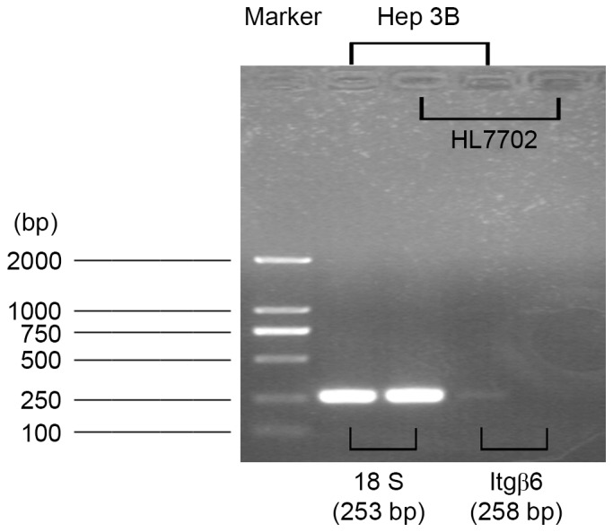
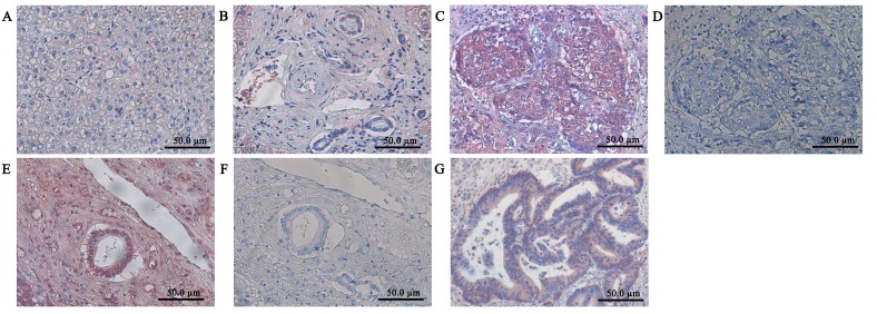
Immunoreactivity for Itgβ6 was absent in normal liver tissue, liver cells, and intrahepatic bile ducts, which was similar to the negative controls (Fig. 1A and B). In contrast, strong staining was present in the nests and intrahepatic bile ducts of the HCC samples (Fig. 1C and D); blank controls are presented in Fig. 1E and F. Itgβ6 immunoreactivity was present in the positive control colorectal carcinoma sections (Fig. 1G) and detected in hepatic metastases adenoma samples (data not shown). The expression of Itgβ6 was measured at mRNA levels in the human normal hepatocyte HL-7702 and human hepatic cancer Hep-3B cell lines. Itgβ6 mRNA was detected in Hep-3B cells, but Itgβ6 expression was not detected in HL-7702 cells (Fig. 2).
Figure 1.
Itgβ6 immunohistochemistry in normal liver, hepatocellular carcinoma, and colorectal carcinoma. (A) Normal liver tissue. (B) Normal intrahepatic bile ducts. (C and D) Hepatocellular carcinoma nests. (E and F) Intrahepatic bile ducts of the HCC. (G) colorectal carcinoma. (D and F) Negative controls in the (D) nests and (F) intrahepatic bile ducts of the HCC. Itgβ6 staining was absent from normal livers, while it is clearly visible in HCC and colorectal carcinoma cells. Representative images are presented at magnification, ×200. Itgβ6, integrin β6.
Figure 2.

Itgβ6 mRNA was detected in Hep-3B cells, but not detectable in HL-7702 cells. Itgβ6, integrin β6.
No clear association between Itgβ6 immunoreactivity and the grade of HCC
No clear association between Itgβ6 immunoreactivity and the CLIP grade was observed. There was also no significant correlation between Itgβ6 expression and tumor grade (r=−0.284) observed, probably due to the small group size (n=23; Table II).
Table II.
Integrin β6 expression in HCC.
| Integrin β6 expression, n (%) | ||||
|---|---|---|---|---|
| Variables | – | + | ++ | +++ |
| HCC (n=23) | 0 | 3 (13) | 9 (39) | 11 (48) |
Negative (−), 0% of cytoplasmic stain; weak (+), <20%; moderate (++), 20–50%, strong (+++), >50%. HCC, hepatocellular carcinoma.
TGF-β1 and LPA induce Itgβ6 expression in Hep-3B cells
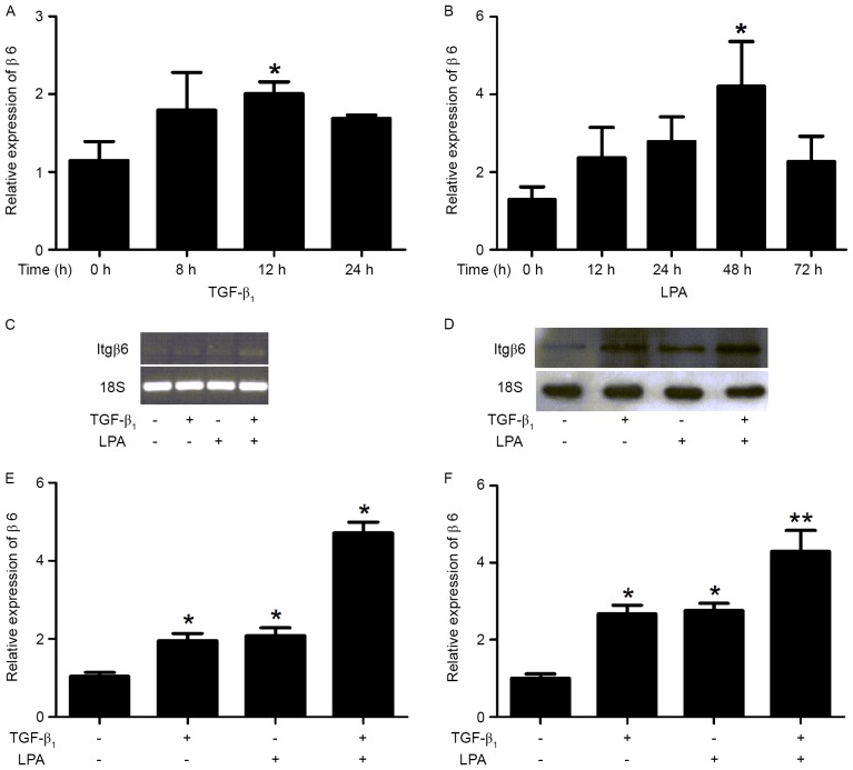
The association between the Itgβ6 mRNA levels and TGF-β1/LPA was evaluated in Hep-3B cells treated with or without TGF-β1 or LPA for the time periods indicated (Fig. 3A and B). TGF-β1 and LPA induced an increase in Itgβ6 expression at mRNA and protein levels in Hep-3B cells (Fig. 3C and D). When compared with the non-treated cells, TGF-β1, LPA and TGF-β1+LPA treatments increased β6 mRNA levels by 1.87-, 2.00- and 4.53-fold, respectively (Fig. 3E), and protein levels by 2.68-, 2.76- and 4.29-fold, respectively (Fig. 3F).
Figure 3.
TGF-β1 and LPA induces the Itgβ6 expression in Hep-3B cells. Itgβ6 expression was examined using reverse transcription PCR and western blot analysis in Hep-3B cells. Hep-3B cells were serum-starved for 24 h, then treated with either TGF-β1 or LPA for various time periods. Hep-3B cells were serum-starved for 24 h, then treated with either TGF-β (5 ng/ml) for 8 h, LPA (10 µM) for 24 h, TGF-β (5 ng/ml) for 8 h + LPA (10 µM) for 24 h, or DMEM/0.1% BSA alone (serving as a negative control). (A) TGF-β1- and (B) LPA-induced Itgβ6 expression for various time periods in Hep-3B cells. (C) Itgβ6 and 18s mRNA expression by PCR that included 28 cycles. Ethidium bromide staining of β6 and 18s electrophoresis gels was included to indicate equal loading among the same set of RNA samples. (D) Hep-3B cells were serum-starved for 24 h then treated with either TGF-β (5 ng/ml) for 14 h, LPA (10 µM) for 30 h or TGF-β (5 ng/ml) for 14 h+ LPA (10 µM) for 30 h, or DMEM/0.1% BSA alone (serving as a negative control), followed by western blot analysis with the indicated antibodies. (E) When compared with non-treated cells, the increased β6 mRNA expression induced by TGF-β1, LPA or TGF-β1+LPA was 1.87-, 2.00- and 4.53-fold, respectively. (F) When compared to non-treated cells, the increased β6 protein expression induced by TGF-β1, LPA or TGF-β1+LPA was 2.68-, 2.76- and 4.29-fold, respectively. Data are presented as the mean ± standard error of the mean for three independent experiments performed in triplicate with similar results. *P<0.05 and **P<0.01 vs. control. TGF-β1, transforming growth factor-β1; LPA, lysophosphatidic acid; Itgβ6/β6, integrin β6; PCR, polymerase chain reaction.
TGF-β1 and LPA activate the Itgβ6 gene promoter
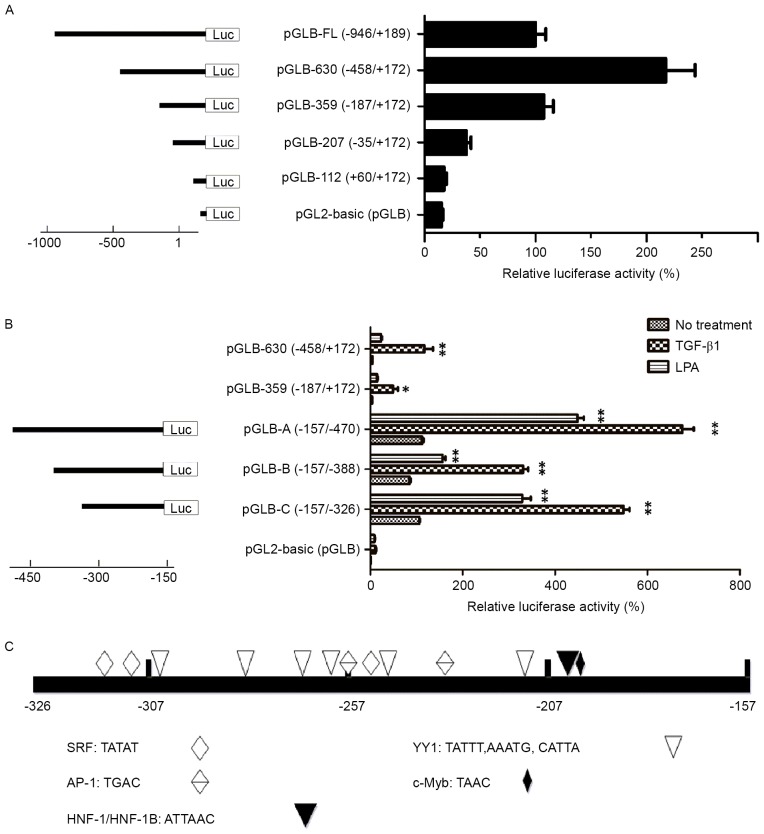
To confirm that TGF-β1 and LPA induce transcriptional activation of the human Itgβ6 gene, the Itgβ6 promoter was cloned and luciferase reporter gene assays measured the activity by the generation of a series of Itgβ6 promoter deletion mutants. Hep-3B cells were transiently transfected with pGLB-112, pGLB-207, pGLB-359, pGLB-630, pGLB-FL, and pGL2-basic (pGLB, control). The pGLB-630 (−458/+172 bp region) was identified to stimulate the maximal luciferase activity (Fig. 4A). Sequence deletion from −458 to −187 bp resulted in an ~50% reduction in luciferase activity. To investigate the role of the specific regions in stimulation of Itgβ6 promoter activity, the region from −470 to −157 bp (containing −458 to −187 bp) was amplified, and additional deletion constructs were generated for the region and named pGLB-A (−470/-157 bp), pGLB-B (−388/-157 bp), and pGLB-C (−326/-157 bp). Hep-3B cells were transiently transfected with the plasmids containing these deletion constructs. The transfected cells treated with TGF-β1 or LPA markedly increased luciferase activity (Fig. 4B), thus confirming that TGF-β1 and LPA trigger transcriptional activation of the Itgβ6 gene promoter. Notably, the −326 to −157 bp proximal fragment was identified to be the minimum sequence that retained the full response to TGF-β1 or LPA. Transcriptional Element Search System (TESS; http://www.cbil.upenn.edu/tess) analysis indicated that the cloned fragment contained several well-defined transcription factor-binding sites, including Activator protein (AP-1), transcriptional activator (c-Myb), hepatocyte nuclear factor (HNF)-1/HNF-1B, serum response factor (SRF), and Yin Yang 1 (YY1) that have been implicated in hepatocarcinogenesis (Fig. 4C).
Figure 4.
TGF-β1 and LPA activates the Itgβ6 gene promoter. (A) Deletion analysis of the Itgβ6 gene promoter: Hep-3B cells were transfected with luciferase reporter constructs containing different lengths of the β6 promoter as indicated. In the right column, luciferase activity was converted to percent activity normalized to cells transfected with the construct containing the full-length Itgβ6 promoter (−946 to +189 bp), defined as 100%. The pGLB-630 (−458/+172 bp region) was identified to exhibit maximum luciferase activity. Data are the mean ± SEM of triplicate assays, representative of three independent experiments. (B) Hep-3B cells were transfected with luciferase reporter constructs containing the −470 to −157 bp fragment (pGLB-A) or its deletion forms, pGLB-B (−388/-157 bp) and pGLB-C (−326/-157 bp). Fold increases in luciferase activity induced by TGF-β1 and LPA in transfected cells were determined using one-way analysis of variance with *P<0.05 and **P<0.01 vs. control (no treatment). Data are the mean ± SEM of triplicate assays, representative of two independent experiments. (C) TESS analysis indicated that the cloned fragment harbored certain well-defined transcription factor-binding sites: AP-1, c-Myb, HNF-1/HNF-1B, SRF and YY1; these were implicated in hepatocarcinogenesis. AP-1, Activator protein; c-Myb, transcriptional activator; HNF-1/-1B, hepatocyte nuclear factor −1/-1B; SRF serum response factor; YY1, Yin Yang 1; TGF-β1, transforming growth factor-β1; LPA, lysophosphatidic acid; Itgβ6/β6, integrin β6; SEM, standard error of the mean.
Discussion
In the present study, the expression levels of Itgβ6 were evaluated using hepatocellular carcinoma. Itgβ6 expression was increased in HCC cell lines and tissue samples compared with normal liver cells and liver tissue; TGF-β1 and LPA induced Itgβ6 expression and activated the Itgβ6 gene promoter. The positive regulatory region of the promoter at −326 to −157 bp was identified by deletion analysis.
Itgβ6 was expressed in human HCC tissues and the Hep-3B hepatocellular carcinoma cell line, while Itgβ6 expression was absent in human normal liver tissue and the normal HL-7702 hepatocyte cell line. These results contrast the results demonstrated by Patsenker et al (14), which demonstrated the complete absence of Itgβ6 in HCC in samples obtained from the Department of Visceral Surgery and Medicine, University of Bern (Bern, Switzerland). The difference in the expression of Itgβ6 may be due to race and hepatitis infection status. For example, hepatitis C virus is involved in the development of HCC in the Swiss population, while hepatitis B virus-associated HCC occurs at a higher rate in China (31,32).
A previous study indicated that the expression of Itgβ6 was associated with poor prognosis and survival in colorectal carcinoma (33). However, in non-small lung carcinoma, expression of Itgβ6 was predominantly observed in well-differentiated tumors, and considered to be an indicator of good prognosis (34). In the present study, there was no significant correlation between Itgβ6 immunoreactivity and CLIP stage in patients with HCC, which may be due to the small group size (n=23).
TGF-β1 and LPA treatment in Hep-3B cells increased Itgβ6 mRNA and protein expression. Notably, the additive stimulatory effect of the combination of TGF-β1 and LPA suggests a potential for multiple mechanisms regulating Itgβ6 expression. These results are consistent with previous studies that demonstrated TGF-β1 and LPA may facilitate the development of human HCC (22–26).
Although it has been well-established that Itgβ6 is implicated in numerous aspects of cancer cell biology (13–19,35–37), the transcription factors and signal transduction pathways involved remain poorly understood. The present study describes experiments investigating the Itgβ6 promoter. Truncation experiments indicated that the cloned human Itgβ6 promoter region (−946/+189, 1,135 bp) contained a positive regulatory region from −326 to −157 bp. TESS analysis demonstrated that this region harbored certain well-defined transcription factor-binding sites that are associated with hepatocarcinogenesis, including AP-1, c-Myb, HNF-1/HNF-1B, SRF, and YY1.
Additionally, activation of Itgβ6 by TGF-β1 and LPA were revealed to significantly increase Itgβ6 promoter activity, therefore confirming that TGF-β1 and LPA triggered transcriptional activation of the Itgβ6 gene promoter. This observation was in accordance with induced increases in Itgβ6 mRNA and protein levels by TGF-β1 and LPA in Hep-3B cells, and highlights the possibility that TGF-β1 and LPA may contribute to the transcriptional activation of Itgβ6 through the regulation of the aforementioned transcription factor-binding sites. In conclusion, to the best of our knowledge, the present study is the first to identify Itgβ6 expression in HCC. TGF-β1 and LPA induced the Itgβ6 expression in Hep-3B cells and activated the Itgβ6 gene promoter. Future studies will include examination of therapeutic strategies for hepatocellular carcinoma by Itgβ6 genetic intervention.
Acknowledgements
Not applicable.
Funding
The present study was supported by the National Natural Science Foundation of China (grant no. 30900661).
Availability of data and materials
The datasets used and/or analyzed during the current study are available from the corresponding author on reasonable request.
Authors' contributions
RX, MX, GC and YF made substantial contributions to conception and design, and designed the outline and revised the manuscript. RX, MX, XD, HH, XC and WH contributed to the acquisition and analysis of data for the study. All authors have read and approved the final manuscript.
Ethics approval and consent to participate
The Ethical Committees of the Second Affiliated Hospital of Shantou Medical College and the Medical College of Shantou University approved the present study, and all patients provided written informed consent.
Consent for publication
Not applicable.
Competing interests
The authors declare that they have no competing interests.
References
- 1.Wilson JF. Liver cancer on the rise. Ann Intern Med. 2005;142:1029–1032. doi: 10.7326/0003-4819-142-12_Part_1-200506210-00024. [DOI] [PubMed] [Google Scholar]
- 2.Llovet JM, Ricci S, Mazzaferro V, Hilgard P, Gane E, Blanc JF, de Oliveira AC, Santoro A, Raoul JL, Forner A, et al. Sorafenib in advanced hepatocellular carcinoma. N Engl J Med. 2008;359:378–390. doi: 10.1056/NEJMoa0708857. [DOI] [PubMed] [Google Scholar]
- 3.Sherman M. Hepatocellular Carcinoma: Epidemiology, surveillance, and diagnosis. Semin Liver Dis. 2010;30:3–16. doi: 10.1055/s-0030-1247128. [DOI] [PubMed] [Google Scholar]
- 4.El-Serag HB, Rudolph KL. Hepatocellular carcinoma: Epidemiology and molecular carcinogenesis. Gastroenterology. 2007;132:2557–2576. doi: 10.1053/j.gastro.2007.04.061. [DOI] [PubMed] [Google Scholar]
- 5.Nordenstedt H, White DL, El-Serag HB. The changing pattern of epidemiology in hepatocellular carcinoma. Dig Liver Dis. 2010;42(Suppl 3):S206–S214. doi: 10.1016/S1590-8658(10)60507-5. [DOI] [PMC free article] [PubMed] [Google Scholar]
- 6.Hynes RO. Integrins: A family of cell surface receptors. Cell. 1987;48:549–554. doi: 10.1016/0092-8674(87)90233-9. [DOI] [PubMed] [Google Scholar]
- 7.Giancotti FG, Ruoslahti E. Integrin signaling. Science. 1999;285:1028–1032. doi: 10.1126/science.285.5430.1028. [DOI] [PubMed] [Google Scholar]
- 8.Hynes RO. Integrins: Bidirectional, allosteric signaling machines. Cell. 2002;110:673–687. doi: 10.1016/S0092-8674(02)00971-6. [DOI] [PubMed] [Google Scholar]
- 9.Patsenker E, Popov Y, Stickel F, Jonczyk A, Goodman SL, Schuppan D. Inhibition of integrin alphavbeta6 on cholangiocytes blocks transforming growth factor-beta activation and retards biliary fibrosis progression. Gastroenterology. 2008;135:660–670. doi: 10.1053/j.gastro.2008.04.009. [DOI] [PMC free article] [PubMed] [Google Scholar]
- 10.Breuss JM, Gallo J, DeLisser HM, Klimanskaya IV, Folkesson HG, Pittet JF, Nishimura SL, Aldape K, Landers DV, Carpenter W, et al. Expression of the beta 6 integrin subunit in development, neoplasia and tissue repair suggests a role in epithelial remodeling. J Cell Sci. 1995;108:2241–2251. doi: 10.1242/jcs.108.6.2241. [DOI] [PubMed] [Google Scholar]
- 11.Breuss JM, Gillett N, Lu L, Sheppard D, Pytela R. Restricted distribution of integrin beta 6 mRNA in primate epithelial tissues. J Histochem Cytochem. 1993;41:1521–1527. doi: 10.1177/41.10.8245410. [DOI] [PubMed] [Google Scholar]
- 12.Busk M, Pytela R, Sheppard D. Characterization of the integrin alpha v beta 6 as a fibronectin-binding protein. J Biol Chem. 1992;267:5790–5796. [PubMed] [Google Scholar]
- 13.Yang GY, Xu KS, Pan ZQ, Zhang ZY, Mi YT, Wang JS, Chen R, Niu J. Integrin alpha v beta 6 mediates the potential for colon cancer cells to colonize in and metastasize to the liver. Cancer Sci. 2008;99:879–887. doi: 10.1111/j.1349-7006.2008.00762.x. [DOI] [PMC free article] [PubMed] [Google Scholar]
- 14.Patsenker E, Wilkens L, Banz V, Osterreicher CH, Weimann R, Eisele S, Keogh A, Stroka D, Zimmermann A, Stickel F. The alphavbeta6 integrin is a highly specific immunohistochemical marker for cholangiocarcinoma. J Hepatol. 2010;52:362–369. doi: 10.1016/j.jhep.2009.12.006. [DOI] [PubMed] [Google Scholar]
- 15.Ahmed N, Pansino F, Clyde R, Murthi P, Quinn MA, Rice GE, Agrez MV, Mok S, Baker MS. Overexpression of alpha(v)beta6 integrin in serous epithelial ovarian cancer regulates extracellular matrix degradation via the plasminogen activation cascade. Carcinogenesis. 2002;23:237–244. doi: 10.1093/carcin/23.2.237. [DOI] [PubMed] [Google Scholar]
- 16.Hecht JL, Dolinski BM, Gardner HA, Violette SM, Weinreb PH. Overexpression of the alphavbeta6 integrin in endometrial cancer. Appl Immunohistochem Mol Morphol. 2008;16:543–547. doi: 10.1097/PAI.0b013e31816bc5ee. [DOI] [PubMed] [Google Scholar]
- 17.Zhang ZY, Xu KS, Wang JS, Yang GY, Wang W, Wang JY, Niu WB, Liu EY, Mi YT, Niu J. Integrin alphanvbeta6 acts as a prognostic indicator in gastric carcinoma. Clin Oncol (R Coll Radiol) 2008;20:61–66. doi: 10.1016/j.clon.2007.09.008. [DOI] [PubMed] [Google Scholar]
- 18.Azare J, Leslie K, Al-Ahmadie H, Gerald W, Weinreb PH, Violette SM, Bromberg J. Constitutively activated Stat3 induces tumorigenesis and enhances cell motility of prostate epithelial cells through integrin beta 6. Mol Cell Biol. 2007;27:4444–4453. doi: 10.1128/MCB.02404-06. [DOI] [PMC free article] [PubMed] [Google Scholar]
- 19.Marsh D, Dickinson S, Neill GW, Marshall JF, Hart IR, Thomas GJ. alpha vbeta 6 Integrin promotes the invasion of morphoeic basal cell carcinoma through stromal modulation. Cancer Res. 2008;68:3295–3303. doi: 10.1158/0008-5472.CAN-08-0174. [DOI] [PubMed] [Google Scholar]
- 20.Xu MY, Porte J, Knox AJ, Weinreb PH, Maher TM, Violette SM, McAnulty RJ, Sheppard D, Jenkins G. Lysophosphatidic acid induces alphavbeta6 integrin-mediated TGF-beta activation via the LPA2 receptor and the small G protein G alpha(q) Am J Pathol. 2009;174:1264–1279. doi: 10.2353/ajpath.2009.080160. [DOI] [PMC free article] [PubMed] [Google Scholar]
- 21.Geng H, Lan R, Singha PK, Gilchrist A, Weinreb PH, Violette SM, Weinberg JM, Saikumar P, Venkatachalam MA. Lysophosphatidic acid increases proximal tubule cell secretion of profibrotic cytokines PDGF-B and CTGF through LPA2- and Gαq-mediated Rho and αvβ6 integrin-dependent activation of TGF-β. Am J Pathol. 2012;181:1236–1249. doi: 10.1016/j.ajpath.2012.06.035. [DOI] [PMC free article] [PubMed] [Google Scholar]
- 22.Wu K, Ding J, Chen C, Sun W, Ning BF, Wen W, Huang L, Han T, Yang W, Wang C, et al. Hepatic transforming growth factor beta gives rise to tumor-initiating cells and promotes liver cancer development. Hepatology. 2012;56:2255–2267. doi: 10.1002/hep.26007. [DOI] [PubMed] [Google Scholar]
- 23.Balzarini P, Benetti A, Invernici G, Cristini S, Zicari S, Caruso A, Gatta LB, Berenzi A, Imberti L, Zanotti C, et al. Transforming growth factor-beta1 induces microvascular abnormalities through a down-modulation of neural cell adhesion molecule in human hepatocellular carcinoma. Lab Invest. 2012;92:1297–1309. doi: 10.1038/labinvest.2012.94. [DOI] [PubMed] [Google Scholar]
- 24.Kitisin K, Ganesan N, Tang Y, Jogunoori W, Volpe EA, Kim SS, Katuri V, Kallakury B, Pishvaian M, Albanese C, et al. Disruption of transforming growth factor-beta signaling through beta-spectrin ELF leads to hepatocellular cancer through cyclin D1 activation. Oncogene. 2007;26:7103–7110. doi: 10.1038/sj.onc.1210513. [DOI] [PMC free article] [PubMed] [Google Scholar]
- 25.Zhang R, Zhang Z, Pan X, Huang X, Huang Z, Zhang G. ATX-LPA axis induces expression of OPN in hepatic cancer cell SMMC7721. Anat Rec (Hoboken) 2011;294:406–411. doi: 10.1002/ar.21324. [DOI] [PubMed] [Google Scholar]
- 26.Park SY, Jeong KJ, Panupinthu N, Yu S, Lee J, Han JW, Kim JM, Lee JS, Kang J, Park CG, et al. Lysophosphatidic acid augments human hepatocellular carcinoma cell invasion through LPA1 receptor and MMP-9 expression. Oncogene. 2011;30:1351–1359. doi: 10.1038/onc.2010.517. [DOI] [PubMed] [Google Scholar]
- 27.Mills GB, Moolenaar WH. The emerging role of lysophosphatidic acid in cancer. Nat Rev Cancer. 2003;3:582–591. doi: 10.1038/nrc1143. [DOI] [PubMed] [Google Scholar]
- 28.A new prognostic system for hepatocellular carcinoma: A retrospective study of 435 patients: The cancer of the liver Italian program (CLIP) investigators. Hepatology. 1998;28:751–755. doi: 10.1002/hep.510280322. [DOI] [PubMed] [Google Scholar]
- 29.Livak KJ, Schmittgen TD. Analysis of relative gene expression data using real-time quantitative PCR and the 2(-Delta Delta C(T)) method. Methods. 2001;25:402–408. doi: 10.1006/meth.2001.1262. [DOI] [PubMed] [Google Scholar]
- 30.Knoche K, Kephart D. Cloning blunt-end PfuDNA Polymerase-generated PCR fragments into pGEM®-T vector systems. Promega Notes. 1999;71:10. [Google Scholar]
- 31.Kuske L, Mensen A, Müllhaupt B, Negro F, Semela D, Moradpour D, Male PJ, Heim MH, Malinverni R, Cerny A, Dufour JF. Swiss Hepatitis C Cohort Study: Characteristics of patients with chronic hepatitis C who develop hepatocellular carcinoma. Swiss Med Wkly. 2012;142:w13651. doi: 10.4414/smw.2012.13651. [DOI] [PubMed] [Google Scholar]
- 32.Tan YJ. Hepatitis B virus infection and the risk of hepatocellular carcinoma. World J Gastroentero. 2011;17:4853–4857. doi: 10.3748/wjg.v17.i44.4853. [DOI] [PMC free article] [PubMed] [Google Scholar]
- 33.Bates RC, Bellovin DI, Brown C, Maynard E, Wu B, Kawakatsu H, Sheppard D, Oettgen P, Mercurio AM. Transcriptional activation of integrin beta6 during the epithelial-mesenchymal transition defines a novel prognostic indicator of aggressive colon carcinoma. J Clin Invest. 2005;115:339–347. doi: 10.1172/JCI200523183. [DOI] [PMC free article] [PubMed] [Google Scholar]
- 34.Smythe WR, LeBel E, Bavaria JE, Kaiser LR, Albelda SM. Integrin expression in non-small cell carcinoma of the lung. Cancer Metastasis Rev. 1995;14:229–239. doi: 10.1007/BF00690294. [DOI] [PubMed] [Google Scholar]
- 35.Li X, Yang Y, Hu Y, Dang D, Regezi J, Schmidt BL, Atakilit A, Chen B, Ellis D, Ramos DM. Alphavbeta6-Fyn signaling promotes oral cancer progression. J Biol Chem. 2003;278:41646–41653. doi: 10.1074/jbc.M306274200. [DOI] [PubMed] [Google Scholar]
- 36.Thomas GJ, Nyström ML, Marshall JF. Alphavbeta6 integrin in wound healing and cancer of the oral cavity. J Oral Pathol Med. 2006;35:1–10. doi: 10.1111/j.1600-0714.2005.00374.x. [DOI] [PubMed] [Google Scholar]
- 37.Ramos DM, Dang D, Sadler S. The role of the integrin alpha v beta6 in regulating the epithelial to mesenchymal transition in oral cancer. Anticancer Res. 2009;29:125–130. [PubMed] [Google Scholar]
Associated Data
This section collects any data citations, data availability statements, or supplementary materials included in this article.
Data Availability Statement
The datasets used and/or analyzed during the current study are available from the corresponding author on reasonable request.


