Abstract
Objective
The aim of this study was to evaluate the accuracy and reliability of cephalometric measurements using iPad apps called CephNinja and SmartCeph Pro. The measurements were compared with the measurements obtained using Dolphin Imaging computer software.
Methods
Twenty digital cephalometric radiographs were randomly selected from the archives and traced using the CephNinja app, SmartCeph Pro app, and Dolphin Imaging software. Twenty-one landmarks and 16 measurements were performed in each program. The statistical analysis was conducted using the Bland-Altman analysis at a significance level of 0.05.
Results
For the CephNinja app, there were seven measurements that were in accordance with Dolphin Imaging software. For the SmartCeph Pro app, six measurements were in accordance with Dolphin software. Both apps gave better results for angular measurements than linear ones.
Conclusion
These findings indicate that, although they are user-friendly, orthodontic apps for cephalometrics are not equal with Dolphin software now and need to be developed to be more reliable for most of the measurements.
Keywords: Cephalometrics, smartphones, iPad, apps
INTRODUCTION
Lateral cephalometry is an essential diagnostic tool that orthodontists use to plan treatments, determine the results of a treatment, or predict growth. Rapid advances in technology have led to broad applications in cephalometry, such as computer-aided cephalometric analyses, which have recently been introduced. Many cephalometric computer programs have been developed that benefit image storage, the accuracy of assessments, data sharing, short-term growth prediction, and quick superimposition (1,2).
The last few years have seen an increase in the use of new technology in all areas of our lives, including smartphones or tablet PCs. This is especially true for smartphones, which are no longer used for just phone calls. By using software, or applications (apps), smartphones or tablets can act as calculators, address books, calendars, etc. As in all aspects of our lives, smartphones and tablets are helping people in the medical field as well (3).
Apps are specialized programs downloaded to mobile phones and tablet PCs. Recently, many dental and orthodontic apps have been developed. Some of these apps have been preferred by clinicians, and some by patients (4). For example, cephalometric analysis apps for the iPad have been introduced for orthodontists to measure the skeletal, dental, and soft tissue values by tracing the cephalograms. Using an iPad with cephalometric apps may be the next step in orthodontics for cephalometric tracing.
The aim of this study was to assess the reliability of the CephNinja app and SmartCeph Pro app by comparing them with the commonly used digital cephalometric tracing program, Dolphin Imaging software (Dolphin Imaging and Management Solutions; Los Angeles, CA, USA).
METHODS
Twenty cephalometric radiographs were taken randomly from the archives of the Bezmialem Vakıf University Department of Orthodontics and used in this retrospective study. The exclusion criteria were the following: unerupted or missing incisors, unerupted teeth overlying incisor apices, poor quality of radiographs (with artifacts), craniofacial deformity, excess soft tissue that could prevent locating landmarks, and non-permanent dentition with impacted or missing teeth.
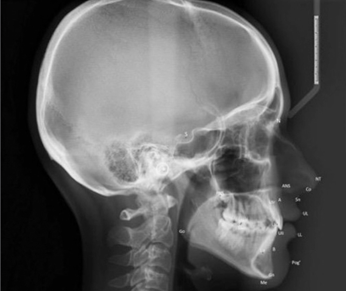
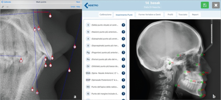
All of the radiographs were taken using a Proline digital cephalometer (Planmeca; Helsinki, Finland) set at 1×25 magnification, as recommended by the manufacturer. The digital images (20 cephalograms) were placed in the same manufacturer’s program (Dimaxis; Planmeca, Helsinki, Finland) imported into Dolphin Imaging software and traced in a computer. A total of 21 anatomical landmarks were defined on each radiograph, and 16 variables were calculated (Figure 1, Table 1). All of these measurements were performed by the same investigator (EG). Similarly, digital radiographs were imported to the CephNinja app (version 3.3) and SmartCeph Pro app (version 1.1) on an iPad Pro and traced with Apple Pencil (Apple Inc.; CA, USA). A ruler was used to calibrate each cephalogram (Figure 2). In all cephalometric programs, the observer could enhance brightness, contrast, and magnification and zoom in/out.
Figure 1.

Cephalometric landmarks used in the study
S: sella; N: nasion; Co: condylion; ANS: anterior nasal spine; PNS: posterior nasal spine; NT: tip of nose; Go: gonion; Me: menton; Gn: gnathion; B: B point; Pog′: Pogonion soft tissue; A: A point; U1r: upper incisor root; U1i: upper incisor incisal; L1r: lower incisor root; L1i: lower incisor incisal; U6O: upper first molar occlusal; L6O: lower first molar occlusal
Table 1.
Definition of the cephalometric measurements
| SNA (°) | Angle between S-N and N-A |
| SNB (°) | Angle between S-N and N-B |
| ANB (°) | Angle between A-N and N-B |
| Max Mand Plane (°) | Angle between ANS-PNS and Go-Gn |
| U1/Max Plane (°) | Angle between ANS-PNS and line joining crown tip and apex of upper incisor |
| L1/Mand Plane (°) | Angle between Go and Gn and line joining crown tip and apex of lower incisor |
| U1-L1 (°) | Internal angle between upper and lower incisors |
| Lower Lip/E Line (mm) | Perpendicular distance from the lower lip point to E line |
| Upper Lip/E Line (mm) | Perpendicular distance from the upper lip point to E line |
| Ant Cranial Base (°) | Distance between S and N points |
| Wits (mm) | Point A and B projected to occlusal plane and the difference measured |
| FMA (°) | Angle between Frankfort and mandibular Planes |
| Saddle (°) | Angle between N, S, and Articulare (Ar) points |
| Articular (°) | Angle between S, Ar, and Go |
| Gonial (°) | Angle between Ar, Go, and Gn points |
| Sum of angles (°) | Total amount of Saddle, Articular, and Gonial angles |
Figure 2.
An example of interfaces in both apps (a) CephNinja, (b) SmartCeph Pro
Statistical Analysis
The statistical analyses were performed using SPSS version 20.0 (IBM Corp.; Armonk, NY, USA). In order to determine the method error, seven randomly selected radiographs were retraced one week later, after the first measurements and intra-class correlation coefficients were defined. Descriptive statistics were conducted to evaluate the normal distribution of the measurements for the two cephalometric programs (Table 2). Finally, the statistical significances of the measurements between Dolphin-CephNinja and Dolphin-SmartCeph Pro were evaluated using the Bland-Altman analysis.
Table 2.
Descriptive statistics of the cephalometric measurements
| Cephalometric program | Minimum | Maximum | Mean | SD | |
|---|---|---|---|---|---|
| Skeletal measurements | |||||
| SNA (°) | D | 73.9 | 88.6 | 80.6 | 3.50 |
| S | 74 | 88 | 80.4 | 3.54 | |
| C | 73.3 | 86.7 | 79.9 | 3.67 | |
| SNB (°) | D | 68.9 | 87.6 | 76.7 | 3.84 |
| S | 68 | 87.0 | 76.4 | 3.96 | |
| C | 67.7 | 86.1 | 75.81 | 3.98 | |
| ANB (°) | D | −0.7 | 8.4 | 3.83 | 2.65 |
| S | 1.00 | 9 | 4.1 | 2.55 | |
| C | −0.8 | 8.9 | 4.09 | 2.76 | |
| Max Mand Plane (°) | D | 14.3 | 37.2 | 26.6 | 5.39 |
| S | 14 | 35 | 25.7 | 5.43 | |
| C | 14.1 | 36.6 | 23.71 | 5.54 | |
| Ant Cranial Base (°) | D | 59.1 | 68.7 | 63.6 | 3.56 |
| S | 57.8 | 69.3 | 63.0 | 2.97 | |
| C | 58.5 | 68 | 63.12 | 3.11 | |
| Wits (mm) | D | −3.7 | 6.7 | 1.13 | 0.45 |
| S | −3.6 | 5.4 | 2.25 | 1.11 | |
| C | −4.4 | 8.1 | 1.79 | 1.02 | |
| FMA (°) | D | 16.2 | 34.9 | 27 | 3.23 |
| S | 14 | 36 | 24.8 | 4.12 | |
| C | 15.4 | 35.7 | 27.5 | 4.28 | |
| Saddle (°) | D | 114.6 | 133.7 | 124.8 | 10.33 |
| S | 117 | 134 | 125.9 | 9.45 | |
| C | 116.3 | 131.2 | 124.2 | 12.61 | |
| Articular (°) | D | 132 | 157.4 | 145.3 | 14.21 |
| S | 134 | 151 | 141.9 | 17.82 | |
| C | 134.3 | 155.7 | 144.5 | 16.05 | |
| Gonial (°) | D | 115.6 | 141.8 | 126.5 | 14.88 |
| S | 118 | 138 | 128.2 | 13.28 | |
| C | 114 | 138.5 | 127.3 | 19.96 | |
| Sum of Angles (°) | D | 386.9 | 404.9 | 396.7 | 18.77 |
| S | 386 | 405 | 386 | 19.05 | |
| C | 386 | 405 | 396.1 | 14.49 | |
| Dental measurements | |||||
| U1/Max Plane (°) | D | 99.9 | 121.5 | 110.7 | 5.51 |
| S | 97 | 115 | 109.2 | 4.81 | |
| C | 95.2 | 115.2 | 108.48 | 4.86 | |
| L1/Mand Plane (°) | D | 87.8 | 107.9 | 97.1 | 6.31 |
| S | 85 | 108 | 96.9 | 7.34 | |
| C | 84.5 | 112.4 | 96.7 | 7.33 | |
| U1-L1 (°) | D | 108.9 | 143.1 | 125.47 | 8.58 |
| S | 118 | 141 | 129 | 8.09 | |
| C | 115.4 | 146.6 | 128.94 | 8.27 | |
| Soft tissue measurements | |||||
| Lower Lip/E Line (mm) | D | −5.3 | 3.6 | −0.90 | 2.34 |
| S | −6.1 | 4.3 | −0.72 | 2.10 | |
| C | −4.9 | 3.5 | −0.77 | 2.29 | |
| Upper Lip/E Line (mm) | D | −7.1 | 0.9 | −2.89 | 2.40 |
| S | −7.7 | 0.8 | −2.9 | 2.38 | |
| C | −7.4 | 1.1 | −2.80 | 2.41 | |
D: Dolphin Imaging software; C: CephNinja app; S: SmartCeph Pro app; SD: standard deviation
RESULTS
For the method errors, the correlation coefficients were found to be above 0.900 for all parameters, and the descriptive statistics for each group are shown in Table 2. According to the Bland-Altman analysis, in total, seven measurements were in accordance with Dolphin for CephNinja whereas six measurements were in accordance with Dolphin for SmartCeph Pro (Table 3, 4).
Table 3.
Bland-Altman analyses of the cephalometric measurements comparison between Dolphin and CephNinja
| Bias | Lower limit | Upper limit | p | |
|---|---|---|---|---|
| Skeletal measurements | ||||
| SNA (°) | 0.67 | 0.32 | 1.01 | 0.001* |
| SNB (°) | 0.97 | 0.75 | 1.18 | 0.000* |
| ANB (°) | −0.26 | −0.48 | −0.04 | 0.023* |
| Max Mand Plane (°) | 0.96 | 0.49 | 1.43 | 0.000* |
| Ant Cranial Base | 0.32 | 0.12 | 0.67 | 0.011* |
| Wits | 0.15 | 0.01 | 1.02 | 0.03* |
| FMA | −0.44 | −0.2 | 0.55 | 0.62 |
| Saddle | 0.13 | 0.02 | 0.3 | 0.13 |
| Articular | 0.23 | 0.11 | 0.78 | 0.052 |
| Gonial | 0.41 | −0.02 | 0.70 | 0.07 |
| Sum of angles | 0.18 | 0.03 | 0.41 | 0.06 |
| Dental measurements | ||||
| U1/Max Plane (°) | 2.25 | 0.93 | 3.57 | 0.002* |
| L1/Mand Plane (°) | 0.40 | −0.90 | 1.70 | 0.528 |
| U1-L1 (°) | −3.47 | −5.15 | −1.79 | 0.000* |
| Soft tissue measurements | ||||
| Lower Lip/E Line (mm) | −0.13 | −0.22 | −0.03 | 0.011* |
| Upper Lip/E Line (mm) | −0.09 | −0.20 | 0.016 | 0.089 |
Significant at p≤0.05
Table 4.
Bland-Altman analyses of the cephalometric measurements comparison between Dolphin and SmartCeph Pro
| Bias | Lower limit | Upper limit | p | |
|---|---|---|---|---|
| Skeletal measurements | ||||
| SNA (°) | 0.4 | −0.7 | 0.08 | 0.93 |
| SNB (°) | 0.3 | −0.7 | 0.81 | 0.048* |
| ANB (°) | 0.11 | 0.03 | 1.15 | 0.045* |
| Max Mand Plane (°) | −0.037 | −0.01 | −0.21 | 0.39 |
| Ant Cranial Base | 0.34 | 0.1 | 0.78 | 0.48 |
| Wits | 0.91 | −0.32 | 1.7 | 0.02* |
| FMA | 2.4 | −0.1 | 3.3 | 0.032* |
| Saddle | 0.51 | 0.12 | 1.10 | 0.04* |
| Articular | 0.43 | 0.33 | 1.04 | 0.005* |
| Gonial | 0.24 | 0.02 | 0.89 | 0.040* |
| Sum of angles | 0.06 | −0.2 | 0.61 | 0.036* |
| Dental measurements | ||||
| U1/Max Plane (°) | 0.19 | −0.9 | 1.0 | 0.29 |
| L1/Mand Plane (°) | −0.03 | −0.1 | 1.3 | 0.30 |
| U1-L1 (°) | 0.2 | −2.6 | 2.3 | 0.02* |
| Soft tissue measurements | ||||
| Lower Lip/E Line (mm) | −0.12 | −0.01 | 0.2 | 0.01* |
| Upper Lip/E Line (mm) | −0.04 | −0.01 | 0.32 | 0.08 |
Significant at p≤0.05
Skeletal Measurements
For CephNinja, statistically significant differences were found between the two groups, with the exception of Ant Cranial Base, FMA, Articular, Gonial, and Sum of angles. For the SNA and SNB measurements, CephNinja and SmartCeph Pro gave lower values than Dolphin software. For SmartCeph Pro, SNA, Max Mand plane, and Ant Cranial Base angles were in accordance with Dolphin software.
Dental Measurements
The statistical analyses showed significant differences, except in the L1/mandibular plane. Generally, the CephNinja app gave lower means than Dolphin Imaging software. For SmartCeph Pro, U1/Max Plane and L1/Mand Plane were in accordance with Dolphin.
Soft Tissue Measurements
Only the upper lip/E line showed no difference in the analysis for CephNinja and for SmartCeph Pro. Lower lip/E line revealed significantly lower values than Dolphin software.
DISCUSSION
Lateral cephalometry is an essential material to diagnose the anteroposterior and vertical discrepancies and to evaluate the relationship between soft tissue and dental structures (5). Developments in technology have led to the rising use of digital cephalometric analysis systems, which have several advantages: radiation doses are reduced, data storage is improved, and images are easily manipulated (6). Regardless of whether the chosen technique is digital or a smartphone app, it is essential that it be reliable, safe, precise, and have a high rate of reproducibility (7). There are several studies in the orthodontic literature testing Dolphin Imaging software’s reliability and reproducibility, and it has been found to be reliable and used more often than other digital cephalometric software (7–9). The present study evaluated the reliability of the cephalometric apps CephNinja and SmartCeph Pro by comparing them with Dolphin Imaging software.
One of the most significant causes of tracing error is uncertainty in landmark identification, which requires skills dependent on an examiner’s experience (10). It has been reported that there are significant differences in landmark identification between trained and untrained operators. Additionally, it is well known that sufficient knowledge in digital cephalometrics decreases errors and improves reliability (11). Our operator (EG) had 3 years of experience in orthodontics. Landmark identification, tracing, measuring, and magnification errors are all major sources of errors in cephalometrics (12). In order to minimize these sources of errors, the CephNinja and SmartCeph Pro apps allow the clinician to zoom in, zoom out, move the point, and reposition to choose the ideal place for a landmark.
There are two kinds of cephalometric software programs: completely automated programs and semiautomatic programs with hand-operated landmark determination (13). The three software programs in the current study use the latter technique. First, the landmarks are manually located; then, the computer system performs the cephalometric analysis. This can cause fewer measurement errors than the conventional (manual) cephalometric analysis. Overall, errors resulting from drawing and measuring with a ruler and a protractor may be eliminated by these computer programs (14).
Generally, the differences in most of the measurements were statistically significant between these groups. For example, the differences in the linear measurements were greater than those of the angular parameters, which may result from calibration or image distortion. This is in accordance with a previous study (7). In both apps, angular measurements were more reliable than linear ones. CephNinja and SmartCeph Pro were not reliable for Wits appraisal measurements. Regardless of the method used, it has been reported that the gonion, porion, orbitale, lower incisor apex, and menton were the most unreliable and inconsistent points (6,15). Additionally, the nasion, menton, and posterior nasal spine were also sources of mistakes (16). In our study, the measurements related to these points revealed significant differences, except in the L1/mandibular plane angles. Similarly, the SNB and ANB angles were not in accordance with Dolphin’s results for both apps. Celik et al. (7) found a difference in the lower lip/E line, but we found no difference in our current study for both apps.
There are many smartphone applications in the Android Market and Apple Store, with more than 70 orthodontically relevant apps in 2014, and this number is rising daily. Searches for orthodontic apps online give results for only the iPhone, iPad, and Android smartphones and tablets, which is a reflection of the market impact of these two operating systems. The apps for orthodontic clinicians are related to orthodontic journals, meetings, products, diagnosis, and practice management; however, the current diagnostic orthodontic tooth apps only help with tooth size measurements and cephalometrics (17). In cephalometrics, SmartCeph Pro and CephNinja are the major user-friendly apps available for iPad. SmartCeph Pro is in Italian and has tools to find ideal place for cephalometric points but includes only Jarabak and MBT analysis whereas CephNinja has more types of analysis.
CONCLUSION
In the CephNinja app and SmartCeph Pro app, seven and six measurements, respectively, were found to be similar to those from Dolphin Imaging software based on 22 measurements. Therefore, improvements in these apps that provide similar results to those from Dolphin Imaging software are required.
Footnotes
Ethics Committee Approval: Authors declared that the research was conducted according to the principles of the World Medical Association Declaration of Helsinki “Ethical Principles for Medical Research Involving Human Subjects”, (amended in October 2013).
Informed Consent: N/A.
Peer-review: Externally peer-reviewed.
Author Contributions: Concept - S.A., H.Y., E.G., S.I.R.; Design - S.A., H.Y., E.G., S.I.R.; Supervision - S.A., H.Y., E.G., S.I.R.; Resources - H.Y., E.G.; Materials - S.A., E.G.; Data Collection and/or Processing - H.Y., E.G.; Analysis and/or Interpretation - S.A., S.I.R.; Literature Search - S.A., H.Y., E.G., S.I.R.; Writing Manuscript - S.A.; Critical Review - S.A., H.Y., S.I.R.
Conflict of Interest: No conflict of interest was declared by the authors.
Financial Disclosure: The authors declared that this study has received no financial support.
REFERENCES
- 1.Naslund EB, Kruger M, Petersson A, Hansen K. Analysis of low-dose digital lateral cephalometric radiographs. Dentomaxillofac Radiol. 1998;27:136–9. doi: 10.1038/sj/dmfr/4600333. https://doi.org/10.1038/sj.dmfr.4600333. [DOI] [PubMed] [Google Scholar]
- 2.Kızıldağ Ö, Toroğlu MS, Kayalıoğlu M, Uzel İ. Kısa dönemli büyüme tahmininde bilgisayar programının güvenirliğinin değerlendirilmesi. Turkish J Orthod. 2006;19:241–51. https://doi.org/10.13076/1300-3550-19-3-241. [Google Scholar]
- 3.Wallace S, Clark M, White J. ‘It’s on my iPhone’: attitudes to the use of mobile computing devices in medical education, a mixed-methods study. BMJ Open. 2012;2:1–7. doi: 10.1136/bmjopen-2012-001099. https://doi.org/10.1136/bmjopen-2012-001099. [DOI] [PMC free article] [PubMed] [Google Scholar]
- 4.Singh P. Orthodontic apps for smartphones. J Orthod. 2013;40:249–55. doi: 10.1179/1465313313Y.0000000052. https://doi.org/10.1179/1465313313Y.0000000052. [DOI] [PubMed] [Google Scholar]
- 5.Keser Eİ, Kocadereli İ. Geçmişten günümüze ortodontide kraniyofasiyal görüntüleme. Turkish J Orthod. 2004;17:116–25. https://doi.org/10.13076/1300-3550-17-1-116. [Google Scholar]
- 6.Chen YJ, Chen SK, Chang HF, Chen KC. Comparison of landmark identification in traditional versus computer-aided digital cephalometry. Angle Orthod. 2000;70:387–92. doi: 10.1043/0003-3219(2000)070<0387:COLIIT>2.0.CO;2. [DOI] [PubMed] [Google Scholar]
- 7.Celik E, Polat-Ozsoy O, Toygar Memikoglu TU. Comparison of cephalometric measurements with digital versus conventional cephalometric analysis. Eur J Orthod. 2009;31:241–6. doi: 10.1093/ejo/cjn105. https://doi.org/10.1093/ejo/cjn105. [DOI] [PubMed] [Google Scholar]
- 8.Nouri M, Hamidiaval S, Akbarzadeh Baghban A, Basafa M, Fahim M. Efficacy of a newly designed cephalometric analysis software for mcnamara analysis in comparison with dolphin software. J Dent (Tehran) 2015;12:60–9. [PMC free article] [PubMed] [Google Scholar]
- 9.Prabhakar R, Rajakumar P, Karthikeyan MK, Saravanan R, Vikram NR, Reddy A. A hard tissue cephalometric comparative study between hand tracing and computerized tracing. J Pharm Bioallied Sci. 2014;6:101–6. doi: 10.4103/0975-7406.137401. https://doi.org/10.4103/0975-7406.137401. [DOI] [PMC free article] [PubMed] [Google Scholar]
- 10.Arponen H, Elf H, Evalahti M, Waltimo-Siren J. Reliability of cranial base measurements on lateral skull radiographs. Orthod Craniofac Res. 2008;11:201–10. doi: 10.1111/j.1601-6343.2008.00431.x. https://doi.org/10.1111/j.1601-6343.2008.00431.x. [DOI] [PubMed] [Google Scholar]
- 11.Gliddon MJ, Xia JJ, Gateno J, Wong HT, Lasky RE, Teichgraeber JF, et al. The accuracy of cephalometric tracing superimposition. J Oral Maxillofac Surg. 2006;64:194–202. doi: 10.1016/j.joms.2005.10.028. https://doi.org/10.1016/j.joms.2005.10.028. [DOI] [PubMed] [Google Scholar]
- 12.Houston WJ. The analysis of errors in orthodontic measurements. Am J Orthod. 1983;83:382–90. doi: 10.1016/0002-9416(83)90322-6. https://doi.org/10.1016/0002-9416(83)90322-6. [DOI] [PubMed] [Google Scholar]
- 13.Sommer T, Ciesielski R, Erbersdobler J, Orthuber W, Fischer-Brandies H. Precision of cephalometric analysis via fully and semiautomatic evaluation of digital lateral cephalographs. Dentomaxillofac Radiol. 2009;38:401–6. doi: 10.1259/dmfr/85543699. https://doi.org/10.1259/dmfr/85543699. [DOI] [PubMed] [Google Scholar]
- 14.Liu JK, Chen YT, Cheng KS. Accuracy of computerized automatic identification of cephalometric landmarks. Am J Orthod Dentofacial Orthop. 2000;118:535–40. doi: 10.1067/mod.2000.110168. https://doi.org/10.1067/mod.2000.110168. [DOI] [PubMed] [Google Scholar]
- 15.Chen YJ, Chen SK, Yao JC, Chang HF. The effects of differences in landmark identification on the cephalometric measurements in traditional versus digitized cephalometry. Angle Orthod. 2004;74:155–61. doi: 10.1043/0003-3219(2004)074<0155:TEODIL>2.0.CO;2. [DOI] [PubMed] [Google Scholar]
- 16.Lagravere MO, Low C, Flores-Mir C, Chung R, Carey JP, Heo G, et al. Intraexaminer and interexaminer reliabilities of landmark identification on digitized lateral cephalograms and formatted 3-dimensional cone-beam computerized tomography images. Am J Orthod Dentofacial Orthop. 2010;137:598–604. doi: 10.1016/j.ajodo.2008.07.018. https://doi.org/10.1016/j.ajodo.2008.07.018. [DOI] [PubMed] [Google Scholar]
- 17.Baheti MJ, Toshniwal N. Orthodontic apps at fingertips. Prog Orthod. 2014;15:36. doi: 10.1186/s40510-014-0036-y. https://doi.org/10.1186/s40510-014-0036-y. [DOI] [PMC free article] [PubMed] [Google Scholar]


