Abstract
Approximately one in ten (approximately 500,000) pregnancies results in preterm birth (PTB) annually in the United States. Although we have seen a slight decrease in the U.S. PTB rate between 2007 to 2014, data from 2014 to 2015 shows the preterm birth rate has slightly increased. It is even more intriguing to note that the rate of PTB has not significantly decreased since the 1980s. In order to decrease the rate of spontaneous preterm birth (sPTB), it is imperative that we improve our understanding of normal and abnormal reproductive tissue structure and function and how these tissues interact with each other at a cellular and biochemical level. Since other chapters in this issue will be focusing on the myometrium and fetal membranes, the goal of this chapter is to focus on the compartment of the cervix. We will review the current literature on normal and abnormal human cervical tissue remodeling and identify gaps in knowledge. Our goal is also to introduce a revised paradigm of normal cervical tissue structure and function which will provide novel research opportunities that may ultimately lead to developing safe and effective interventions to significantly decrease the rate and complications of prematurity.
Keywords: cervix, premature cervical remodeling, cervical insufficiency, preterm birth
Introduction
Approximately one in ten (approximately 500,000) pregnancies results in preterm birth (PTB) annually in the United States. (1) Since premature babies are at significant risk of neonatal demise, complicated and prolonged NICU stays, lifelong disabilities, and chronic illnesses, the problem of PTB not only presents an emotionally challenging time for families impacted by a preterm birth, but it is also an enormous financial burden on US Healthcare. Caughey et al recently reported that based on cost estimates in 2006, the cost of PTB in 2015 was approximately $31 billion. (2) Although we have seen a slight decrease in the US PTB rate between 2007 to 2014, data from 2014 to 2015 shows the preterm birth rate has slightly increased. (1,3) It is even more intriguing to note that the rate of PTB has not significantly decreased since the 1980s. (4,5)
Our field of Obstetrics has not been able to make an impactful and lasting dent in the rate of PTB in part for the following reasons. First, the etiology of sPTB likely involves extremely complex interactions between numerous factors including but not limited to genetics, hormones, the immune system, reproductive tissue properties (uterus, cervix, fetal membranes, placenta), vascular system, nervous system, maternal anatomy (geometric factors), our microbiome and the environment. In order to understand the impact of all these factors on pregnancy, we must start to look at the problem of preterm birth from a multidisciplinary perspective. Second, definitions of preterm birth (i.e. gestational age cutoffs) and outcomes of interest have not been standardized in studies making generalizability and comparability difficult. Along these same lines, there are several “phenotypes” of sPTB (i.e. those that start with premature activation of the fetal membranes leading to premature membrane rupture vs premature activation of the myometrium leading to preterm labor vs premature cervical remodeling leading to cervical failure) and we cannot assume that these phenotypes should be lumped into one category when analyzing sPTB. (6) Third, because pregnancy is a “protected state”, progress in our field has been thwarted by the lack of ability to study gestational-age matched normal and abnormal human reproductive tissues (i.e. placenta, myometrium, fetal membranes, cervical tissue) at various time points in pregnancy. The ideal progression in science is first to understand normal tissue structure and physiology, then characterize how normal tissue structure and function goes awry in disease states. Once we are able to detect these changes, we can then identify targets to alter the mechanistic pathways to either prevent or revert pathology back to the normal state. Since it is challenging, if not impossible, to obtain and test normal and gestational age-matched abnormal tissues in pregnancy, our field is left to make certain assumptions regarding mechanistic pathways leading to sPTB. Could this be one reason why our current therapies are not always effective?
Although the inciting factors leading to sPTB vary, the final pathway to sPTB inevitably must involve premature remodeling / softening / shortening of the cervix leading to dilation of the cervix and delivery of the fetus. This point is exemplified by the fact that if the fetal membranes were to prematurely rupture or the patient went into preterm labor, the fetus would not be delivered if the cervix remained closed. In order to decrease the rate of sPTB, it is imperative that we improve our understanding of normal and abnormal reproductive tissue structure and function and how these tissues interact with each other at a cellular and biochemical level. Since other chapters in this issue will be focusing on the myometrium and fetal membranes, the goal of this chapter is to focus on the compartment of the cervix. We will review the current literature on normal and abnormal human cervical tissue remodeling and identify gaps in knowledge. Our goal is also to introduce a revised paradigm of normal cervical tissue structure and function which will provide novel research opportunities that may ultimately lead to developing safe and effective interventions to significantly decrease the rate and complications of prematurity.
Normal human cervical tissue structure and function – the prevailing paradigm
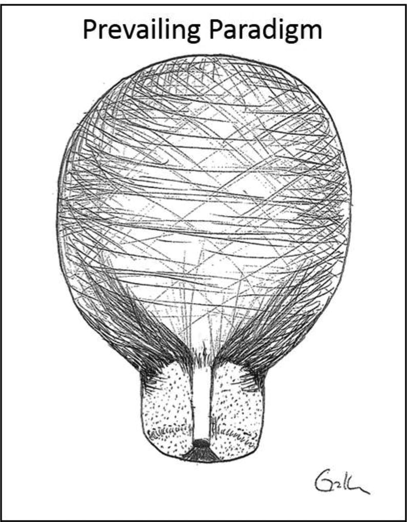
The cervix is the mechanical barrier located at the bottom of the uterus that keeps the uterus closed during pregnancy. As a pregnancy grows, the cervix must withstand tissue stresses generated from a complex set of forces from the weight of the growing pregnancy and pull of the uterine wall. (7) Our understanding of human cervical tissue structure was established in 1940s when Danforth et al reported that the cervix is a predominantly homogenous, hydrated, collagenous structure (about 85–90% collagen/extracellular matrix [ECM] that contains proteoglycans, glycosaminoglycans, elastin, matricellular proteins) and a minimal amount of cells (about 10–15%) which includes immune cells, fibroblasts, smooth muscle cells, and glandular/vascular cells. (8–10) In addition, over time, an assumption has been made that there may be a distinct line that demarcates where the uterus (smooth muscle body) ends and the cervix (collagen/ECM) begins. (Figure 1; [11])
Figure 1.
Image adapted from Vink et al (11) showing the prevailing paradigm of cervical tissue structure. This paradigm states the cervix is mainly a homogenous collagenous structure with minimal cellular content. It also suggests a border may exist between the muscular body of the uterus and collagenous structure of the cervix.
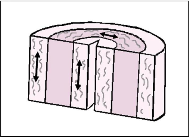
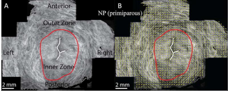
Similar to other load-bearing tissues in the body (i.e. bone, tendon), the mechanical strength of cervix is thought to rely mainly in part on the collagen network in the ECM. Studies in the 1970s reported that the collagen network in the cervix is comprised of three distinct zones – one zone of collagen in the middle of the stroma that is oriented circumferentially around the endocervical canal (which provides resistance to cervical dilation) and an inner (toward the endocervical canal) and outer (towards the vagina) zone of collagen fibers that run parallel to the endocervical canal These out and inner zones of collagen likely attach the cervix to the uterus. (6,12,13; Figure 2) Recently, Yao et al used optical coherence tomography (OCT) to measure the orientation of collagen fibers and their distribution in the upper cervix using hydrated unfixed samples, and the study reported in general there are two radial zones and the outer zone contains collagen fibers that are circumferentially oriented. (14; Figure 3) Reusch et al used second harmonic generation microscopy to show that collagen fibers in the cervix form interweaving zones that change as one moves from the external to internal os. (15) In addition to the overall structure of the collagen network, the strength of a collagen network also depends on collagen fiber alignment and the degree and type of cross-linking that is present between collagen fibrils.(16,17) Recent work by Zork et al has shown that collagen crosslinks can be measured in the human cervix and that the internal os exhibits more collagen crosslink heterogeneity compared to the external os.(18) Both the work by Reusch et al and Zork et al stress the point that unlike the prevailing paradigm, the cervix is not a homogeneous structure and that regional differences in tissue composition do exist between the internal and external os.
Figure 2.
Three zones of collagen organization – an outer and inner zone of fibers that run parallel to the endocervical canal and a middle zone of fibers that are circumferentially organized around the endocervical canal. (12,13)
Figure 3.
Images adapted from Yao et al. Optical coherence tomography image of a transverse slice of human nonpregnant cervical tissue at level of internal os showing there is an outer zone of circumferentially oriented collagen fibers. (14)
Other important components of ECM that influence its overall mechanical strength, include proteoglycans/glycosaminoglycans and matricellular proteins. Proteoglycans (ie decorin, versican, fibromodulin, biglycan, asporin) are important for organizing collagen fibrils and glycosaminoglycans (ie hyaluronic acid) influence tissue hydration and thus tissue compliance. (16,19–25) Matricellular proteins (i.e. tenascins, thrombospondins, SPARC proteins) are rapidly turned over during the remodeling process and can regulate matrix formation and cell-matrix interactions. (16, 26)
In addition to the stromal ECM, normal cervical tissue architecture includes the endocervical canal, which is lined by a single layer of mucin-secreting columnar epithelium, which form deep clefts or mucinous glands. The ectocervix (vaginal portion of the cervix) is covered with nonkeratinized stratified squamous epithelium. The junction between the endo- and ecto-cervical cells is the squamocolumnar junction. (27) The endo- and ectocervical cells are thought to play an important barrier function to protect against ascending infection from the vagina. (28,29)
Although most of the previous work evaluating cervical tissue structure has focused on the ECM, we also know the cervix is vascular and blood flow to the cervix provides a means of oxygenation and communication (i.e. via hormones, immune cells, chemokines/cytokines etc) from the rest of the body. The blood supply to the cervix is provided by branches of the uterine artery. A recent study by Bereza et al showed that in the nonpregnant state, there are four distinct “zones” of vessels in the vaginal and supravaginal apsects of the cervix. There is an outer zone which contains large arteries and veins, a middle zone (in the stroma) that contains arterioles and venules, an endocervical mucosal capillary zone and a zone parallel to the endocervical canal that contains small capillaries and veins. (30–34) How or if the cervical vasculature changes throughout pregnancy remains unknown. Even less is known about cervical tissue lymphatic changes in pregnancy.
Another interesting and important avenue of communication from the rest of the body to the cervix is via the nervous system. Although it is unclear why this is the case, the cervix is one of the most densely innervated structures of all the reproductive organs. Innervation to the pelvic organs is predominantly autonomic with sympathetic innervation provided by the superior hypogastric plexus (presacral nerve) which originates from spinal levels T10-L2. Parasympathetic innervation is derived from neurons originating from spinal levels S2 – S4 and axons from these spinal nerves combine to form the pelvic splanchnic nerves. The hypogastric (sympathetic) and pelvic splanchnic (parasympathetic) nerves combine to form the inferior hypogastric plexus (or the pelvic plexus) which further divides into branches that innervate the various pelvic organs including the cervix. The cervix also contains afferent sensory nerve fibers which transmit painful stimuli through the pelvic splanchnic nerves to the sacral nerves (S2–4). (35) Although this is still an emerging area of research that is ripe for further investigation, a few studies have shown that the human cervix is more densely innervated than the uterus and in pregnancy that uterine innervation decreases significantly 30-fold compared to non-pregnant uterine tissue) while the degree of innervation in the cervix remains the same. (36–42)
Cervical tissue remodeling in normal pregnancy
For most of pregnancy, the cervix must remain strong and closed to keep the fetus in utero. Then in a dramatic reversal of roles, at the end of pregnancy, the cervix must be compliant enough to dilate and allow for delivery of the fetus. Once the fetus is delivered, the cervix goes from being fully dilated to having a closed internal os – a process than can occur within minutes. Notably, while the internal os closes, the external os usually remains loose and open. In subsequent pregnancies, the external os may continue to remain soft and dilated while the internal os is firmly closed. This common finding is usually described by clinicians as a “multiparous cervix”.
Clinically, we can feel the cervix start to soften (Hegar sign) and see increased cyanosis as early as one month after conception. (43) Studies evaluating the mechanical properties of human cervical tissue in the non-pregnant, pregnant and postpartum state have confirmed our clinical findings that cervical tissue does indeed soften as pregnancy progresses. (44–47). Unfortunately, studies that evaluate how the human cervix changes at a biochemical level throughout human pregnancy is lacking as it is challenging to obtain cervical tissue (whole cervix tissue or biopsies) at various time points throughout pregnancy. Thus, the data that does exist regarding how the cervix remodels in pregnancy at a biochemical level have resulted from rodent studies.
This review provides a brief overview of the phases of cervical remodeling that has been shown to occur in rodents. For comprehensive details, please refer to the excellent reviews by Word et al (25), House et al (44), Nallasamy et al (48), Mahendroo et al (49) and Timmons et al (50). In short, cervical remodeling in rodents has been characterized to consist of four overlapping phases. The first phase is “cervical softening” which is described as a prolonged, progressive softening phase that starts early in pregnancy. This is a finding that we see clinically with the Hegar sign that starts within the first month of pregnancy. Although the cervix softens, the cervix stays closed because the mechanical forces acting on it are not large enough to dilate it. This delicate equilibrium balance most likely differs for every patient depending on cervical stroma consistency, the weight of the fetus, and maternal anatomy. (47) The cervical softening phase in rodents has been shown to involve decreases in collagen solubility (which is an indirect method to evaluate collagen crosslinking) and a shift to include more immature (weaker) collagen crosslinks vs mature (stronger) collagen crosslinks. (16, 17, 51) Yoshida et al reported that these alterations in the ratio of mature to immature collagen crosslinks directly relates to changes in cervical tissue compliance. (17) Akins et al reported that the cervical softening phase also includes decreases in matricellular proteins, thrombospondin 2 and tenascin-C. (16)
As rodent pregnancies approach term, the cervix undergoes a more rapid “ripening” phase, which results in increased cervical tissue compliance or weakening to prepare for cervical dilation. (25, 44, 49, 50) At a biochemical level, this phase is characterized by changes in the organization, shape and size of collagen fibers and an increase in matrix metalloproteinases (MMPs) which are enzymes that degrade ECM. (52,53) There is also an increase in the amount of hyaluronic acid, which is thought to increase tissue hydration and may be involved in disrupting or weakening of the collagen network. (25,29,49–51,54) However, preliminary studies by Akgul et al found that HA is not essential to normal cervical ripening. Due to the fact that HA has such a dramatic shift in content nearing term, the biomechanical role of HA still needs to be elucidated. (29)
Several studies have suggested that normal cervical ripening may be a sterile inflammatory state characterized by an influx of immune cells into the cervix. (55–65) However, although studies have shown an influx of immune cells into cervical tissue, it is unclear what triggers the immune cells to travel to the cervix. In addition, evidence from other studies suggest that immune cell activation is not vital for cervical ripening. (66–69). Interestingly, several studies have investigated the role of the nervous system in cervical remodeling and parturition and these studies propose that nerves in the cervix may play a role in inducing a “sterile” neurogenic inflammatory state. (70–72) Given that the cervix remains densely innervated throughout pregnancy, this is a tantalizing hypothesis that is in dire need of further investigation. Once the cervix is “ripe” it can now dilate as the uterus goes into labor and allow for delivery of the fetus. The last phase of cervical remodeling is “postpartum repair”. (25, 49) To date, detailed data on how the cervix dramatically goes from a fully dilated structure to have a closed internal os in a matter of minutes is lacking.
Although extensive and extremely detailed work has evaluated the phases of cervical remodeling in rodents, we must keep in mind that rodents do not share the same anatomical structure of their reproductive organs with humans and rodents are quadrupedal mammals which undergo different gravitational forces on their pregnant pelvic organs compared to bipedal humans that stand upright. In addition, hormonal (estrogen and progesterone) changes that are seen in rodents also do not perfectly mimic changes seen in humans. Regardless, since obtaining cervical tissue during human pregnancy is challenging, insight obtained from rodent studies are vital to establishing a guide as to what may possibly be occurring in human cervical remodeling.
Current knowledge on the pathophysiology of human premature cervical remodeling
To date, studies that have evaluated the mechanisms leading to cervical “failure” in pregnancy have been inconclusive in part because diagnostic criteria to define a “weak” cervix remain elusive. Traditionally, we have used the clinical term “cervical insufficiency” or “an incompetent cervix” to define a condition where the cervix tends to asymptomatically dilate repeatedly in the mid-trimester leading to a spontaneous abortion or periviable/premature delivery. (73) Due to recent studies that have shown that premature cervical shortening significantly increases a woman’s risk for sPTB, many now consider premature cervical shortening as a sign of “cervical insufficiency”. (74)
Although studies have identified risk factors for “cervical insufficiency” (73), studies that have obtained and evaluated human cervical tissue to determine the underlying biochemical cause of “weak cervical tissue” in pregnancy remain inconclusive. Table 1 lists the studies that have evaluated human cervical tissue in women with “cervical insufficiency” and sheds light on several issues. (75–82) First, there is a paucity of data evaluating human cervical tissue in women with cervical insufficiency. Second, some of these studies defined “cervical insufficiency” differently and methods for identifying those with “cervical insufficiency” in some studies may not be ideal or accurate (i.e. by ICD-10 codes). Third, cervical biopsies were not obtained in the same location. As studies are beginning to show, the cervix is not a homogenous structure and biopsy location does matter. Fourth, timing of cervical biopsy in pregnancy were different. In most studies, cervical biopsies were obtained in the nonpregnant state or postpartum after the cervix has undergone cervical dilation and delivery of the fetus. Some studies obtained biopsies during pregnancy in patients with “cervical insufficiency” but the control patients were either nonpregnant women undergoing benign hysterectomy or not gestational-age matched controls in pregnancy. These reasons might influence why we have not been able to find conclusive results regarding if there is an ECM defect in cervical tissue from women who exhibit premature cervical failure.
Table 1.
Studies that have obtained and evaluated human cervical tissue in women with a history of cervical insufficiency (CI)
| Author | Date | Patient Groups | Definition of CI | Biopsy Location |
Type of Biopsy |
Biopsy Timing |
Reported Findings |
|---|---|---|---|---|---|---|---|
| Roddick et al (75) | 1961 | Case report of 2 patients with CI |
|
Posterior lip of external os (3cm × 2cm) | Wedge biopsy | After delivery | Connective tissue was scant but muscular tissue was abundant. |
| Buckingham et al (76) | 1965 |
|
Painless dilation of the cervix to 3cm or more in the middle trimester followed by PPROM and delivery. | Posterior lip of external os/ lower aspect of cervix | Some patients had a wedge biopsy, some had punch biopsy |
|
Patients with CI had lower % collagen and higher % muscle in their cervical tissue. |
| Leppert et al (77) | 1987 |
|
Evidence of progressive, painless dilation without uterine contraction, and effacement of the cervix in the late first or second trimester, a history of previous painless dilation of the cervix and/or history of cervical trauma, and a cerclage in place before time of delivery |
|
Wedge biopsy |
|
Patients with CI had absent or decreased elastin fibers (n=6) and decreased desmosine content (n=1) |
| Rechberger et al (78) | 1988 |
|
1 + second trimester losses without painful contractions and short cervix (1cm or less) | Distal aspect of the cervix | Scissors |
|
|
| Petersen et al (79) | 1996 |
|
No other cause of cervical incompetence could be deduced from the history and where the first pregnancy sustained beyond the 12th week of pregnancy ended with a late SAB or preterm delivery. | Anterior lip of external os | Forceps |
|
Women with CI had lower hydroxyproline concentration and increased collagen extractability vs parous controls. |
| Oxlund et al (80) | 2010 |
|
Identified by ICD-10 diagnosis of CI as well as by “cerclage” or short cervical length | 3, 6, 12 o’clock at the external os | Punch biopsy (core) |
|
|
| Gedikbasi et al (81) | 2016 |
|
History of painless cervical dilation after the first trimester with subsequent expulsion of the pregnancy in the second trimester, before 24 weeks, without contractions or labor in the absence of other clear pathology such as bleeding, infection or ruptured membranes | Unclear | Unclear |
|
|
| Sundtoft et al (82) | 2017 |
|
ICD-10 codes for CI, suspected preterm birth and/or vaginal and abdominal cerclage |
|
Forceps |
|
|
An updated paradigm of cervical tissue structure and function
With the introduction of cervical length measurement, it has become clinically evident that when cervices start to shorten in pregnancy, this process is usually characterized by funneling or dilation/failure of the internal os while the external os remains closed. (83) In addition, we know that after a vaginal delivery of a fetus, the internal os rapidly closes while the external os stays dilated (sometimes for weeks or months or even into subsequent pregnancies). Recently, our lab re-evaluated whole transverse slices of normal, nonpregnant cervical tissue from the internal os, mid-cervix and external os to further understand why these regional differences in cervical tissue function exist. (11)
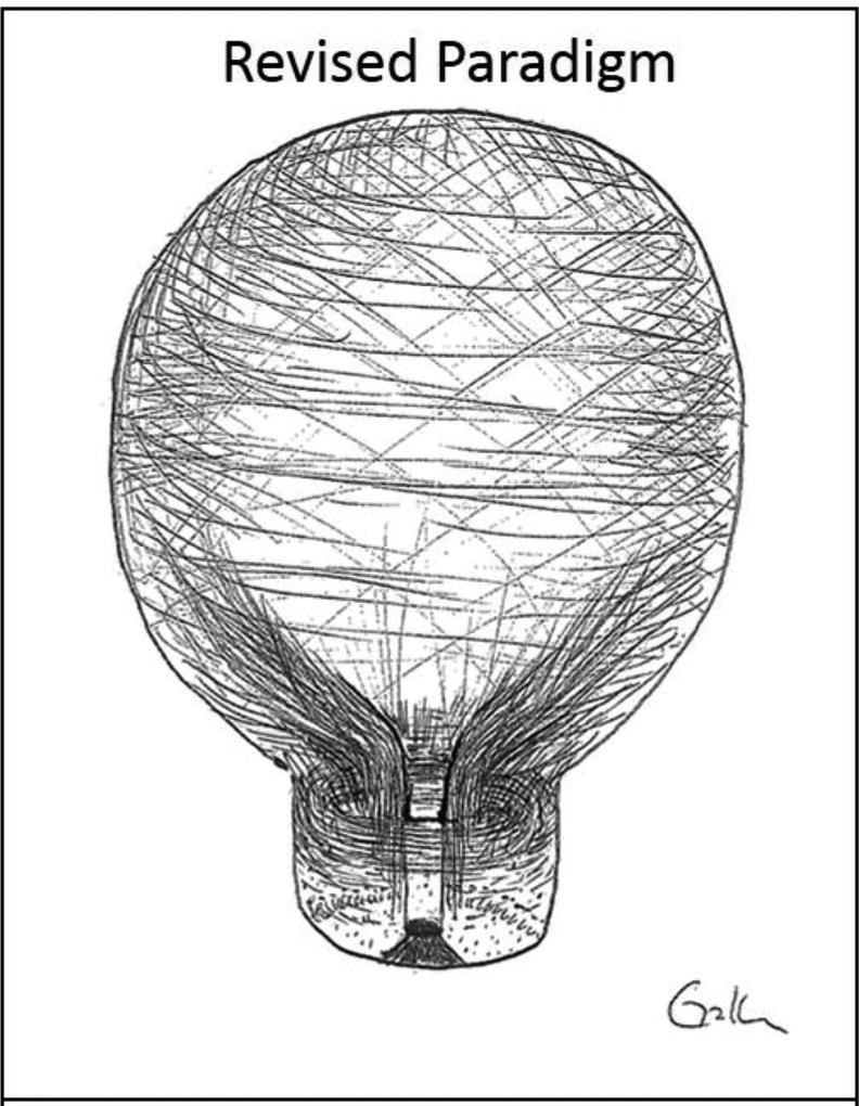
Unlike the prevailing paradigm which characterizes the human cervix as a mostly homogenous, collagenous structure with minimal cellular content, we found that there is an abundant amount of smooth muscle at the internal os (about 50–60% of the tissue) and that there are bundles of smooth muscle that circumferentially oriented around the periphery of the cervix. (11) It is interesting to note that this muscle structure mimics the collagen network “zone” that was reported by Yao et al. (14) This smooth muscle orientation persists as one moves down to the mid-cervix. The smooth muscle content then gradually decreases in the lower half of the cervix. At the external os, smooth muscle can be found scattered in the tissue and accounts for about 10% of the tissue content. (Figure 4, [11]) In addition, we found that whole slices of cervical tissue from the internal os are more contractile than tissue obtained from the external os in functional organ baths. (11) Recent rodent studies have also shown that the rodent cervix contains a significant amount of smooth muscle and that the cervix does contract in response to contractile agonists. (84)
Figure 4.
Image adapted from Vink et al showing the revised paradigm of cervical tissue structure. This revised paradigm shows the upper half of the cervix contains a significant amount of smooth muscle with bundles that are circumferentially oriented around the endocervical canal. (11)
Given these findings, as well as the fact that the cervix remains highly innervated in pregnancy while uterine innervation decreases, and that the internal os is more densely innervated than the external os, it is intriguing to consider the possibility that the smooth muscle at the internal os may form a specialized sphincter. If the internal os is a sphincter, it is then reasonable to ask, “is cervical funneling or dilation of the internal os in pregnancy actually ‘sphincter failure’”? Along the same line, when the internal os closes rapidly after vaginal delivery, is the sphincter resuming is original shape? Going one step farther, some studies have shown cesarean sections done in the second stage of labor may increase the rate of sPTB in subsequent pregnancies. (85) If the sPTB in subsequent pregnancies are due premature cervical failure, could it be the “sphincter” was damaged or altered at time of cesarean section? Given the structure and clinical function of the internal os, these concepts make sense biologically but further studies are needed.
The concept of a sphincter in the cervix is actually not new. Several decades ago, studies questioned why smooth muscle cell existed in the cervix. In the 1950s, a study showed in response to contractile agonists, the pregnant cervix is able to contract separate from the uterus.(86) Several studies showed the cervix exhibits electromyography (EMG) activity and cervical EMG activity correlates to the length of the latent phase of labor. These studies suggested the cervix can contract and prolong the length of the latent phase of labor. (87–93) Unfortunately, over time (and likely due to the assumption that the cervix was a mostly homogenous, collagenous structure), evaluating cervical smooth muscle function fell out of favor as more attention was given to evaluating if cervical failure in pregnancy was due to defects in the ECM.
Where do we go now?
This review introduces a revised paradigm of cervical tissue structure and function that includes the possibility of a specialized sphincter in the cervix. This paradigm states the cervix is not a passive, homogenous, collagenous structure and resident smooth muscle cells in the stroma may play an active role in cervical function. This paradigm opens new doors for exploration which include but are not limited to the following. 1) What is the contractile role of cervical smooth muscle cells at the internal os in normal pregnancies and pregnancies complicated by premature cervical failure? Similarly, cervical smooth muscle cells are contiguous with uterine smooth muscle cells and likely have the ability to send signals to the uterus. As such, could they have a role in orchestrating or propagating uterine contractions? 2) We know that smooth muscle cells from other body systems (i.e. vascular and uterine smooth muscle cells) can produce ECM and enzymes involved in remodeling ECM. (94–97) If cervical smooth muscle cells share similar capabilities, what is their role in maintaining ECM homeostasis in the cervix? 3) As the fetus grows, the weight of the pregnancy stretches the cervix, particularly at the internal os. (98,99) Towards the end of a normal pregnancy, Braxton-Hicks and labor contractions occur which further stretch the cervix. Given that i) tissue stress and stretch are an important mechanical factors in pregnancy, ii) smooth muscle cells can produce ECM and enzymes involved in remodeling ECM (94–97) and iii) uterine smooth muscle stretch can influence myometrial tissue remodeling (95–97,100), could cervical smooth muscle stretch influence cervical remodeling? Further, if cervical smooth muscle cells have altered ‘stretch’ sensors, could this trigger premature remodeling? Lastly, areas that also have largely been forgotten but are in dire need of investigation are the role of the vascular and nervous system in normal and abnormal cervical tissue remodeling.
As we embark on studies investigating these novel areas, it is imperative that we move forward with clear and standardized terminology. Since “cervical insufficiency” can be a confusing clinical term, recently it was proposed to adopt the term “premature cervical remodeling” as it does not rely on clinical history but focuses on the physical changes in the cervix. (6) In order to use this terminology, however, we must establish objective detection and quantification methods to diagnose premature cervical remodeling. Currently, several groups are working on developing non-invasive methods to quantify cervical remodeling and these are discussed in the second issue of this series in Seminars in Perinatology. It is also imperative that we analyze normal and abnormal human tissues in pregnancy and that our in vitro models mimic the pregnant state (i.e. the hormonal milieu of pregnancy). Lastly, but most importantly, it is essential that we remember that each “compartment” (i.e. uterus, cervix, fetal membranes, immune/vascular/nervous systems, environment etc.) does not operate in isolation. Future studies will need to take into account interactions between “compartments”. Moving forward, the path to success is one that involves active, multidisciplinary collaboration and communication between experts in various fields that include but are not limited to geneticists, epidemiologists, basic scientists, clinicians, systems biologists and engineers. With such an approach, we may finally identify novel and effective interventions to significantly decrease the rate and complications of prematurity.
Acknowledgments
The research included in this review was supported financially by the Society for Maternal Fetal Medicine/ American Association of Obstetricians & Gynecologists Foundation Scholarship, The Louis J. Gerstner, Jr. Foundation, March of Dimes Prematurity Research Center at the University of Pennsylvania, and K08HD088758 from the Eunice Kennedy Shriver National Institute of Child Health and Human Development. The content is solely the responsibly of the authors and does not necessarily represent the official views of the funding agencies listed here. The authors thank Christine Hendon, Ph.D. from the Department of Electrical Engineering at Columbia University for the use of the optical coherence images used in Figure 3.
Footnotes
Publisher's Disclaimer: This is a PDF file of an unedited manuscript that has been accepted for publication. As a service to our customers we are providing this early version of the manuscript. The manuscript will undergo copyediting, typesetting, and review of the resulting proof before it is published in its final citable form. Please note that during the production process errors may be discovered which could affect the content, and all legal disclaimers that apply to the journal pertain.
Disclosure: The authors have nothing to disclose.
References
- 1.Centers for Disease Control and Prevention. [Accessed May 13, 2017]; at http://www.cdc.gov/reproductivehealth//MaternalInfantHealth/PretermBirth.htm.
- 2.Caughey AB, Zupancic JA, Greenberg JM, Garfield SS, Thung SF, Iams JD. Clinical and Cost Impact Analysis of a Novel Prognostic Test for Early Detection of Preterm Birth. AJP Rep. 2016 Oct;6(4):e407–e416. doi: 10.1055/s-0036-1593866. [DOI] [PMC free article] [PubMed] [Google Scholar]
- 3.Gyamfi-Bannerman C, Ananth CV. Trends in spontaneous and indicated preterm delivery among singleton gestations in the United States. Obstet Gynecol 2005–2012. 2014;124:1069–74. doi: 10.1097/AOG.0000000000000546. [DOI] [PubMed] [Google Scholar]
- 4.Martin B, Hamilton S, Ventura M, Osterman T. Mathews, Births: Final data for 2011. Natl Vital Stat Rep. 2013;62(1):1–90. [PubMed] [Google Scholar]
- 5.Schoen CN, Tabbah S, Iams JD, Caughey AB, Berghella V. Why the united states preterm birth rate is declining, American journal of obstetrics and gynecology. Am J Obstet Gynecol. 2015 Aug;213(2):175–80. doi: 10.1016/j.ajog.2014.12.011. Epub 2014 Dec 12. [DOI] [PubMed] [Google Scholar]
- 6.Vink J, Feltovich H. Cervical etiology of spontaneous preterm birth. Semin Fetal Neonatal Med. 2016 Apr;21(2):106–12. doi: 10.1016/j.siny.2015.12.009. Epub 2016 Jan 14. [DOI] [PMC free article] [PubMed] [Google Scholar]
- 7.Myers KM, Elad D. Biomechanics of the human uterus. Wiley Interdiscip Rev Syst Biol Med. 2017 May 12; doi: 10.1002/wsbm.1388. [DOI] [PubMed] [Google Scholar]
- 8.Danforth D. The fibrous nature of the human cervix and its relation to the isthmic segment in gravid and nongravid uteri. Proc Inst Med Chic. 1947 Jan 15;16(10):295. [PubMed] [Google Scholar]
- 9.Danforth D. The fibrous nature of the human cervix, and its relation to the isthmic segment in gravid and nongravid uteri. Am J Obstet Gynecol. 1947 Apr;53(4):541–60. doi: 10.1016/0002-9378(47)90273-1. [DOI] [PubMed] [Google Scholar]
- 10.Danforth DN. The morphology of the human cervix. Clin Obstet Gynecol. 1983 Mar;26(1):7–13. doi: 10.1097/00003081-198303000-00005. [DOI] [PubMed] [Google Scholar]
- 11.Vink JY, Qin S, Brock CO, Zork NM, Feltovich HM, Chen X, Urie P, Myers KM, Hall TJ, Wapner R, Kitajewski JK, Shawber CJ, Gallos G. A new paradigm for the role of smooth muscle cells in the human cervix. Am J Obstet Gynecol. 2016 Oct;215(4):478.e1–478.e11. doi: 10.1016/j.ajog.2016.04.053. [DOI] [PubMed] [Google Scholar]
- 12.Dubrauszky V, Schwalm H, Fleischer M. Fibre system of connective tissue in childbearing age, menopause and pregnancy. Archiv Gynakol. 1971;210:276–92. doi: 10.1007/BF00667740. [DOI] [PubMed] [Google Scholar]
- 13.Aspden RM. Collagen organization in the cervix and its relation to mechanical function. Coll Relat Res. 1988;8:103–12. doi: 10.1016/s0174-173x(88)80022-0. [DOI] [PubMed] [Google Scholar]
- 14.Yao W, Gan Y, Myers KM, Vink JY, Wapner RJ, Hendon CP. Collagen Fiber Orientation and Dispersion in the Upper Cervix of Non-Pregnant and Pregnant Women. PLoS One. 2016 Nov 29;11(11):e0166709. doi: 10.1371/journal.pone.0166709. eCollection 2016. [DOI] [PMC free article] [PubMed] [Google Scholar]
- 15.Reusch LM, Feltovich H, Carlson LC, et al. Nonlinear optical microscopy and ultrasound imaging of human cervical structure. J Biomed Opt. 2013;18:031110. doi: 10.1117/1.JBO.18.3.031110. [DOI] [PMC free article] [PubMed] [Google Scholar]
- 16.Akins ML, Luby-Phelps K, Bank RA, Mahendroo M. Cervical softening during pregnancy: regulated changes in collagen cross-linking and composition of matricellular proteins in the mouse. Biol Reprod. 2011 May;84(5):1053–62. doi: 10.1095/biolreprod.110.089599. Epub 2011 Jan 19. [DOI] [PMC free article] [PubMed] [Google Scholar]
- 17.Yoshida K, Jiang H, Kim M, et al. Quantitative evaluation of collagen crosslinks and corresponding tensile mechanical properties in mouse cervical tissue during normal pregnancy. PLoS One. 2014;9:e112391. doi: 10.1371/journal.pone.0112391. [DOI] [PMC free article] [PubMed] [Google Scholar]
- 18.Zork NM, Myers KM, Yoshida K, et al. A systematic evaluation of collagen cross-links in the human cervix. Am J Obstet Gynecol. 2015;212:321.e1–8. doi: 10.1016/j.ajog.2014.09.036. [DOI] [PMC free article] [PubMed] [Google Scholar]
- 19.Leppert PC. Anatomy and physiology of cervical ripening. Clin Obstet Gynecol. 1995 Jun;38(2):267–79. doi: 10.1097/00003081-199506000-00009. [DOI] [PubMed] [Google Scholar]
- 20.Danforth DN, Veis A, Breen M, Weinstein HG, Buckingham JC, Manalo P. The effect of pregnancy and labor on the human cervix: changes in collagen, glycoproteins, and glycosaminoglycans. Am J Obstet Gynecol. 1974;120:641–651. doi: 10.1016/0002-9378(74)90608-5. [DOI] [PubMed] [Google Scholar]
- 21.Norman M, Ekman G, Ulmsten U, Barchan K, Malmstrom A. Proteoglycan metabolism in the connective tissue of pregnant and non-pregnant human cervix. An in vitro study. Biochem J. 1991;275(Pt 2):515–52. doi: 10.1042/bj2750515. [DOI] [PMC free article] [PubMed] [Google Scholar]
- 22.Osmers R, Rath W, Pflanz MA, Kuhn W, Stuhlsatz HW, Szeverenyi M. Glycosaminoglycans in cervical connective tissue during pregnancy and parturition. Obstet Gynecol. 1993;81:88–92. [PubMed] [Google Scholar]
- 23.Westergren-Thorsson G, Norman M, Bjornsson S, Endresen U, Stjernholm Y, Ekman G, Malmstrom A. Differential expressions of mRNA for proteoglycans, collagens and transforming growth factor-beta in the human cervix during pregnancy and involution. Biochim Biophys Acta. 1998;1406:203–213. doi: 10.1016/s0925-4439(98)00005-2. [DOI] [PubMed] [Google Scholar]
- 24.Akgul Y, Holt R, Mummert M, Word A, Mahendroo M. Dynamic changes in cervical glycosaminoglycan composition during normal pregnancy and preterm birth. Endocrinology. 2012 Jul;153(7):3493–503. doi: 10.1210/en.2011-1950. Epub 2012 Apr. [DOI] [PMC free article] [PubMed] [Google Scholar]
- 25.Word RA, Li XH, Hnat M, Carrick K. Dynamics of cervical remodeling during pregnancy and parturition: mechanisms and current concepts. Semin Reprod Med. 2007 Jan;25(1):69–79. doi: 10.1055/s-2006-956777. [DOI] [PubMed] [Google Scholar]
- 26.Paul Bornstein. Matricellular proteins: an overview. J Cell Commun Signal. 2009 Dec;3(3–4):163–5. doi: 10.1007/s12079-009-0069-z. Epub 2009 Sep 25. [DOI] [PMC free article] [PubMed] [Google Scholar]
- 27.Cunningham G, Leveno K, Bloom S, Spong CY, Dashe J, Hoffman B, Casey B, Sheffield J, editors. Williams Obstetrics. Columbus, OH: McGraw-Hill Education; 2014. Maternal Anatomy; p. 26. [Google Scholar]
- 28.Nold C, Anton L, Brown A, Elovitz M. Inflammation promotes a cytokine response and disrupts the cervical epithelial barrier: a possible mechanism of premature cervical remodeling and preterm birth. Am J Obstet Gynecol. 2012 Mar;206(3):208.e1–7. doi: 10.1016/j.ajog.2011.12.036. Epub 2012 Feb 12. [DOI] [PubMed] [Google Scholar]
- 29.Akgul Y, Word RA, Ensign LM, Yamaguchi Y, Lydon J, Hanes J, Mahendroo M. Hyaluronan in cervical epithelia protects against infection-mediated preterm birth. J Clin Invest. 2014 Dec;124(12):5481–9. doi: 10.1172/JCI78765. Epub 2014 Nov 10. [DOI] [PMC free article] [PubMed] [Google Scholar]
- 30.Bereza T, Skrzat J, Brzozowska I, Maduzia D, Matuszyk A, Chmielewski P, Klimek-Piotrowska W, Tomaszewski K. Blood supply of human uterine cervix - a SEM study. Folia Med Cracov. 2013;53(4):5–12. [PubMed] [Google Scholar]
- 31.Bereza T, Tomaszewski KA, Walocha J, Mizia E, Bachul P, Chmielewski P. Vascular architecture of the human uterine cervix, as assessed in light- and scanning electron microscopy. Folia Morphol (Warsz) 2012 Aug;71(3):142–7. [PubMed] [Google Scholar]
- 32.Bereza T, Tomaszewski KA, Bałajewicz-Nowak M, Mizia E, Pasternak A, Walocha J. The vascular architecture of the supravaginal and vaginal parts of the human uterine cervix: a study using corrosion casting and scanning electron microscopy. J Anat. 2012 Oct;221(4):352–7. doi: 10.1111/j.1469-7580.2012.01550.x. Epub 2012 Jul 30. [DOI] [PMC free article] [PubMed] [Google Scholar]
- 33.Walocha JA, Litwin JA, Bereza T, Klimek-Piotrowska W, Miodoński AJ. Vascular architecture of human uterine cervix visualized by corrosion casting and scanning electron microscopy. Hum Reprod. 2012 Mar;27(3):727–32. doi: 10.1093/humrep/der458. Epub 2012 Jan 16. [DOI] [PubMed] [Google Scholar]
- 34.deSouza NM, Hawley IC, Schwieso JE, Gilderdale DJ, Soutter WP. The uterine cervix on in vitro and in vivo MR images: a study of zonal anatomy and vascularity using an enveloping cervical coil. AJR Am J Roentgenol. 1994 Sep;163(3):607–12. doi: 10.2214/ajr.163.3.8079853. [DOI] [PubMed] [Google Scholar]
- 35.Cunningham G, Leveno K, Bloom S, Spong CY, Dashe J, Hoffman B, Casey B, Sheffield J, editors. Williams Obstetrics. Columbus, OH: McGraw-Hill Education; 2014. Maternal Anatomy; pp. 28–29. [Google Scholar]
- 36.Stjernholm Y, Sennström M, Granström L, Ekman G, Liang Y, Johansson O. Neurochemical and cellular markers in human cervix of late pregnant, postpartal and non-pregnant women. Acta Obstet Gynecol Scand. 2000 Jul;79(7):528–37. [PubMed] [Google Scholar]
- 37.Sahlin L, Wang H, Stjernholm Y, Lundberg M, Ekman G, Holmgren A, Eriksson H. The expression of glutaredoxin is increased in the human cervix in term pregnancy and immediately post-partum, particularly after prostaglandin-induced delivery. Mol Hum Reprod. 2000 Dec;6(12):1147–53. doi: 10.1093/molehr/6.12.1147. [DOI] [PubMed] [Google Scholar]
- 38.Tingåker BK, Johansson O, Cluff AH, Ekman-Ordeberg G. Unaltered innervation of the human cervix uteri in contrast to the corpus during pregnancy and labor as revealed by PGP 9.5 immunohistochemistry. Eur J Obstet Gynecol Reprod Biol. 2006 Mar 1;125(1):66–71. doi: 10.1016/j.ejogrb.2005.07.020. Epub 2005 Sep 26. [DOI] [PubMed] [Google Scholar]
- 39.Tingaker BK, Ekman-Ordeberg G, Forsgren S. Presence of sensory nerve corpuscles in the human corpus and cervix uteri during pregnancy and labor as revealed by immunohistochemistry. Reprod Biol Endocrinol. 2006 Aug 29;4:45. doi: 10.1186/1477-7827-4-45. [DOI] [PMC free article] [PubMed] [Google Scholar]
- 40.Di Tommaso S, Cavallotti C, Malvasi A, Vergara D, Rizzello A, De Nuccio F, Tinelli A. A Qualitative and Quantitative Study of the Innervation of the Human Non Pregnant Uterus. Curr Protein Pept Sci. 2016;18(2):140–148. doi: 10.2174/1389203717666160330105341. [DOI] [PubMed] [Google Scholar]
- 41.Bryman I, Norström A, Dahlström A, Lindblom B. Immunohistochemical evidence for preserved innervation of the human cervix during pregnancy. Gynecol Obstet Invest. 1987;24(2):73–9. doi: 10.1159/000298782. [DOI] [PubMed] [Google Scholar]
- 42.Vera PL, Haase EB, Schramm LP. Origins of the sympathetic innervation of the cervical end of the uterus in the rat. Brain Res. 1997 Jan 30;747(1):140–3. doi: 10.1016/s0006-8993(96)01107-9. [DOI] [PubMed] [Google Scholar]
- 43.Hegar A. Diagnose de Fruhesten Schwangerschaftsperiode. Dtsch med Wochenschr. 1985;21:565. [Google Scholar]
- 44.House M, Kaplan DL, Socrate S. Relationships between mechanical properties and extracellular matrix constituents of the cervical stroma during pregnancy. Semin Perinatol. 2009 Oct;33(5):300–7. doi: 10.1053/j.semperi.2009.06.002. [DOI] [PMC free article] [PubMed] [Google Scholar]
- 45.Badir S, Mazza E, Zimmermann R, Bajka M. Cervical softening occurs early in pregnancy: characterization of cervical stiffness in 100 healthy women using the aspiration technique. Prenat. Diagn. 2013b;33:737–741. doi: 10.1002/pd.4116. [DOI] [PubMed] [Google Scholar]
- 46.Parra-Saavedra M, Gómez L, Barrero A, Parra G, Vergara F, Navarro E. Prediction of preterm birth using the cervical consistency index. Ultrasound Obstet. Gynecol. 2011;38:44–51. doi: 10.1002/uog.9010. [DOI] [PubMed] [Google Scholar]
- 47.Myers KM, Feltovich H, Mazza E, Vink J, Bajka M, Wapner RJ, Hall TJ, House M. The mechanical role of the cervix in pregnancy. J Biomech. 2015 Jun 25;48(9):1511–23. doi: 10.1016/j.jbiomech.2015.02.065. [DOI] [PMC free article] [PubMed] [Google Scholar]
- 48.Nallasamy S, Mahendroo M. Distinct Roles of Cervical Epithelia and Stroma in Pregnancy and Parturition. Semin Reprod Med. 2017 Mar;35(2):190–200. doi: 10.1055/s-0037-1599091. [DOI] [PubMed] [Google Scholar]
- 49.Mahendroo M. Cervical remodeling in term and preterm birth: insights from an animal model. Reproduction. 2012 Apr;143(4):429–38. doi: 10.1530/REP-11-0466. [DOI] [PubMed] [Google Scholar]
- 50.Timmons B, Akins M, Mahendroo M. Cervical remodeling during pregnancy and parturition. Trends Endocrinol Metab. 2010 Jun;21(6):353–61. doi: 10.1016/j.tem.2010.01.011. [DOI] [PMC free article] [PubMed] [Google Scholar]
- 51.Read CP, Word RA, Ruscheinsky MA, Timmons BC, Mahendroo MS. Cervical remodeling during pregnancy and parturition: molecular characterization of the softening phase in mice. Reproduction. 2007;134:327–340. doi: 10.1530/REP-07-0032. [DOI] [PubMed] [Google Scholar]
- 52.Gonzalez JM, Franzke CW, Yang F, Romero R, Girardi G. Complement activation triggers metalloproteinases release inducing cervical remodeling and preterm birth in mice. Am J Pathol. 2011 Aug;179(2):838–49. doi: 10.1016/j.ajpath.2011.04.024. [DOI] [PMC free article] [PubMed] [Google Scholar]
- 53.Stygar D, Wang H, Vladic YS, Ekman G, Eriksson H, Sahlin L. Increased level of matrix metalloproteinases 2 and 9 in the ripening process of the human cervix. Biol Reprod. 2002 Sep;67(3):889–94. doi: 10.1095/biolreprod.102.005116. [DOI] [PubMed] [Google Scholar]
- 54.Holt R, Timmons B, Akgul Y, Akins M, Mahendroo M. The molecular mechanisms of cervical ripening differ between term and preterm birth. Endocrinology. 2011;152:1036–1046. doi: 10.1210/en.2010-1105. [DOI] [PMC free article] [PubMed] [Google Scholar]
- 55.Dubicke A, Ekman-Ordeberg G, Mazurek P, Miller L, Yellon SM. Density of Stromal Cells and Macrophages Associated With Collagen Remodeling in the Human Cervix in Preterm and Term Birth. Reprod Sci. 2016 May;23(5):595–603. doi: 10.1177/1933719115616497. [DOI] [PMC free article] [PubMed] [Google Scholar]
- 56.Dobyns AE, Goyal R, Carpenter LG, Freeman TC, Longo LD, Yellon SM. Macrophage gene expression associated with remodeling of the prepartum rat cervix: microarray and pathway analyses. PLoS One. 2015 Mar 26;10(3):e0119782. doi: 10.1371/journal.pone.0119782. [DOI] [PMC free article] [PubMed] [Google Scholar]
- 57.Payne KJ, Clyde LA, Weldon AJ, Milford TA, Yellon SM. Residency and activation of myeloid cells during remodeling of the prepartum murine cervix. Biol Reprod. 2012 Nov 1;87(5):106. doi: 10.1095/biolreprod.112.101840. [DOI] [PMC free article] [PubMed] [Google Scholar]
- 58.Mackler AM, Iezza G, Akin MR, McMillan P, Yellon SM. Macrophage trafficking in the uterus and cervix precedes parturition in the mouse. Biol Reprod. 1999 Oct;61(4):879–83. doi: 10.1095/biolreprod61.4.879. [DOI] [PubMed] [Google Scholar]
- 59.Yellon SM, Mackler AM, Kirby MA. The role of leukocyte traffic and activation in parturition. J Soc Gynecol Investig. 2003 Sep;10(6):323–38. doi: 10.1016/s1071-5576(03)00116-3. [DOI] [PubMed] [Google Scholar]
- 60.Yellon SM, Ebner CA, Sugimoto Y. Parturition and recruitment of macrophages in cervix of mice lacking the prostaglandin F receptor. Biol Reprod. 2008 Mar;78(3):438–44. doi: 10.1095/biolreprod.107.063404. [DOI] [PMC free article] [PubMed] [Google Scholar]
- 61.Liggins GC. Cervical ripening as an inflammatory reaction. In: Ellwood D, Anderson ABM, editors. The Cervix in Pregnancy and Labour, Clinical and Biochemical Investigation. Edinburgh: Churchill Livingston; 1981. [Google Scholar]
- 62.Junqueira LC, Zugaib M, Montes GS, Toledo OM, Krisztán RM, Shigihara KM. Morphologic and histochemical evidence for the occurrence of collagenolysis and for the role of neutrophilicpolymorphonuclear leukocytes during cervical dilation. Am J Obstet Gynecol. 1980 Oct 1;138(3):273–81. doi: 10.1016/0002-9378(80)90248-3. [DOI] [PubMed] [Google Scholar]
- 63.Luque EH, Munoz de Toro MM, Ramos JG, Rodriguez HA, Sherwood OD. Role of relaxin and estrogen in the control of eosinophilic invasion and collagen remodeling in rat cervical tissue at term. Biology of Reproduction. 1998;59:795–800. doi: 10.1095/biolreprod59.4.795. [DOI] [PubMed] [Google Scholar]
- 64.Young A, Thomson AJ, Ledingham M, Jordan F, Greer IA, Norman JE. Immunolocalization of proinflammatory cytokines in myometrium, cervix, and fetal membranes during human parturition at term. Biology of Reproduction. 2002;66:445–449. doi: 10.1095/biolreprod66.2.445. [DOI] [PubMed] [Google Scholar]
- 65.Osman I, Young A, Ledingham MA, Thomson AJ, Jordan F, Greer IA, Norman JE. Leukocyte density and pro-inflammatory cytokine expression in human fetal membranes, decidua, cervix and myometrium before and during labour at term. Molecular Human Reproduction. 2003;9:41–45. doi: 10.1093/molehr/gag001. [DOI] [PubMed] [Google Scholar]
- 66.Hirsch E, Filipovich Y, Mahendroo M. Signaling via the type I IL-1 and TNF receptors is necessary for bacterially induced preterm labor in a murine model. American Journal of Obstetrics and Gynecology. 2006;194:1334–1340. doi: 10.1016/j.ajog.2005.11.004. [DOI] [PubMed] [Google Scholar]
- 67.Gonzalez JM, Xu H, Chai J, Ofori E, Elovitz MA. Preterm and term cervical ripening in CD1 Mice (Musmusculus): similar or divergent molecular mechanisms? Biol Reprod. 2009 Dec;81(6):1226–32. doi: 10.1095/biolreprod.108.075309. [DOI] [PubMed] [Google Scholar]
- 68.Timmons BC, Fairhurst AM, Mahendroo MS. Temporal changes in myeloid cells in the cervix during pregnancy and parturition. J Immunol. 2009 Mar 1;182(5):2700–7. doi: 10.4049/jimmunol.0803138. [DOI] [PMC free article] [PubMed] [Google Scholar]
- 69.Timmons BC, Mahendroo MS. Timing of neutrophil activation and expression of proinflammatory markers do not support a role for neutrophils in cervical ripening in the mouse. Biol Reprod. 2006 Feb;74(2):236–45. doi: 10.1095/biolreprod.105.044891. [DOI] [PubMed] [Google Scholar]
- 70.Mowa CN. Uterine Cervical Neurotransmission and Cervical Remodeling. Curr Protein Pept Sci. 2016;18(2):120–124. doi: 10.2174/1389203717666160322150224. [DOI] [PubMed] [Google Scholar]
- 71.Mowa CN, Papka RE. The role of sensory neurons in cervical ripening: effects of estrogen and neuropeptides. J Histochem Cytochem. 2004 Oct;52(10):1249–58. doi: 10.1177/002215540405201001. [DOI] [PubMed] [Google Scholar]
- 72.Collins JJ, Usip S, McCarson KE, Papka RE. Sensory nerves and neuropeptides in uterine cervical ripening. Peptides. 2002 Jan;23(1):167–83. doi: 10.1016/s0196-9781(01)00593-9. [DOI] [PubMed] [Google Scholar]
- 73.ACOG Practice Bulletin No. 142: Cerclage for the management of cervical insufficiency. American College of Obstetricians and Gynecologists. Obstet Gynecol. 2014 Feb;123(2 Pt 1):372–9. doi: 10.1097/01.AOG.0000443276.68274.cc. [DOI] [PubMed] [Google Scholar]
- 74.Iams JD, Goldenberg RL, Meis PJ, Mercer BM, Moawad A, Das A, Thom E, McNellis D, Copper RL, Johnson F, Roberts JM. The length of the cervix and the risk of spontaneous premature delivery. National Institute of Child Health and Human Development Maternal Fetal Medicine Unit Network. N Engl J Med. 1996 Feb 29;334(9):567–72. doi: 10.1056/NEJM199602293340904. [DOI] [PubMed] [Google Scholar]
- 75.Roddick JW, Jr, Buckingham JC, Danforth DN. The muscular cervix--a cause of incompetency in pregnancy. Obstet Gynecol. 1961 May;17:562–5. [PubMed] [Google Scholar]
- 76.Buckingham JC, Buethe RA, Jr, Danforth DN. Collagen-Muscle Ratio In Clinically Normal And Clinically Incompetent Cervices. Am J Obstet Gynecol. 1965 Jan 15;91:232–7. doi: 10.1016/0002-9378(65)90205-x. [DOI] [PubMed] [Google Scholar]
- 77.Leppert PC, Yu SY, Keller S, Cerreta J, Mandl I. Decreased elastic fibers and desmosine content in incompetent cervix. Am J Obstet Gynecol. 1987 Nov;157(5):1134–9. doi: 10.1016/s0002-9378(87)80277-6. [DOI] [PubMed] [Google Scholar]
- 78.Rechberger T, Uldbjerg N, Oxlund H. Connective tissue changes in the cervix during normal pregnancy and pregnancy complicated by cervical incompetence. Obstet Gynecol. 1988 Apr;71(4):563–7. [PubMed] [Google Scholar]
- 79.Petersen LK, Uldbjerg N. Cervical collagen in non-pregnant women with previous cervical incompetence. Eur J Obstet Gynecol Reprod Biol. 1996 Jul;67(1):41–5. doi: 10.1016/0301-2115(96)02440-2. [DOI] [PubMed] [Google Scholar]
- 80.Oxlund BS, Ørtoft G, Brüel A, Danielsen CC, Oxlund H, Uldbjerg N. Cervical collagen and biomechanical strength in non-pregnant women with a history of cervical insufficiency. Reprod Biol Endocrinol. 2010 Jul 30;8:92. doi: 10.1186/1477-7827-8-92. [DOI] [PMC free article] [PubMed] [Google Scholar]
- 81.Gedikbasi A, Yücel B, Arslan O, Giris M, Gedikbasi A, Abbasoglu SD. Dynamic collagen changes in cervix during the first trimester and decreased collagen content in cervical insufficiency. J Matern Fetal Neonatal Med. 2016 Sep;29(18):2968–72. doi: 10.3109/14767058.2015.1109623. [DOI] [PubMed] [Google Scholar]
- 82.Sundtoft I, Langhoff-Roos J, Sandager P, Sommer S, Uldbjerg N. Cervical collagen is reduced in non-pregnant women with a history of cervical insufficiency and a short cervix. Acta Obstet Gynecol Scand. 2017 Apr 4; doi: 10.1111/aogs.13143. [DOI] [PubMed] [Google Scholar]
- 83.Mancuso MS, Szychowski JM, Owen J, Hankins G, Iams JD, Sheffield JS, Perez-Delboy A, Berghella V, Wing DA, Guzman ER. Vaginal Ultrasound Trial Consortium. Cervical funneling: effect on gestational length and ultrasound-indicated cerclage in high-risk women. Am J Obstet Gynecol. 2010 Sep;203(3):259.e1–5. doi: 10.1016/j.ajog.2010.07.002. [DOI] [PMC free article] [PubMed] [Google Scholar]
- 84.Ferland DJ, Darios ES, Watts SW. The persistence of active smooth muscle in the female rat cervix through pregnancy. Am J Obstet Gynecol. 2015 Feb;212(2):244.e1–8. doi: 10.1016/j.ajog.2014.08.001. [DOI] [PubMed] [Google Scholar]
- 85.Berghella Vincenzo, Gimovsky Alexis C, Levine Lisa D, Vink Joy. Cesarean in the second stage: a possible risk factor for subsequent spontaneous preterm birth. Accepted for publication in Am J Obstet Gynecol. 2017 doi: 10.1016/j.ajog.2017.04.019. [DOI] [PMC free article] [PubMed] [Google Scholar]
- 86.Schild Ho, Fitzpatrick Rj, Nixon Wc. Activity of the human cervix and corpus uteri. Their response to drugs in early pregnancy. Lancet. 1951 Feb 3;1(6649):250–3. doi: 10.1016/s0140-6736(51)93313-2. [DOI] [PubMed] [Google Scholar]
- 87.Pajntar M, Leskosek B, Rudel D, Verdenik I. Contribution of cervical smooth muscle activity to the duration of latent and active phases of labor. Br J Obstet Gynaecol. 2001;108:533–538. doi: 10.1111/j.1471-0528.2001.00109.x. [DOI] [PubMed] [Google Scholar]
- 88.Rudel D, Pajntar M. Active contractions of the cervix in the latent phase of labor. Br J Obstet Gynaecol. 1999;106:446–452. doi: 10.1111/j.1471-0528.1999.tb08298.x. [DOI] [PubMed] [Google Scholar]
- 89.Pajntar M, Verdenik I, Pusenjak S, Rudel D, Leskosek B. Activity of smooth muscles in human cervix and uterus. Eur J Obstet Gynecol Reprod Biol. 1998;79:199–204. doi: 10.1016/s0301-2115(98)00048-7. [DOI] [PubMed] [Google Scholar]
- 90.Pajntar M, Verdenik I. Electromyographic activity in cervices with very low Bishop score during labor. Int J Gynaecol Obstet. 1995;49:277–281. doi: 10.1016/0020-7292(95)02374-l. [DOI] [PubMed] [Google Scholar]
- 91.Pajntar M, Rudel D. Changes in electromyographic activity of the cervix after stimulation of labor with oxytocin. Gynecol Obstet Invest. 1991;31:204–207. doi: 10.1159/000293159. [DOI] [PubMed] [Google Scholar]
- 92.Pajntar M, Roskar E, Rudel D. Longitudinally and circularly measured EMG activity in the human uterine cervix during labor. Acta Physiol Hung. 1988;71:497–502. [PubMed] [Google Scholar]
- 93.Pajntar M, Roskar E, Rudel D. Electromyographic observations on the human cervix during labor. Am J Obstet Gynecol. 1987;156:691–697. doi: 10.1016/0002-9378(87)90080-9. [DOI] [PubMed] [Google Scholar]
- 94.Owens GK1, Kumar MS, Wamhoff BR. Molecular regulation of vascular smooth muscle cell differentiation in development and disease. Physiol Rev. 2004 Jul;84(3):767–801. doi: 10.1152/physrev.00041.2003. [DOI] [PubMed] [Google Scholar]
- 95.Shynlova O, Dorogin A, Lye SJ. Stretch-induced uterine myocyte differentiation during rat pregnancy: involvement of caspase activation. Biol Reprod. 2010 Jun;82(6):1248–55. doi: 10.1095/biolreprod.109.081158. [DOI] [PubMed] [Google Scholar]
- 96.Shynlova O, Williams SJ, Draper H, White BG, MacPhee DJ, Lye SJ. Uterine stretch regulates temporal and spatial expression of fibronectin protein and its alpha 5 integrin receptor in myometrium of unilaterally pregnant rats. Biol Reprod. 2007 Nov;77(5):880–8. doi: 10.1095/biolreprod.107.062356. [DOI] [PubMed] [Google Scholar]
- 97.Nguyen TT, Shynlova O, Lye SJ. Matrix Metalloproteinase Expression in the Rat Myometrium During Pregnancy, Term Labor, and Postpartum. Biol Reprod. 2016 Jul;95(1):24. doi: 10.1095/biolreprod.115.138248. [DOI] [PMC free article] [PubMed] [Google Scholar]
- 98.Westervelt AR, Fernandez M, House M, Vink J, Nhan-Chang CL, Wapner R, Myers KM. A Parameterized Ultrasound-Based Finite Element Analysis of the Mechanical Environment of Pregnancy. J Biomech Eng. 2017 May 1;139(5) doi: 10.1115/1.4036259. [DOI] [PMC free article] [PubMed] [Google Scholar]
- 99.Myers KM, Hendon CP, Gan Y, Yao W, Yoshida K, Fernandez M, Vink J, Wapner RJ. A continuous fiber distribution material model for human cervical tissue. J Biomech. 2015 Jun 25;48(9):1533–40. doi: 10.1016/j.jbiomech.2015.02.060. [DOI] [PMC free article] [PubMed] [Google Scholar]
- 100.Shynlova O, Kwong R, Lye SJ. Mechanical stretch regulates hypertrophic phenotype of the myometrium during pregnancy. Reproduction. 2010 Jan;139(1):247–53. doi: 10.1530/REP-09-0260. [DOI] [PubMed] [Google Scholar]


