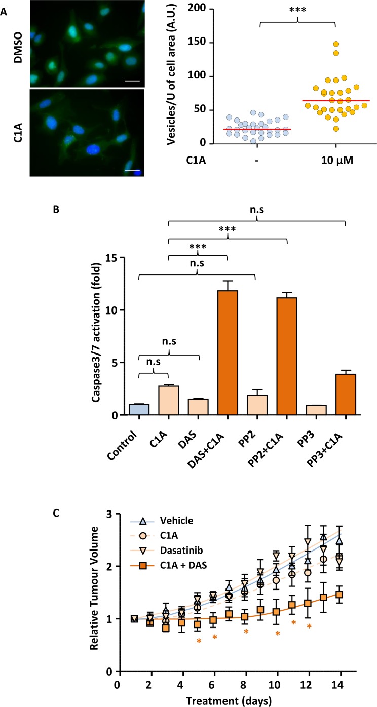
Figure 6. Autophagy inhibition enables dasatinib to reduce tumour volume in A549 mice xenografts.
(A) C1A inhibits autophagy in vitro. U2OS-LC3-GFP cells were treated or not with 10 μM C1A for 24 h and the accumulation of autophagocytic vesicles imaged by fluorescent microscopy and quantified in Image J. Images shown are representative of n = 30/condition. (B) C1A promotes the induction of apoptosis by dasatinib in vitro. A549 cells treated with or without C1A (10 μM), dasatinib (1 μM), PP2 (10 μM) or PP3 (10 μM) for 24 h and subjected to a luminometric caspase3/7 activity assay. (C) Co-administration of C1A and dasatinib prevents A549 xenografts tumour growth in nude mice. A549 cells were injected subcutaneously in nude mice and treatment initiated when tumours reached 50-100 mm3 with or without 20 mg/kg C1A and 75 mg/kg dasatinib by daily oral gavage for 2 weeks. Tumour volumes were determined by caliper measurement. N = 5 per condition. (A–C) Results are representative of experiments performed in triplicate. Statistical analysis by ANOVA with *p < 0.05, **p < 0.01, ***p < 0.005.

