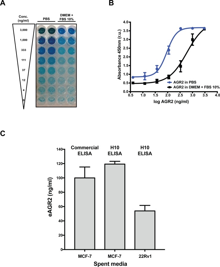
Figure 3. Characterization of a novel in vitro AGR2 ELISA.
(A) Colorimetric detection of AGR2 by H10 ELISA with a commercial antibody in PBS or DMEM with 10% FBS. (B) Quantification of the dynamic range of the AGR2 ELISA in PBS or DMEM with 10% FBS. (C) Detection of eAGR2 from spent media derived from MCF-7 and 22Rv1 cell lines. Comparison of H10 based ELISA and commercial AGR2 ELISA (USCN SEC285Hu) to compare performance of each assay. All data represent three independent biological replicates.

